Fungal classification or taxonomy is usually based on the sexual spores produced by the organisms. This is because the asexual forms of most fungi especially those that are of medical importance are not well known; and those groups of fungi that are of medical importance are sexual spore producers. The kingdom fungi comprise of four (4) or more distinct divisions (i.e. phylum) or groups of fungal organisms classified based on their sexual cycle, spore formation and/or mode of reproduction (i.e. sexual and asexual reproduction).
Kingdom fungi contain diverse species of organisms (including yeasts, moulds and mushrooms amongst others) that have enormous medical, industrial and economic applications. The main fungal classes or divisions are zygomycetes, ascomycetes, basidiomycetes and deuteromycetes. Only deuteromycetes produce conidia (i.e. asexual spores) but sexual spores are produced by fungi in the phylum or division’s zygomycetes, ascomycetes and basidiomycetes. Generally, the classification of fungi is based on:
- The hyphal or hyphae morphology of the fungi (septate or non-septate).
- The type of spore formed by the fungi (conidia or sexual spores).
ZYGOMYCETES (ZYGOMYCOTA)
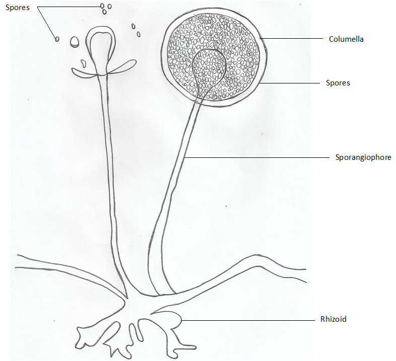
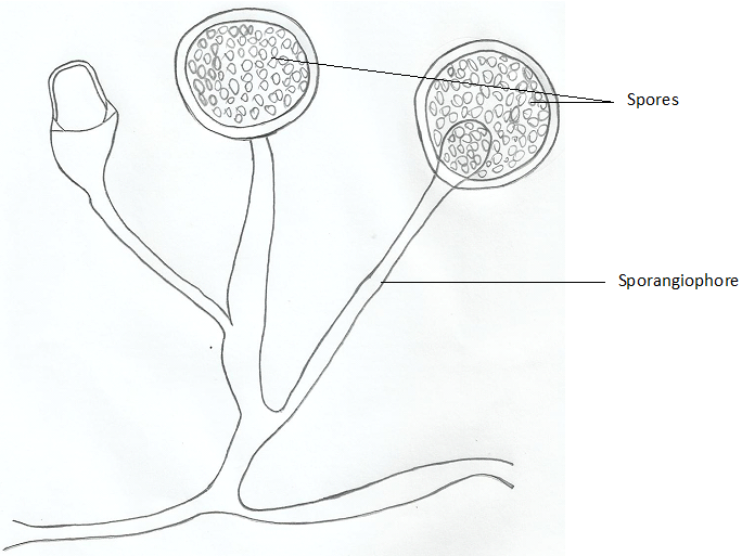
Zygomycetes are commonly known as the bread moulds; and fungi in this division or phylum include Rhizopus species, Mucor species, Absidia and Pilobolus. Fungi in the division Zygomycota undergo both sexual and asexual reproduction. They are usually characterized by the presence of sexual spores, asexual sporangiospores and the presence of septate or aseptate hyphae. Zygomycetes are largely saprophytes, and they live on decaying plant and animal matter in the soil. The hyphae of fungi in the division Zygomycota are mostly aseptate (i.e. without cross-walls) and they are said to be coenocytic or multinucleated in form.
Fungi in this division form asexual spores or conidia through the formation of sporangia (singular: sporangium) which contain numerous sporangiospores but sexual spores known as zygospores are formed via the mating of two haploid nuclei or fusion of morphologically similar gametangia that divide by the process of meiosis and mitosis. Generally, the zygomycetes parasitize plants, animals and humans but fungi in this division are mainly known for their food spoilage activities. Zygomycetes such as Rhizopus species (Figure 1) and Mucor species (Figure 2) are important food spoilage fungi; and they are fast growing fungi with numerous economic importance.
ASCOMYCETES (ASCOMYCOTA)
Ascomycetes are fungi that reproduce sexually via the formation of endogenous ascospores (i.e. sexual spores) normally enclosed in a sac known as the ascus sac. The asci (singular: ascus) are known to contain about four or eight ascospores. Fungi in this division are commonly known as sac fungi, and they contain a large and diverse group of fungi with various economic importance. They also reproduce asexually to produce conidia. Ascomycetes form well developed branching septate hyphae (i.e. hyphae with cross-walls).
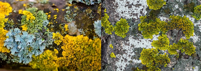
They are ubiquitously found in the soil as saprophytes, and a handful of them parasitize plants, humans and animals. Some ascomycetes are unicellular cells while others exhibit dimorphism. The division Ascomycota includes moulds, mildews and yeasts; and representative fungi in this group are Microsporum, Trichophyton, Piedraia, Blastomyces, Aspergillus, Saccharomyces cerevisiae (baker’s yeast)and Histoplasma species amongst others. Majority of the ascomycetes are causative agents of dermatophytosis in humans (e.g. ringworm).
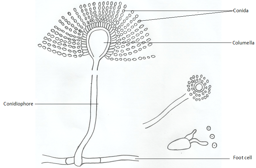
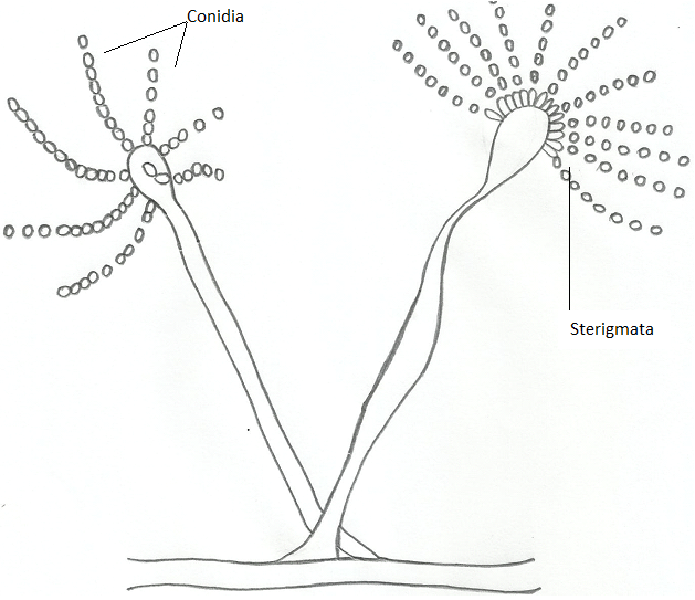
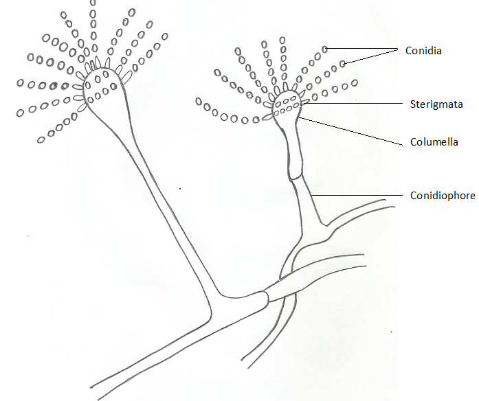
Claviceps purpurea is an ascomycete that parasitizes plants to cause ergot disease; thus causing ergotism (a psychotic and neurological disease) in humans and animals who eat crop plants infested by the fungus. Aspergillus species including A. niger (Figure 3), A. flavus (Figure 4) and A. fumigatus (Figure 5) are important fungal organisms of the division Ascomycota, and some species of Aspergillus (A. flavus in particular) are known to produce potent toxins known as aflatoxinswhich are cancer-causing in nature. A. flavus and other fungi that produce aflatoxins are common contaminants of food especially grains which they infest to produce the potent toxin.
BASIDIOMYCETES (BASIDIOMYCOTA)
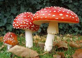
Basidiomycetes are fungi that form sexual spores known as basidiospores. The basidiospores which are usually four in number are generally carried on a club-shaped structure known as the basidium (plural: basidia). Fungi in this division are commonly known as club fungi. Basidiomycetes have septate hyphae like the ascomycetes but their hyphae are more complex in nature. Their basidiospores are enclosed within fruiting bodies known as basidiocarps. Examples of fungi that are basidiomycetes include toadstools, shelf fungi, mushrooms, smut fungi, Cryptococcus neoformans, rust fungi and puffballs amongst others.Fungi in the division basidiomycota possess tremendous economic importance.
For example, the mushrooms in the Agaricus species such as A. campestris and A. bisporus are amongst the important mushrooms used as a source of human food. Mushroom cultivation either for domestic or commercial use is an important aspect of microbiology; and all over the world, mushroom is cultivated and sold because of their perceived medical and health importance. Basidiomycetes are ubiquitously found in the natural environment as saprophytes and parasites of animals and humans as well as plants. Though a handful of the basidiomycetes such as the Agaricus mushrooms are edible and can be eaten as food, some group of mushrooms especially the Amanita species are poisonous and can cause severe clinical condition when eaten.
DEUTEROMYCETES (DEUTEROMYCOTA)
Deuteromycetes are generally known as “imperfect fungi” unlike the ascomycetes, zygomycetes and basidiomycetes which are “true fungi” because they have a sexual cycle of reproduction. This division contains fungi whose teleomorphs (i.e. sexual reproducing forms) discovery is still in the continuum; and they contain a variety of heterogeneous fungal organisms. Deuteromycetes are characterized by an anamorphic stage in which asexual spores or conidia are produced. Fungi in the division deuteromycetes have no known sexual cycle of reproduction, and they mainly reproduce via the formation of conidia or asexual spores.
The division deuteromycota contain many important human pathogens including Candida, Histoplasma, Coccidioides and Paracoccidioides and other dematiaceous fungi. Other imperfect fungi such as Penicillium are important to humans. For example, the antibiotic penicillin is naturally soured from Penicillium species such as P. chrysogenum and P. notatum. It is noteworthy that the division deuteromycetes is only but an artificial classification of fungi (i.e. it is a non-phylogenetic class); and as the sexual stage of organisms in this group is being discovered, the fungi will be regrouped into the correct fungal division or phylum. Generally, the deuteromycetes are ubiquitously found in the environment especially in the soil, and they are important pathogens of plants, animals and humans.
OTHER FUNGAL DIVISIONS
Other fungal divisions include the Chytridiomycetes and Glomeromycetes groups. Chytridiomycetes are simple true fungi commonly known as chytrids; and they include many free-living fungi and those that parasitize plants and animals and as well as protists in the natural environment. Chytrid fungi are ubiquitous in the environment, and they reproduce asexually to form motile zoospores. Sexual reproduction in chytridiomycetes results in the formation of zygote that eventually becomes a sporangium. Species of fungi in the division chytridiomycetes include Allomyces and Batrachochytrium which causes mycoses in amphibians especially frogs.
Glomeromycetes are known for their symbiotic associations with plant roots. They form endomycorrhizae (a fungal-plant root association) with herbaceous plants; and their main role in this association is to help the plant roots to absorb nutrients and water from the soil. Glomeromycetes only form asexual spores; and the penetration of the plant cell walls by the fungal hyphae lead to the formation of arbuscules or swollen structures around the plant roots, a phenomenon characteristic of the glomeromycetes fungi-plant relationship.
References
Anaissie E.J, McGinnis M.R, Pfaller M.A (2009). Clinical Mycology. 2nd ed. Philadelphia, PA: Churchill Livingstone Elsevier. London.
Beck R.W (2000). A chronology of microbiology in historical context. Washington, D.C.: ASM Press.
Black, J.G. (2008). Microbiology: Principles and Explorations (7th ed.). Hoboken, NJ: J. Wiley & Sons.
Brooks G.F., Butel J.S and Morse S.A (2004). Medical Microbiology, 23rd edition. McGraw Hill Publishers. USA.
Brown G.D and Netea M.G (2007). Immunology of Fungal Infections. Springer Publishers, Netherlands.
Calderone R.A and Cihlar R.L (eds). Fungal Pathogenesis: Principles and Clinical Applications. New York: Marcel Dekker; 2002.
Chakrabarti A and Slavin M.A (2011). Endemic fungal infection in the Asia-Pacific region. Med Mycol, 9:337-344.
Champoux J.J, Neidhardt F.C, Drew W.L and Plorde J.J (2004). Sherris Medical Microbiology: An Introduction to Infectious Diseases. 4th edition. McGraw Hill Companies Inc, USA.
Chemotherapy of microbial diseases. In: Chabner B.A, Brunton L.L, Knollman B.C, eds. Goodman and Gilman’s The Pharmacological Basis of Therapeutics. 12th ed. New York, McGraw-Hill; 2011.
Chung K.T, Stevens Jr., S.E and Ferris D.H (1995). A chronology of events and pioneers of microbiology. SIM News, 45(1):3–13.
Germain G. St. and Summerbell R (2010). Identifying Fungi. Second edition. Star Pub Co.
Ghannoum MA, Rice LB (1999). Antifungal agents: Mode of action, mechanisms of resistance, and correlation of these mechanisms with bacterial resistance. Clin Microbiol Rev, 12:501–517.
Gillespie S.H and Bamford K.B (2012). Medical Microbiology and Infection at a glance. 4th edition. Wiley-Blackwell Publishers, UK.
Larone D.H (2011). Medically Important Fungi: A Guide to Identification. Fifth edition. American Society of Microbiology Press, USA.
Levinson W (2010). Review of Medical Microbiology and Immunology. Twelfth edition. The McGraw-Hill Companies, USA.
Madigan M.T., Martinko J.M., Dunlap P.V and Clark D.P (2009). Brock Biology of Microorganisms, 12th edition. Pearson Benjamin Cummings Inc, USA.
Mahon C. R, Lehman D.C and Manuselis G (2011). Textbook of Diagnostic Microbiology. Fourth edition. Saunders Publishers, USA.
Discover more from Microbiology Class
Subscribe to get the latest posts sent to your email.