
Digital PCR (dPCR) is a method of quantifying nucleic acid (DNA or RNA) targets without standard curves by dividing the bulk reaction into thousands of smaller, independent reactions. It is an advanced molecular biology method that measures DNA, RNA, or cDNA by splitting a single sample into thousands of tiny, separate reaction spaces.
Polymerase Chain Reaction (PCR) is one of the most transformative molecular biology techniques ever developed. Since its invention in the 1980s, PCR has revolutionized how scientists detect and quantify nucleic acids. Over the decades, PCR technology has evolved through three main generations: conventional PCR, quantitative real-time PCR (qPCR), and digital PCR (dPCR). Each successive generation has improved on precision, quantification accuracy, and sensitivity.
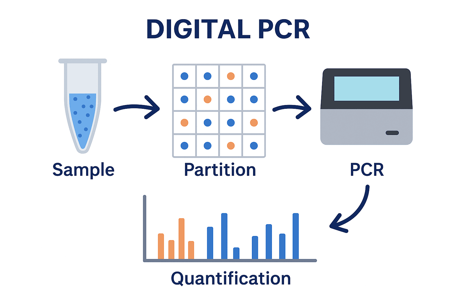
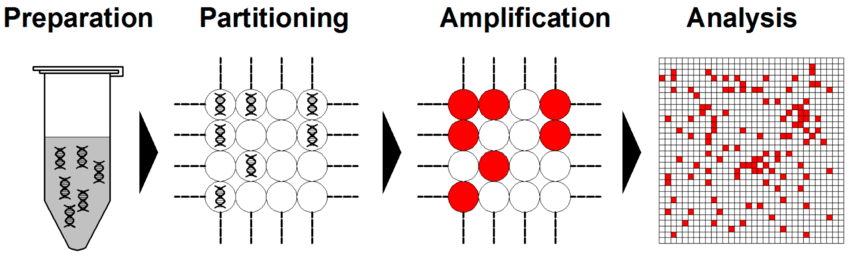
dPCR represents the third generation of PCR-based quantification methods (Figure 1). It enables absolute quantification of nucleic acids without the need for calibration curves or reference standards as is the case with qPCR. Unlike qPCR, which measures the accumulation of fluorescence during amplification, dPCR divides a sample into thousands to millions of partitions and performs PCR in each partition separately. The partitions are classified as positive(if amplification occurs) or negative (if no amplification occurs). The number of target DNA molecules in the original sample is then determined statistically, using Poisson distribution principles.
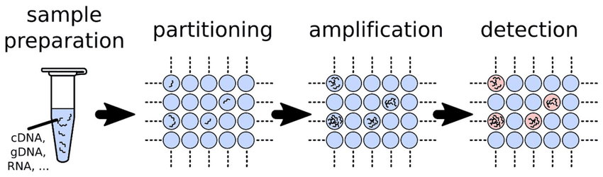
In terms of its working principle, dPCR works by partitioning a DNA sample into thousands of individual reactions, such as oil droplets or microwells, followed by PCR amplification within each partition. After amplification, each partition is analysed for a fluorescent signal indicating the presence or absence of the target sequence. The number of positive and negative partitions is then analysed using Poisson statistics to account for partitions containing more than one target molecule, enabling direct and highly precise absolute quantification of the target DNA molecules in the original sample.
The main benefits of dPCR are its absolute quantification without the need for a standard curve, high sensitivity for detecting rare targets or very low-abundance genetic material, and improved robustness and accuracy in samples containing PCR inhibitors that can compromise conventional qPCR assays.
This innovation in dPCR offers extremely precise and sensitive nucleic acid quantification which allows for the detection of single-copy targets or rare mutations among a vast excess of wild-type DNA. Over the last decade, dPCR has gained traction in clinical diagnostics, environmental monitoring, cancer research, infectious disease detection, and genetic studies.
Principle of digital PCR
The concept underlying digital PCR (dPCR) is simple yet powerful. Unlike conventional PCR and quantitative PCR (qPCR), which amplify and quantify DNA in a bulk reaction, dPCR partitions a DNA sample into thousands of physically separated micro-reactions before amplification. Ideally, each partition contains either no target DNA molecule or one or more copies of the target sequence. Each partition then functions as an independent PCR micro-reactor, in which amplification proceeds under the same principles as conventional PCR.
Following amplification, partitions are classified as positive or negative according to the presence or absence of the target-specific fluorescence signal. The proportion of positive and negative partitions is then used to calculate the absolute concentration of the target DNA using Poisson statistics, which account for the possibility that some partitions contain more than one target molecule. dPCR therefore does not require a standard curve for quantification, unlike qPCR.
This partitioning-based approach provides several advantages for environmental and microbial applications. Because quantification is based on endpoint detection and the number of positive partitions, dPCR can provide highly precise absolute quantification of low-abundance targets, including antibiotic resistance genes (ARGs), even when target concentrations are close to the detection limit. It is also comparatively less sensitive to amplification-efficiency differences and certain matrix effects than qPCR, which is particularly relevant for complex environmental samples such as agricultural soils that may contain PCR inhibitors.
History of dPCR: from molecular counting to digital precision
dPCR emerged from a simple but transformative idea: instead of measuring DNA amplification as one bulk reaction, divide a sample into thousands of tiny reactions and determine which partitions contain the target. The conceptual roots of this approach appeared in the early 1990s, when researchers demonstrated that nucleic-acid molecules could be quantified through limiting dilution and endpoint amplification.
A major advance came in 1999, when Bert Vogelstein and Kenneth Kinzler introduced the term digital PCR and demonstrated its potential for detecting rare genetic alterations (Figure 1). Vogelstein and Kinzler both formally introduced the technique and coined the exact term “Digital PCR” in a landmark 1999 paper published in the Proceedings of the National Academy of Sciences (PNAS). Their innovative work established a distinctive principle: molecular concentration could be inferred from the proportion of positive and negative reactions, using Poisson statistics rather than conventional amplification curves. This transformed PCR from an analogue measurement into a molecular counting technique.
During the 2000s, technological development made dPCR increasingly practical. Microfluidic systems, nanowell arrays, and improved fluorescence chemistry enabled researchers to partition samples reproducibly and analyze large numbers of individual reactions. These innovations gave rise to commercial platforms capable of performing highly sensitive nucleic-acid measurements.
The field expanded rapidly as dPCR demonstrated advantages in applications where small differences mattered. Researchers began using it for rare-variant detection, copy-number analysis, pathogen quantification, gene-expression studies, and environmental DNA measurement. Unlike quantitative PCR, dPCR does not require a standard curve for absolute quantification and can provide greater tolerance to certain amplification inhibitors.
In the 2010s, droplet-based systems further accelerated adoption by partitioning reactions into thousands of water-in-oil droplets. By the 2020s, dPCR had matured into a precision molecular technology used in research, diagnostics, biotechnology, and quality control. The history of dPCR reflects a broader shift in molecular biology from estimating population-level signals toward counting individual molecular events with increasing statistical and analytical precision.
Partitioning of the sample in dPCR experiment
In dPCR, the sample mixture containing the nucleic acid template, primers, probes, nucleotides, and polymerase is divided into many individual partitions, usually numbering from 10,000 to several million (Figure 2). Partitioning can be achieved using droplets, microfluidic chambers, or microwells. The objective of partitioning is to distribute DNA molecules randomly across partitions, following a Poisson distribution, such that most partitions contain zero or onetarget molecule. The partitioning step in dPCR is crucial because it converts the analog fluorescence signal (as in qPCR) into binary digital signals either “1” (target detected) or “0” (no target).
Cao et al. (2020). Digital PCR as an Emerging Tool for Monitoring of Microbial Biodegradation. Molecules, 25(3), 706. Quan et al. dPCR: A Technology Review. Sensors 2018, 18, 1271.
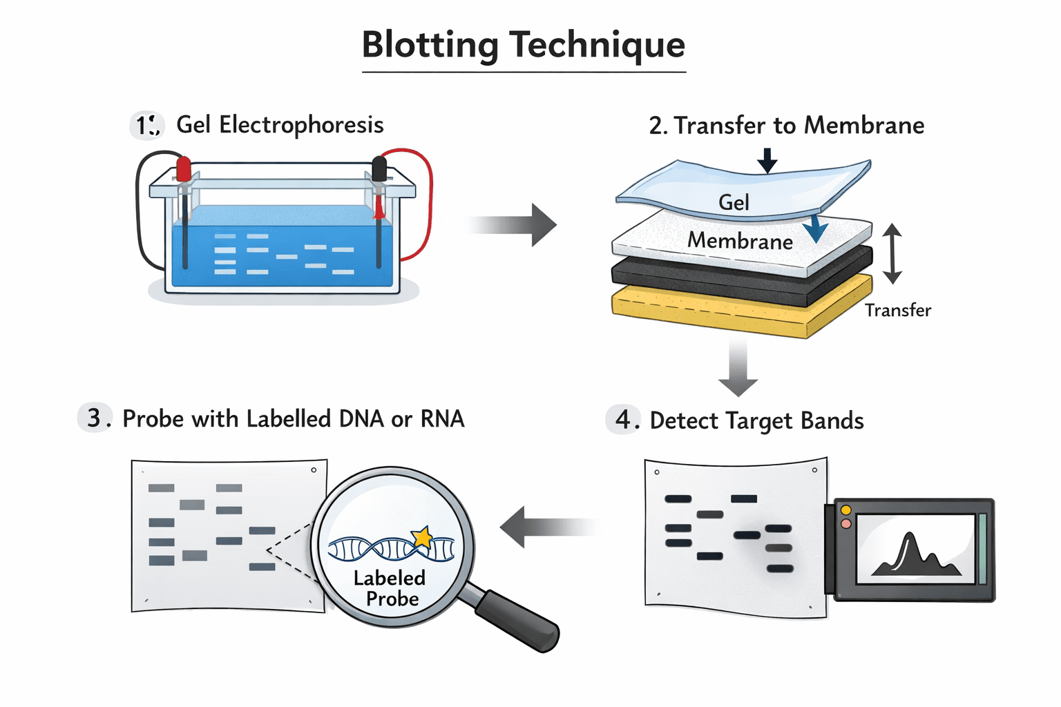
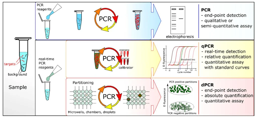
In conventional PCR, the amplification products are analyzed at the end of the reaction (end-point PCR) by gel electrophoresis and detected after fluorescent staining (Figure 3). qPCR and dPCR use the same amplification reagents and fluorescent labeling systems. In qPCR, the amount of amplified DNA is measured at each cycle during the PCR reaction, i.e., in real-time. The ‘absolute’ quantity of target sequence is interpolated using a standard curve generated with a calibrator.
But in dPCR, the sample is first partitioned into many sub-volumes (in microwells, chambers or droplets) such that each partition contains either a few or no target sequences. After PCR, the proportion of amplification-positive partitions serves to calculate the concentration of the target sequence using Poisson’s statistics. dPCR is a method of absolute nucleic acid quantification that hinges on the detection of end-point fluorescent signals and the enumeration of binomial events (absence (0) or presence (1) of fluorescence in a partition).
This statistical foundation permits to identify the parameters that constraint the performance metrics of this analytical method. dPCR is theoretically advantageous over qPCR given effective means to perform sample partitioning and target amplification of single molecules.
Quan et al. dPCR: A Technology Review. Sensors 2018, 18, 1271. https://doi.org/10.3390/s18041271
PCR amplification process in dPCR
Following partitioning, each reaction compartment in dPCR functions as an independent microreaction vessel and undergoes repeated cycles of denaturation, primer annealing, and DNA extension. Although the thermal cycling principle is comparable to conventional PCR, dPCR distinguishes itself by physically separating the reaction mixture into thousands of individual partitions before amplification.
During thermal cycling, double-stranded DNA is first denatured into single strands. Primers then anneal to complementary sequences within the target region, allowing the DNA polymerase to extend the primers and synthesize new strands. These steps are repeated through multiple cycles, resulting in exponential amplification of the target sequence in partitions that initially received one or more copies of the DNA template.
Target-specific fluorescent probes can be incorporated into the reaction to enable direct detection of amplification. For example, TaqMan probes contain a fluorescent reporter and a quencher. When the probe binds to its complementary target during amplification, the DNA polymerase cleaves the probe through its 5′ nuclease activity. This separates the reporter from the quencher, producing an increase in fluorescence that can be measured after amplification.
Partitions containing the target sequence therefore develop a measurable fluorescence signal and are classified as positive. In contrast, partitions that did not receive the target remain below the fluorescence threshold and are designated negative. The resulting pattern of positive and negative partitions provides the fundamental digital readout of dPCR.
Because each partition is analyzed individually, the technique converts amplification into a binary endpoint measurement rather than relying primarily on the continuous fluorescence curves used in real-time quantitative PCR. The proportion of positive partitions can subsequently be used to determine the absolute concentration of the target nucleic acid through Poisson-based statistical analysis, without requiring a conventional calibration curve.
Detection and quantification process in dPCR
Following the amplification stage, each partition is individually analyzed using an optical detection system that measures the fluorescence generated by the amplified target. The detector distinguishes partitions according to their fluorescence intensity, allowing them to be classified as either positive or negative. Positive partitions exhibit a fluorescence signal above the predefined threshold, indicating the presence of the target nucleic acid, whereas negative partitions show little or no detectable fluorescence, suggesting that the target is absent.
The instrument records the total number of positive and negative partitions and uses these measurements to determine the proportion of partitions containing the target. Because the partitions function as independent reactions, the resulting positive-to-negative distribution provides the basis for estimating the absolute concentration of the target molecule. Statistical analysis, typically based on Poisson distribution, is then applied to account for partitions containing multiple target molecules and calculate the final concentration.
The fraction of positive partitions (p) allows estimation of the average number of target molecules per partition (λ) using the Poisson equation:
λ = – ln (1 – p)
From λ and the known partition volume, the absolute concentration of the target in the original sample can be calculated. This statistical correction accounts for partitions that may contain more than one target molecule.
λ = average number of target molecules per partition
P = Fraction of positive partitions. It is the proportion of partitions showing amplification (fluorescence). P(0) is the probability that a partition received zero molecules
Ln = Natural logarithm. Used to reverse the exponential relationship in the Poisson model.
(1 – p) = Fraction of negative partitions. Represents the proportion of partitions that received zero target molecules.
–ln(1 – p) = Correction factor. Converts the observed positive fraction into an estimate of the average molecules per partition, accounting for multiple occupancy.
dPCR and the Poisson principle: converting partition counts into absolute abundance
dPCR provides a highly precise approach for quantifying low-abundance targets such as antibiotic resistance genes (ARGs). Unlike conventional qPCR, which estimates target abundance from amplification cycles relative to a standard curve, dPCR partitions a DNA sample into thousands of independent microreactions. Following amplification, each partition is classified as positive (target detected) or negative (target not detected). The final concentration is calculated from the distribution of positive and negative partitions.
The statistical basis of dPCR is the Poisson distribution, which describes the probability of randomly distributing a given number of target molecules among a large number of partitions. Importantly, partitioning is random, meaning that some partitions contain no target molecules, some contain one, and some may contain two or more. Therefore, the number of positive partitions does not directly equal the number of target molecules. A partition that is positive only tells us that at least one target molecule was present.
The proportion of negative partitions is particularly informative because the probability of observing zero target molecules follows the Poisson relationship P(0) = e⁻λ, where λ represents the average number of target molecules per partition. Consequently, λ can be estimated from the fraction of negative partitions as λ = −ln(P(0)). This Poisson correction accounts for partitions containing multiple target molecules and converts the observed positive/negative partition pattern into an estimate of the absolute concentration of the target.
This approach is particularly valuable for ARG analysis because it provides absolute copy-number estimates without requiring an external calibration curve. dPCR can therefore detect and quantify small differences in ARG abundance across mixture treatments, doses, soils, and time points, providing an independent quantitative layer that complements sequencing-based relative abundance data.
Digital counting in dPCR
dPCR uses a partition-based quantification of nucleic acid targets unlike other PCR technologies. It introduces a fundamentally different approach to nucleic acid quantification by converting a complex molecular measurement into a binary counting process. During the assay, a reaction mixture is distributed across thousands of discrete partitions, with each partition containing either zero, one, or several copies of the target molecule. Following amplification, every partition is identified as either positive, indicating target detection, or negative, indicating its absence.
The resulting pattern of positive and negative partitions provides the basis for absolute quantification. Rather than measuring fluorescence against a continuously varying standard, dPCR determines target abundance from the proportion of partitions that contain the amplified sequence. Statistical models, particularly Poisson distribution principles, account for the possibility that individual partitions initially contain more than one target molecule. This enables estimation of the original target concentration from the observed partitioning pattern.
A major advantage of this digital framework applicable only to dPCR is its independence from conventional calibration curves as is often the case in qPCR. Quantification does not require a series of external standards with known concentrations, nor does it depend on reference genes to establish relative abundance. Consequently, dPCR can provide direct estimates of target copy number or concentration within the analyzed sample.
This counting-based strategy is particularly valuable when precise measurement of small differences is required. It also supports the detection of low-frequency genetic variants, copy-number alterations, and rare molecular targets. By transforming molecular detection into a statistically interpretable counting system, dPCR provides a robust framework for obtaining absolute measurements of nucleic acid targets with minimal reliance on external quantification references.
Workflow of digital PCR
dPCR is a highly sensitive molecular technique designed to detect and quantify nucleic acids through the physical partitioning of a reaction mixture into numerous independent microreactions. Unlike conventional PCR, which generally measures amplification across a bulk reaction, dPCR determines the presence or absence of target molecules within individual partitions, enabling absolute quantification without requiring a standard curve.
The dPCR workflow begins with careful sample preparation, which is fundamental to obtaining reliable results. Biological samples must be appropriately collected, preserved, and processed to extract DNA or RNA while minimizing degradation and potential inhibitors. The extracted nucleic acid is then assessed for quality and concentration before being incorporated into the reaction mixture. Proper handling at this stage helps maintain consistency between samples and reduces experimental variability.
The next stage involves reaction mixture preparation, where the template, primers, probes or fluorescent dyes, polymerase, nucleotides, buffer components, and other required reagents are combined in appropriate proportions. The prepared mixture is subsequently introduced into a partitioning system that divides the reaction into thousands of discrete compartments. Each partition may contain zero, one, or multiple target molecules.
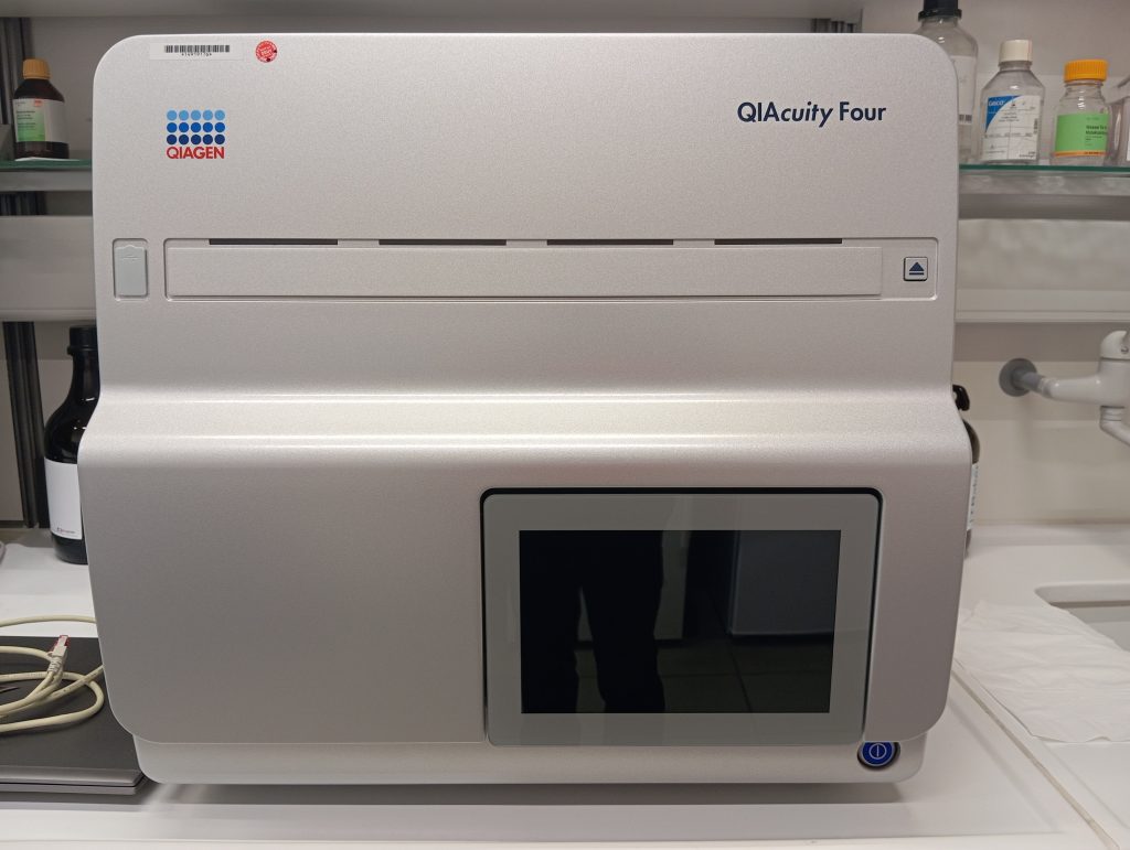
Following partitioning, the reactions undergo thermal cycling in a dPCR machine as seen in Figure 1, allowing target sequences to be amplified. After amplification, fluorescence from individual partitions is measured and classified according to whether the target is detected. Specialized software then interprets the positive and negative partitions using statistical principles, commonly based on Poisson distribution, to calculate the absolute concentration of the target nucleic acid.
The dPCR workflow integrates controlled sample preparation, precise reaction assembly, partition generation, amplification, fluorescence detection, and computational analysis into a sequential process that supports sensitive and reproducible molecular quantification.
Sample preparation and reaction setup for dPCR experiments
Successful dPCR begins with the preparation of a clean, intact, and representative nucleic acid template. DNA or RNA is initially isolated from the biological or environmental material of interest, such as blood, tissue, cultured cells, soil, or water samples, using an appropriate nucleic acid extraction procedure. Commercial extraction systems, including those manufactured by Qiagen and other established suppliers, can provide reproducible recovery and reduce variability between samples.
The quality of the extracted nucleic acid is particularly important for dPCR because inhibitory substances can interfere with enzymatic amplification and compromise accurate target detection. Common contaminants include proteins, phenolic compounds, ethanol residues, polysaccharides, and humic substances derived from environmental samples. These compounds may reduce polymerase efficiency, alter fluorescence signals, or interfere with the generation and interpretation of reaction partitions. Therefore, extracted DNA should be evaluated for concentration, purity, and integrity before proceeding with the dPCR assay. When necessary, additional purification or dilution can be performed to minimize the effect of residual inhibitors.
For RNA-based targets, the extracted RNA cannot generally be amplified directly by conventional DNA-dependent polymerase systems. Instead, RNA is first converted into complementary DNA (cDNA) through a reverse-transcription step. The resulting cDNA serves as the template for digital amplification. When reverse transcription and digital PCR are incorporated into the same analytical workflow, the method is commonly referred to as reverse transcription digital PCR (RT-dPCR). Careful handling of RNA is essential because degradation can reduce template availability and consequently affect quantitative measurements.
In terms of the reaction setup of a dPCR experiment, once the nucleic acid template has been prepared, the dPCR reaction mixture is assembled using components required for target-specific amplification and fluorescence-based detection. A typical reaction contains the DNA or cDNA template, forward and reverse sequence-specific primers, and a fluorescent hydrolysis probe such as a TaqMan-type probe. Additional components include deoxynucleoside triphosphates (dNTPs), magnesium chloride (MgCl₂), DNA polymerase, and an optimized reaction buffer.
Hot-start DNA polymerases are frequently selected because they remain inactive during reaction preparation and become activated under the initial thermal conditions, helping to reduce nonspecific amplification. Although the chemical composition of a dPCR reaction resembles that of a quantitative PCR (qPCR) mixture, the analytical framework differs substantially. dPCR does not depend on a conventional amplification curve, external calibration standards, or a reference dye for absolute target quantification. Instead, the reaction is partitioned into numerous individual microreactions, allowing target molecules to be identified according to the distribution of positive and negative partitions. This partition-based strategy enables direct and highly precise nucleic acid quantification.
Partition generation in dPCR experiment
Partition generation is a fundamental stage in dPCR, transforming a single reaction mixture into a large population of physically separated microreactions. Each partition functions as an independent reaction environment in which target nucleic acid molecules may be present or absent. This compartmentalization enables the binary detection of amplification signals and provides the statistical foundation for absolute target quantification.
Different dPCR platforms employ distinct partitioning strategies. Droplet-based systems use an emulsification process to disperse the PCR mixture into thousands of water-in-oil droplets. Each droplet serves as an isolated reaction chamber, with systems such as the Bio-Rad QX200 producing highly consistent droplet populations. Chip-based systems rely on microfluidic architectures to distribute the reaction mixture across numerous individually sealed microchambers. The QuantStudio Absolute Q, for example, uses an integrated partitioning format to create discrete reaction compartments. Microwell-based systems employ arrays containing thousands of miniature wells, with each well accommodating a defined volume of the reaction mixture and functioning as a separate amplification unit.
The efficiency and consistency of partition generation directly influence dPCR performance. Ideally, partitions should exhibit minimal variation in volume, geometry, and distribution, because substantial heterogeneity can affect the interpretation of fluorescence-positive and fluorescence-negative populations. Efficient partitioning also minimizes cross-contamination between reaction compartments and promotes reliable segregation of individual target molecules.
Following amplification, the proportion of positive and negative partitions is used to estimate the concentration of the target through Poisson-based statistical analysis. Consequently, partition generation is not merely a sample-preparation step; it establishes the physical and statistical framework that allows dPCR to achieve highly sensitive and quantitative molecular measurements. Uniform, well-defined partition populations therefore provide the foundation for accurate absolute quantification across diverse dPCR applications.
Thermal cycling in dPCR
Thermal cycling is a fundamental stage of digital polymerase chain reaction (dPCR), enabling the selective amplification of target DNA molecules within thousands of physically separated partitions. Although the temperature profile resembles that of conventional PCR, dPCR performs amplification independently inside each partition, creating a highly compartmentalized reaction environment.
During the denaturation phase, elevated temperatures disrupt the hydrogen bonds between complementary DNA strands, producing single-stranded templates. The temperature is then reduced to promote primer annealing, allowing sequence-specific primers to bind to their complementary target regions. During extension, the DNA polymerase synthesizes new strands by incorporating complementary nucleotides, thereby duplicating the target sequence.
These three stages denaturation, annealing, and extension are repeatedly cycled to generate sufficient copies of the target DNA. As amplification proceeds, partitions containing the target sequence accumulate detectable fluorescent signals, whereas partitions lacking the target remain non-fluorescent or below the detection threshold. Each partition functions as an autonomous microscopic PCR reactor with its own amplification outcome.
The repeated thermal transitions therefore transform individual DNA molecules into measurable positive or negative signals, providing the molecular basis for dPCR quantification. Careful control of cycle number, temperature, ramp rate, and reaction chemistry is essential for consistent amplification and reliable partition classification.
Fluorescence detection in dPCR
Following target amplification, the partitioned reaction mixture undergoes fluorescence-based interrogation to determine whether individual partitions contain amplified nucleic acid. This optical readout represents a critical stage of dPCR, as the measured fluorescence signal provides the basis for distinguishing amplification-positive partitions from those without detectable target.
In droplet-based dPCR platforms, the generated droplets are transported individually through an optical detection region. As each droplet passes a laser or excitation source, fluorescent reporters emit signals that are captured by dedicated detectors. The resulting measurements are converted into fluorescence intensity values for each partition. In contrast, chip-based dPCR systems generally employ imaging technologies to examine an array of spatially separated reaction chambers simultaneously. A scanner or imaging module records fluorescence across the complete partition array, enabling rapid acquisition of thousands of individual measurements.
The collected fluorescence data are subsequently processed using predefined or algorithmically determined thresholds. Partitions exhibiting fluorescence above the established threshold are designated as amplification-positive, whereas those below it are classified as negative. This binary partitioning transforms continuous fluorescence measurements into discrete molecular counts, which can then be used for absolute target quantification through statistical analysis of partition occupancy.
Data analysis of dPCR output
dPCR converts molecular detection into discrete digital measurements by partitioning a reaction into numerous independent microreactions. Following amplification, each partition is classified according to the presence or absence of the target sequence, generating positive and negative event counts. Specialized analytical software then interprets these binary measurements using Poisson statistical modeling. This correction accounts for the probability that individual partitions may contain more than one target molecule, enabling calculation of the absolute concentration of the analyte without requiring an external calibration curve.
The resulting concentration reflects the estimated number of target molecules within a defined reaction volume and provides a direct quantitative measure of nucleic-acid abundance. Depending on the experimental objective, dPCR data can be normalized and expressed in several forms. Common reporting units include copies per microliter of reaction mixture, copies per nanogram of extracted DNA, and copies per cell in genomic applications. Such normalization facilitates comparison among samples with different DNA inputs and enables quantitative interpretation of target abundance in applications such as pathogen detection, mutation analysis, copy-number determination, and genomic characterization.
Types and advantages of dPCR platforms
dPCR technologies are classified according to the method used to partition PCR mixtures into discrete reaction compartments. The major platforms include droplet digital PCR (ddPCR), chip-based digital PCR, and microwell-array dPCR. In each approach, partitioning enables individual reactions to be analyzed independently, supporting sensitive and precise nucleic-acid quantification.
Droplet Digital PCR (ddPCR) is the most widely adopted dPCR format. The reaction mixture is divided into tens of thousands of nanoliter-sized droplets within a water-in-oil emulsion, with each droplet functioning as an independent PCR reactor. The workflow involves droplet generation, PCR amplification, fluorescence-based droplet reading, and Poisson statistical analysis to determine target concentration. Systems such as the Bio-Rad QX200 and QX One provide high partition numbers, strong analytical precision, and improved tolerance to PCR inhibitors. However, ddPCR requires emulsification and droplet stabilization and generally relies on endpoint fluorescence measurements.
Chip-based digital PCR uses microfluidic technology to distribute samples across thousands of individual reaction chambers within a solid chip or cartridge. Platforms such as Thermo Fisher’s QuantStudio Absolute Q and Stilla’s Naica System provide highly uniform partitions without oil emulsification. This format can simplify sample processing and facilitate multiplex analysis, although specialized chips can be relatively expensive and may offer less scalability than droplet-based systems.
Microwell-array dPCR separates reaction mixtures into physically defined wells, with fluorescence commonly measured through imaging. Systems such as Fluidigm BioMark HD demonstrate the potential of this format for visual partition verification and multiplex analysis. Its limitations include fewer partitions than many droplet systems and higher costs per reaction.
Across these platforms, dPCR provides several analytical advantages over conventional PCR and qPCR. It enables absolute quantification by determining target molecule concentrations without standard curves. Partitioning also enhances sensitivity and precision, allowing detection of rare targets and low-frequency variants. Endpoint measurement can reduce the impact of certain PCR inhibitors, improving performance with complex samples such as blood and environmental materials. In addition, dPCR offers strong reproducibility, supports multiplex detection using multiple fluorescent probes, and is particularly valuable for rare-event analysis, including low-abundance pathogens, viral variants, and rare genetic mutations.
Limitations of dPCR
dPCR offers high analytical sensitivity and precise nucleic-acid quantification, but several practical limitations restrict its broader application. High cost remains a major concern because dPCR instruments, specialized consumables, and partitioning reagents are generally more expensive than conventional qPCR systems. Throughput limitations also make dPCR less suitable for large-scale screening or studies involving extensive sample numbers.
In terms of quantitative capacity, dPCR typically provides a dynamic range of approximately 4-5 orders of magnitude, which is narrower than the 7-8 log range commonly achievable with qPCR. The technique also involves greater operational complexity, as samples must undergo partitioning before fluorescence-based endpoint detection, requiring specialized instrumentation and workflow optimization.
Data processing is computationally intensive compared with standard qPCR analysis. Accurate concentration estimates depend on statistical modeling, particularly Poisson-based correction, to account for the distribution of target molecules across partitions. This necessitates dedicated analytical software and appropriate interpretation of partition-level data. These factors can increase experimental expense, processing time, technical demands, and barriers to implementation, particularly in high-throughput or resource-limited laboratory settings.
Comparison of dPCR with RT-qPCR
The evolution of nucleic acid quantification has introduced dPCR as a complementary approach to established real-time quantitative PCR (RT-qPCR). Although both methods rely on amplification-based detection, they differ substantially in reaction partitioning, signal interpretation, quantification strategy, sensitivity, and tolerance to experimental variation (Table 1).
Table 1. Differences between RT-qPCR and dPCR
| Feature | RT-qPCR | Digital PCR |
| Quantification Basis | Measures fluorescence during the exponential phase of PCR. Quantification is relative to standard curves. | Measures binary fluorescence (positive/negative) at endpoint, followed by Poisson analysis for absolute quantification. |
| Detection | Real-time monitoring using Ct (cycle threshold) values. | Endpoint fluorescence detection after amplification. |
| Calibration | Requires external standard curves. | No calibration required; provides absolute copy number. |
| Quantitative Range | Broad (up to 8 orders of magnitude). | Narrower (typically 4–5 orders of magnitude). |
dPCR enables absolute target quantification through endpoint analysis of partitioned reactions, whereas RT-qPCR generally determines relative or absolute quantities from fluorescence measurements during amplification and calibration against standards. The following table outlines key distinctions between the two platforms, providing a structured comparison of their underlying principles, workflows, analytical characteristics, and major applications.
Applications of dPCR
dPCR has emerged as a versatile platform for highly sensitive and absolute nucleic-acid quantification across biomedical, environmental, forensic, and agricultural research. By partitioning DNA or RNA samples into numerous independent reactions and digitally determining positive and negative partitions, dPCR enables precise target measurement without requiring external reference standards. Its strong analytical performance is particularly advantageous for low-abundance targets, complex samples, and applications affected by PCR inhibitors.
In clinical diagnostics, dPCR supports the detection of rare cancer-associated mutations, including alterations in KRASand EGFR, while enabling sensitive quantification of circulating tumor DNA for minimal residual disease assessment. It also facilitates reliable copy-number analysis in genetic disorders. In infectious disease research, dPCR provides absolute measurements of viral and bacterial loads, including low-level targets that may be difficult to quantify using conventional methods.
Environmental microbiology benefits from dPCR for monitoring pathogens, microbial communities, and antimicrobial-resistance genes in soil, water, and wastewater, particularly when inhibitory substances compromise amplification. In food safety and agriculture, the technique enables trace-level identification of genetically modified organisms, allergens, and foodborne pathogens. Virological applications include sensitive viral RNA quantification and resolution of borderline amplification results. Additionally, its tolerance of degraded or limited DNA makes dPCR valuable in forensic investigations, ancient-DNA research, and biodiversity studies, where conventional quantification can be challenging.
References
Deharvengt, S. J., Petersen, L. M., Jung, H.-S., & Tsongalis, G. J. (2020). Nucleic acid analysis in the clinical laboratory. In W. Clarke & M. A. Marzinke (Eds.), Contemporary practice in clinical chemistry (4th ed., pp. 215–234). Academic Press.
Nazir, S. (2023). Medical diagnostic value of digital PCR (dPCR): A systematic review. Biomedical Engineering Advances, 6, 100092.
Quan, P. L., Sauzade, M., & Brouzes, E. (2018). dPCR: A technology review. Sensors, 18(4), 1271.
Wainman, L. M., Sathyanarayana, S. H., & Lefferts, J. A. (2024). Applications of digital polymerase chain reaction (dPCR) in molecular and clinical testing. The Journal of Applied Laboratory Medicine, 9(1), 124–137.
Wang, H., Liu, B., Bian, F., & Zhao, Y. (2026). Developing digital PCR technologies based on wettable substrates. Analyst, 151(17), 4878–4901.
Vogelstein B, Kinzler KW (1999). Digital PCR. Proc Natl Acad Sci USA, 96(16):9236–9241.
Zhang, L., Parvin, R., Fan, Q., & Ye, F. (2022). Emerging digital PCR technology in precision medicine. Biosensors and Bioelectronics, 211, 114344.
QIAGEN. (n.d.). QIAcuity digital PCR system. QIAGEN. www.qiagen.com/de-de/applications/digital-pcr
Discover more from Microbiology Class
Subscribe to get the latest posts sent to your email.