Blotting is a fundamental molecular biology technique that enables the transfer of biological macromolecules, such as nucleic acids and proteins, from a gel medium onto a solid support, typically a chemically reactive membrane. This process facilitates subsequent detection, quantification, and characterization of the molecules, allowing researchers to study their presence, size, abundance, and interactions with high specificity. The technique is widely used in laboratories worldwide for applications ranging from gene expression analysis to protein identification and diagnostics.
Blotting serves as a bridge between molecular separation and detailed molecular analysis. By enabling the stable transfer of biological molecules from gels to membranes and providing a platform for precise detection, blotting techniques form an essential toolkit in molecular biology. Mastery of these techniques allows researchers to interrogate complex biological systems with accuracy and efficiency, supporting discoveries in genetics, proteomics, and biomedical research.
At its core, blotting involves several key steps. First, the molecules of interest are separated according to size using gel electrophoresis. In this step, nucleic acids (DNA or RNA) or proteins are loaded into a gel matrix commonly agarose for nucleic acids and polyacrylamide for proteins and subjected to an electric field. Smaller molecules migrate faster, while larger molecules move more slowly, creating a size-based separation pattern. Once separation is achieved, the gel containing the resolved molecules is brought into contact with a membrane. This membrane is typically made from nitrocellulose or polyvinylidene difluoride (PVDF), both of which have high affinity for biomolecules and facilitate stable immobilization.
The transfer process, which can be performed via capillary action, vacuum, or electroblotting, ensures that the molecules are effectively moved from the gel onto the membrane without losing their relative positions. Capillary blotting relies on the passive movement of buffer solution from the gel to the membrane, carrying the biomolecules along. Electroblotting, on the other hand, uses an electric field to actively drive charged molecules, such as DNA or proteins, toward the membrane, enabling faster and more efficient transfer. The choice of method depends on the type and size of the molecules being studied, as well as the downstream applications.
Once immobilized on the membrane, the molecules are accessible for a variety of detection methods. Nucleic acids can be probed using complementary labeled DNA or RNA sequences, allowing researchers to identify specific genes or transcripts. Proteins can be detected with specific antibodies in immunoblotting procedures, revealing their expression levels, post-translational modifications, and interactions with other proteins. The high sensitivity of blotting techniques makes them indispensable in both basic research and clinical diagnostics, including the detection of pathogenic viruses, evaluation of protein biomarkers, and verification of gene knockdown or overexpression in experimental systems.
The Historical Development of Blotting Techniques in Molecular Biology
Blotting techniques are among the most significant innovations in molecular biology, with their origins tracing back to the early 1970s. They emerged in response to the growing need for methods that could reliably detect and characterize nucleic acids and proteins after separation by gel electrophoresis. Before the advent of blotting, researchers faced significant challenges in studying individual DNA, RNA, or protein molecules, as gels alone were difficult to handle for downstream analyses. The invention of blotting represented a turning point, enabling the stable transfer of macromolecules from gels to membranes where they could be more easily probed, quantified, and visualized.
The first major breakthrough came in 1975 with the development of Southern blotting, named after its inventor, Edwin Southern. Southern’s method provided a systematic way to transfer DNA fragments from an agarose gel onto a nitrocellulose membrane, where they could be hybridized with labeled complementary DNA probes. This innovation allowed researchers to detect specific DNA sequences within complex mixtures, facilitating the identification of genes, mapping of genomes, and analysis of genetic mutations. Southern blotting quickly became a cornerstone technique in recombinant DNA technology, opening avenues for gene cloning, diagnostics, and genetic engineering.
Following the success of Southern blotting, researchers sought analogous methods for other biomolecules. In 1977, Northern blotting was developed to study RNA molecules. Although conceptually similar to Southern blotting, Northern blotting focused on the detection of RNA transcripts, enabling scientists to investigate gene expression patterns across different tissues and conditions. This technique allowed the temporal and spatial analysis of RNA, providing critical insights into transcriptional regulation and the role of specific genes in development and disease.
Soon after, the need to analyze proteins led to the invention of Western blotting in 1979. This technique transferred proteins from polyacrylamide gels onto membranes, where they could be probed with specific antibodies. Western blotting revolutionized protein analysis by allowing the detection of target proteins, their post-translational modifications, and interactions with other molecules. Over time, it became a standard laboratory technique for molecular biology, immunology, and medical diagnostics, with applications ranging from basic research to the detection of disease biomarkers.
The 1980s and 1990s saw further refinements of blotting methods, including improvements in membrane materials, transfer techniques, and detection systems. Nitrocellulose and polyvinylidene difluoride (PVDF) membranes became standard supports due to their high binding capacity and durability. Transfer methods evolved from capillary action to electroblotting, which increased speed and efficiency, particularly for large molecules. Detection technologies also advanced from radioactive labeling to chemiluminescent and fluorescent probes, enhancing sensitivity and safety in the laboratory.
Blotting techniques have had a profound historical impact on molecular biology. By providing a reliable and reproducible method for transferring and detecting macromolecules, they enabled the rapid growth of recombinant DNA research, gene mapping, and proteomics. The legacy of these methods continues to influence modern molecular diagnostics, biotechnology, and therapeutic research. From Edwin Southern’s original DNA blot to the widespread use of Western and Northern blots, these techniques illustrate how a simple conceptual innovation can transform an entire field of science.
Principles and Mechanism of Blotting Techniques
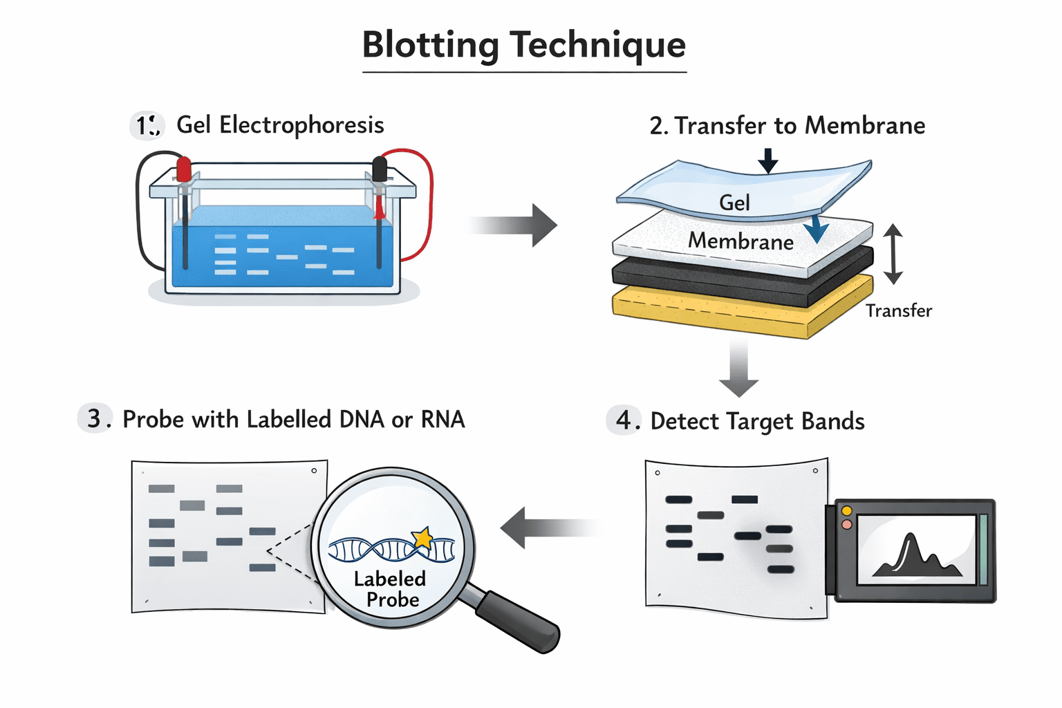
Blotting techniques represent a convergence of electrophoresis and immunological detection methods, providing a powerful approach for the separation and analysis of nucleic acids and proteins. At their core, these techniques allow researchers to isolate specific biomolecules from complex mixtures and transfer them onto a stable support membrane for subsequent detection and characterization. By combining physical separation with molecular recognition, blotting enables precise analysis of DNA, RNA, and protein molecules in research and diagnostic applications. The blotting technique workflow for nucleic acids and protein is shown in Figure 1.

Blotting techniques integrate electrophoretic separation with targeted detection, allowing the precise analysis of nucleic acids and proteins. By transferring molecules from gels onto nitrocellulose membranes through capillary action, researchers gain a reliable and reproducible platform for probing, studying, and quantifying biomolecules. The method’s ability to preserve molecular organization while providing accessibility for downstream analysis has made it an indispensable tool in both fundamental and applied molecular research.
In a typical blotting procedure, molecules are first separated using gel electrophoresis. DNA and RNA fragments are commonly resolved on agarose gels, while proteins are separated using polyacrylamide gels, often under denaturing conditions to ensure uniform migration. Unlike traditional staining methods, which visualize molecules directly in the gel, blotting transfers these unstained molecules onto a nitrocellulose or polyvinylidene difluoride (PVDF) membrane, where they can be more easily accessed and detected.
The transfer process is achieved by assembling the gel and membrane into a “blotting sandwich” arrangement (Figure 2). This setup usually consists of the gel, the membrane, and layers of absorbent paper stacked together so that buffer solution can flow from the gel into the membrane by capillary action. As the buffer moves through the gel, it carries the separated nucleic acid or protein molecules onto the membrane surface, where they become immobilized while retaining the original pattern of separation. This method ensures that the relative positions of the molecules are preserved, facilitating accurate detection and analysis.
Once the molecules are transferred, they can be probed using specific detection strategies. For nucleic acids, labeled complementary sequences hybridize to the target DNA or RNA fragments, allowing their identification. For proteins, immunological detection using specific antibodies enables the visualization and quantification of individual protein species. This combination of physical separation and molecular recognition is what makes blotting such a versatile and widely used technique in molecular biology, biotechnology, and clinical diagnostics.
The blotting sandwich, illustrated in Figure 2, serves as a reliable and permanent platform for transferring proteins and nucleic acid fragments from gels onto a nitrocellulose membrane. This setup ensures that separated biomolecules, whether DNA, RNA, or proteins, are immobilized efficiently for downstream detection and analysis. The method combines physical separation, capillary action, and membrane binding to create a reproducible system for molecular studies.
The process begins with a gel slab that has already undergone electrophoretic separation of the target molecules. The gel is first immersed in an appropriate buffer solution to maintain the stability and mobility of the molecules during transfer. A nitrocellulose or PVDF membrane is then carefully placed directly on top of the gel, ensuring close contact to facilitate efficient binding. The membrane acts as a capture surface, where biomolecules will be immobilized as they migrate out of the gel.
Following this, layers of specialized absorbent paper are stacked on top of the membrane to form the “sandwich.” These papers serve as wicks, drawing buffer through the gel and membrane by capillary action. As the buffer moves upward, it carries denatured nucleic acids and proteins from the gel onto the membrane surface. Importantly, this process preserves the spatial arrangement of the separated molecules, maintaining the resolution achieved during electrophoresis.
The blotting sandwich method is versatile and widely used in Southern, Northern, and Western blotting applications. By providing a stable, reproducible, and accessible platform for molecular transfer, it allows researchers to probe, visualize, and quantify biomolecules with high specificity and sensitivity. The combination of precise molecular separation and efficient immobilization makes the blotting sandwich an essential component of many molecular biology workflows, supporting studies in gene expression, protein characterization, and diagnostic research.
Types of Blotting Techniques in Molecular Biology
Blotting techniques are among the most fundamental tools in molecular biology, enabling researchers to detect, quantify, and analyze nucleic acids and proteins with high specificity. Since their introduction in the 1970s, blotting methods have revolutionized genetic, transcriptomic, and proteomic studies by providing a reliable means to transfer biological molecules from gels onto stable membranes for detailed examination. Among these techniques, three major types are widely employed: Southern blotting, Northern blotting, and Western blotting. Each method has distinct purposes, yet all share the same core principle: separating biomolecules via electrophoresis and transferring them to a membrane where they can be probed or visualized.
Southern, Northern, and Western blotting represent the three principal blotting techniques in molecular biology, each tailored to a specific class of biomolecules. By combining electrophoresis with targeted detection strategies, these methods allow precise analysis of DNA, RNA, and proteins, providing critical information on molecular size, abundance, and functional state. Over the decades, these techniques have not only advanced research in genetics, transcriptomics, and proteomics but also continue to serve as indispensable tools in diagnostics, biotechnology, and molecular medicine. Mastery of these blotting techniques remains essential for any molecular biologist seeking to understand the structure, expression, and regulation of biomolecules in complex biological systems.
Southern Blotting: DNA Analysis
Southern blotting, named after its inventor Edwin Southern, was first developed in 1975 as a method to identify specific DNA sequences within complex mixtures. The technique fundamentally combines gel electrophoresis with nucleic acid hybridization to detect target genes, assess fragment sizes, and quantify their relative abundance in different samples. In a typical Southern blot, genomic DNA is first digested with restriction enzymes to generate fragments of varying lengths. These fragments are then separated by agarose gel electrophoresis according to their size, with smaller fragments migrating faster than larger ones.
Following electrophoresis, the DNA fragments are transferred from the gel onto a nitrocellulose or polyvinylidene difluoride (PVDF) membrane using capillary action or vacuum-assisted transfer. Once immobilized, the DNA is exposed to labeled complementary DNA or RNA probes. These probes hybridize specifically with the target sequences, allowing researchers to detect the gene of interest. Detection methods may include radioactive labeling, chemiluminescence, or fluorescence, each offering varying degrees of sensitivity and resolution.
Southern blotting provides several critical pieces of information. First, it enables the determination of the molecular weight of a restriction fragment, which can reveal structural features of a gene or genomic region. Second, it allows the relative quantification of a gene across different samples, which is useful in studies of gene copy number, polymorphisms, and genetic mapping. For decades, Southern blotting has been instrumental in cloning, diagnostics of genetic diseases, and mapping of genomic sequences, laying the foundation for modern molecular genetics.
Northern Blotting: RNA Analysis
Building on the principles of Southern blotting, Northern blotting was developed in 1977 to study RNA molecules, particularly messenger RNA (mRNA). While Southern blotting focuses on DNA sequences, Northern blotting is used to investigate gene expression by measuring the presence and abundance of specific RNA transcripts in biological samples. Like Southern blotting, the technique relies on gel electrophoresis to separate RNA molecules based on size. RNA is typically resolved on denaturing agarose gels to prevent secondary structures from interfering with migration.
After electrophoretic separation, RNA molecules are transferred onto a nitrocellulose or nylon membrane and immobilized. The membrane is then hybridized with labeled complementary probes designed to recognize the RNA sequence of interest. Detection of the bound probe allows researchers to determine both the molecular weight of the transcript and its relative abundance across samples. Northern blotting has been particularly valuable in studying gene expression patterns during development, cellular differentiation, and disease states. For example, it can reveal whether a gene is upregulated or downregulated in response to environmental stimuli or pathological conditions.
Northern blotting also provides information on transcript integrity and alternative splicing, as multiple bands on the blot may represent different mRNA isoforms. Although newer techniques like quantitative PCR and RNA sequencing have largely supplanted Northern blotting for routine expression analysis, the method remains a gold standard for validating transcript size, assessing RNA quality, and confirming results obtained by other methods.
Western Blotting: Protein Analysis
Western blotting, developed in the late 1970s, extends the blotting concept to protein analysis. Unlike Southern and Northern blotting, which target nucleic acids, Western blotting is used to detect specific proteins within complex mixtures. The technique combines gel electrophoresis with immunodetection, allowing researchers to probe for individual proteins using specific antibodies. Protein samples are first separated based on size through sodium dodecyl sulfate-polyacrylamide gel electrophoresis (SDS-PAGE), which denatures proteins and imparts a uniform negative charge, ensuring that migration is primarily determined by molecular weight.
Following separation, proteins are transferred from the gel onto a nitrocellulose or PVDF membrane. The membrane is then incubated with a primary antibody that specifically binds the protein of interest, followed by a secondary antibody conjugated to a detection enzyme or fluorescent tag. This enables visualization and quantification of the protein. Western blotting allows researchers to determine the molecular weight of the protein, detect post-translational modifications, and measure relative protein levels across different samples.
Western blotting has become indispensable in both basic and applied research. It is widely used for validating gene expression at the protein level, studying signaling pathways, detecting disease biomarkers, and confirming the presence of recombinant proteins in biotechnology applications. Innovations such as chemiluminescent detection, infrared fluorescence, and multiplexing have further enhanced the sensitivity, accuracy, and throughput of Western blotting, making it a cornerstone of proteomics research.
Comparative overview and applications of blotting
Although Southern, Northern, and Western blotting target different biomolecules, they share several key features. All three techniques begin with electrophoretic separation, followed by transfer onto a membrane and detection using a specific probe or antibody. They allow determination of molecular weight, assessment of relative abundance, and qualitative or semi-quantitative analysis of the target molecule. Each technique has distinct applications: Southern blotting for gene identification and structural analysis, Northern blotting for transcript detection and expression profiling, and Western blotting for protein identification and quantification. These methods have historically enabled major advances in molecular biology. Southern blotting contributed to the cloning of genes and characterization of genetic disorders, Northern blotting provided insights into transcriptional regulation, and Western blotting allowed detailed study of protein function and signaling networks. Together, they form a foundational toolkit for investigating the molecular underpinnings of biological systems.
References
Alberts B, Bray D, Lewis J, Raff M, Roberts K and Watson J.D (2002). The molecular Biology of the Cell. Fourth edition. New York, Garland, USA.
Chen I and Dubnau D (2004). DNA uptake during bacterial transformation. Nat. Rev. Microbiol. 2 (3): 241–249.
Cooper G.M and Hausman R.E (2004). The cell: A Molecular Approach. Third edition. ASM Press.
Dale J (2003). Molecular genetics of bacteria. Jeremy W. Dale and Simon Park (4th eds.). John Wiley & Sons Ltd, West Sussex, UK. Pp. 312-313.
Das H.K (2010). Textbook of Biotechnology. Fourth edition. Wiley edition. Wiley India Pvt, Ltd, New Delhi, India.
Lewis R (2004). Human Genetics: Concepts and Applications. Sixth edition. McGraw Hill Publishers, USA.
Lodish H, Berk A, Matsudaira P, Kaiser C.A, Kreiger M, Scott M.P, Zipursky S.L and Darnell J (2004). Molecular Cell Biology. Fifth edition. Scientific American Books, Freeman, New York, USA.
Madigan M.T., Martinko J.M., Dunlap P.V and Clark D.P (2009). Brock Biology of Microorganisms, 12th edition. Pearson Benjamin Cummings Inc, USA.
McPherson M and Moller S (2002). PCR: The Basics. 2nd edition. Taylor and Francis Group. New York, USA.
Sambrook, J., Russell, D.W. (2001). Molecular Cloning: a Laboratory Manual, 3rd edn. Cold Spring Harbor Laboratory Press, New York.
Synder L, Peters J.E, Henkin T.M and Champness W (2013). Molecular Genetics of Bacteria. Fourth edition. American Society of Microbiology Press, USA.
Tamarin Robert H (2002). Principles of Genetics. Seventh edition. Tata McGraw-Hill Publishing Co Ltd, Delhi.
Discover more from Microbiology Class
Subscribe to get the latest posts sent to your email.