
Western blotting, also known as protein immunoblotting, is a widely used analytical technique in molecular biology, biochemistry, and biomedical research for the detection and characterization of specific proteins within a complex biological sample. The method allows researchers to identify proteins based on their molecular weight and their interaction with specific antibodies. Through this approach, scientists can determine not only the presence of a protein but also estimate its relative abundance across different samples. Because of its specificity and reliability, western blotting remains one of the most fundamental techniques used to study protein expression, protein modifications, and cellular responses to environmental or experimental conditions.
The western blotting technique relies on the separation of proteins according to their molecular sizes through gel electrophoresis, followed by their transfer onto a solid support membrane where they are probed using antibodies that specifically recognize the target protein. The use of antibodies distinguishes western blotting from other blotting techniques such as southern blotting and northern blotting. While southern blotting is designed for the detection of DNA fragments and northern blotting is used for RNA analysis, western blotting focuses specifically on proteins. The use of antigen-antibody interactions in western blotting provides a high level of specificity, allowing the detection of a particular protein even in samples containing thousands of different proteins.
At its core, western blotting involves the transfer of proteins from a gel matrix onto a durable membrane, typically made of nitrocellulose or polyvinylidene fluoride (PVDF). Once immobilized on the membrane, the proteins can be probed with antibodies and visualized through a detection system. This process allows molecular biologists to determine the approximate molecular weight of proteins and compare the levels of protein expression in different experimental conditions. As a result, western blotting is frequently used in studies of gene expression, disease biomarkers, signal transduction pathways, and the verification of recombinant protein production.
The process of western blotting begins with the preparation of protein samples. Biological materials such as cultured cells, tissues, microorganisms, or blood samples are first lysed to release their proteins. Lysis buffers are typically used to break open cells and stabilize proteins during extraction. These buffers often contain detergents, salts, and protease inhibitors to prevent protein degradation. The extracted proteins are then quantified using protein assays such as the Bradford assay or bicinchoninic acid (BCA) assay to ensure that equal amounts of protein are loaded into each electrophoresis lane. Accurate protein quantification is essential for meaningful comparison between samples.
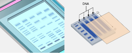
Following protein extraction and quantification, the proteins are separated using sodium dodecyl sulfate-polyacrylamide gel electrophoresis (SDS-PAGE). Unlike agarose gel electrophoresis, which is commonly used for separating nucleic acids such as DNA and RNA, SDS-PAGE utilizes a polyacrylamide gel matrix to resolve proteins with high resolution. Polyacrylamide gels have smaller pore sizes than agarose gels, allowing them to effectively separate proteins that differ slightly in molecular weight. In SDS-PAGE, proteins are treated with the anionic detergent sodium dodecyl sulfate (SDS) prior to electrophoresis. SDS plays a critical role in the process because it denatures proteins by disrupting their secondary and tertiary structures. Additionally, SDS coats the proteins with a uniform negative charge proportional to their length.
This treatment ensures that the migration of proteins through the gel depends primarily on their molecular weight rather than their shape or intrinsic charge. As a result, smaller proteins migrate more quickly through the gel matrix than larger ones. The electrophoresis process occurs in an electric field, where negatively charged proteins move toward the positive electrode (anode). As they travel through the polyacrylamide gel, the proteins become separated according to size, forming distinct bands. Molecular weight markers or protein ladders are typically included in one of the gel lanes to allow estimation of the molecular sizes of separated proteins.
Once the proteins have been separated by SDS-PAGE, the next step in the western blotting procedure is the transfer of proteins from the gel onto a membrane. This step is necessary because polyacrylamide gels are fragile and not suitable for antibody probing. The transfer process immobilizes proteins onto a durable membrane that can be handled and processed for detection. The most commonly used membranes for western blotting are nitrocellulose and PVDF membranes, both of which have strong protein-binding properties. Protein transfer can be achieved using several methods, including wet transfer, semi-dry transfer, and dry transfer techniques.
In the traditional wet transfer method, the gel and membrane are placed together in a transfer sandwich consisting of filter papers and sponges soaked in transfer buffer. An electric current is applied, causing the negatively charged proteins to migrate out of the gel and onto the membrane. This process effectively replicates the banding pattern observed in the gel but transfers it onto the membrane surface. After the proteins have been successfully transferred to the membrane, the membrane undergoes a blocking step. Blocking is essential because the membrane surface has a high capacity for nonspecific protein binding. Without blocking, antibodies used for detection could bind nonspecifically to the membrane, producing background noise that interferes with accurate protein detection.
To prevent this, the membrane is incubated in a blocking solution containing proteins such as bovine serum albumin (BSA) or non-fat dry milk. These proteins occupy the remaining binding sites on the membrane, thereby reducing nonspecific antibody interactions. Following the blocking step, the membrane is incubated with a primary antibody that specifically recognizes the target protein. Primary antibodies are typically raised in animals such as rabbits, mice, or goats and are designed to bind selectively to a particular protein epitope. The specificity of the primary antibody is critical for the success of the western blot, as it ensures that only the protein of interest is detected. After incubation with the primary antibody, the membrane is washed several times to remove any unbound antibody molecules.
The washing step usually involves buffered solutions containing mild detergents such as Tween-20, which help reduce nonspecific interactions while preserving specific antigen-antibody binding. The next step involves the addition of a secondary antibody. The secondary antibody recognizes and binds to the primary antibody. Typically, secondary antibodies are conjugated with enzymes such as horseradish peroxidase (HRP) or alkaline phosphatase (AP). These enzymes allow the detection of the antibody-protein complex through a colorimetric, chemiluminescent, or fluorescent reaction. Because multiple secondary antibodies can bind to a single primary antibody, this step also amplifies the signal and increases detection sensitivity. Once the secondary antibody has been applied and excess antibody washed away, the membrane is exposed to a detection substrate.
In chemiluminescent detection, for example, the enzyme attached to the secondary antibody catalyzes a reaction that produces light. The emitted light can be captured using photographic film or digital imaging systems, revealing bands corresponding to the target protein. The position of each band on the membrane corresponds to the molecular weight of the detected protein, which can be estimated by comparison with the molecular weight markers. The intensity of the band provides a relative measure of the amount of protein present in the sample. Researchers often use software tools to quantify band intensity, allowing semi-quantitative comparisons of protein expression across multiple samples.
Western blotting has numerous applications in biological and medical research. One major application is the analysis of gene expression at the protein level. While techniques such as polymerase chain reaction (PCR) measure gene expression at the RNA level, western blotting provides information about the actual protein products that perform biological functions. This is important because protein abundance does not always correlate directly with mRNA levels due to regulatory mechanisms such as translational control and protein degradation. Another important application of western blotting is the detection of post-translational modifications such as phosphorylation, glycosylation, and ubiquitination.
Specialized antibodies can recognize these modified forms of proteins, allowing researchers to study cellular signaling pathways and regulatory mechanisms. Western blotting is also widely used in medical diagnostics and clinical research. For example, it has historically been used as a confirmatory test in the diagnosis of certain viral infections by detecting specific viral proteins or antibodies. Despite its advantages, western blotting also has several limitations. The technique can be time-consuming and requires careful optimization of experimental conditions. The quality of the results depends heavily on the specificity and performance of the antibodies used.
Poorly characterized antibodies can lead to nonspecific binding or inaccurate detection. Additionally, western blotting is generally considered a semi-quantitative technique rather than a fully quantitative one, although improvements in imaging technologies and analysis software have enhanced its quantitative capabilities. In recent years, alternative methods such as enzyme-linked immunosorbent assays (ELISA), mass spectrometry-based proteomics, and automated capillary electrophoresis systems have been developed to complement or replace traditional western blotting in certain applications. Nevertheless, western blotting remains a cornerstone technique in molecular biology laboratories due to its reliability, relatively low cost, and ability to provide clear visual evidence of protein detection.
Western blotting is a powerful and widely used technique for the detection and analysis of specific proteins within complex biological samples. The method combines protein separation by SDS-PAGE, transfer to a membrane, and antibody-based detection to identify proteins according to their molecular weight and relative abundance. Through careful execution of each step sample preparation, electrophoresis, transfer, blocking, antibody probing, and detection researchers can obtain valuable insights into protein expression and function. As a result, western blotting continues to play an essential role in molecular biology, biotechnology, and biomedical research.
Steps Involved in Western Blotting for Protein Detection
Western blotting is a multistage laboratory technique designed to detect and analyze specific proteins within complex biological mixtures. The procedure relies on the separation of proteins by electrophoresis, their transfer to a membrane, and their identification using antibodies that specifically recognize the target protein. To ensure clarity and reproducibility, the procedure is typically described as a sequence of well-defined steps. Each step plays a crucial role in ensuring that the protein of interest is accurately identified with minimal background interference.
Western blotting involves a systematic series of steps that enable the identification of specific proteins from complex mixtures. Beginning with protein extraction and separation by SDS-PAGE, the procedure continues through electroblotting, blocking of nonspecific binding sites, antibody incubation, and visualization of protein bands. Each step contributes to the accuracy and sensitivity of the method. When properly performed, western blotting provides valuable information about protein identity, molecular weight, and relative expression levels, making it one of the most widely used techniques in molecular biology and biomedical research.
Below are the numbered steps involved in performing western blotting.
1. Preparation of Protein Samples
The first step in western blotting involves preparing protein samples from biological materials such as tissues, cultured cells, microorganisms, or blood samples. Cells are first lysed using specialized buffers that break open the cell membrane and release intracellular proteins. These lysis buffers often contain detergents, salts, buffering agents, and protease inhibitors to prevent degradation of proteins during extraction.
After extraction, the protein concentration of each sample is measured using protein quantification assays such as the Bradford assay or bicinchoninic acid (BCA) assay. Quantifying proteins ensures that equal amounts of protein are loaded into each lane of the gel during electrophoresis. This step is essential for making reliable comparisons between different samples or experimental conditions.
2. Denaturation of Proteins and Sample Preparation
Once proteins are extracted, they are mixed with a loading buffer containing sodium dodecyl sulfate (SDS), a reducing agent such as β-mercaptoethanol or dithiothreitol (DTT), glycerol, and a tracking dye. SDS is a powerful detergent that unfolds proteins by disrupting their secondary and tertiary structures. The reducing agent breaks disulfide bonds within and between protein chains.
This treatment ensures that proteins become linear molecules coated with a uniform negative charge. As a result, their migration during electrophoresis depends primarily on molecular weight rather than their natural shape or charge.
3. Separation of Proteins Using SDS-PAGE
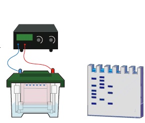
In this step, the prepared protein samples are loaded into wells at the top of a polyacrylamide gel. The gel is placed in an electrophoresis chamber filled with a conductive buffer. When an electric current is applied, the negatively charged proteins migrate toward the positive electrode.
Because the gel matrix acts as a molecular sieve, smaller proteins move through the pores of the gel more rapidly than larger proteins. Over time, this causes the proteins to separate into distinct bands based on their molecular sizes. A molecular weight marker or protein ladder is usually included in one lane of the gel to serve as a reference for estimating the molecular weights of the separated proteins.
This step is critical because accurate separation of proteins is necessary for identifying the target protein in later stages of the western blot.
4. Transfer of Proteins to a Membrane (Electroblotting)
After electrophoresis, the separated proteins must be transferred from the gel onto a solid membrane. This process is known as electroblotting. The transfer is necessary because polyacrylamide gels are fragile and not suitable for direct antibody probing.
To perform electroblotting, the gel is placed in close contact with a nitrocellulose or polyvinylidene fluoride (PVDF) membrane. The gel and membrane are arranged in a transfer “sandwich” consisting of filter papers and sponge pads soaked in transfer buffer. When an electric current is applied across this assembly, the negatively charged proteins migrate out of the gel and onto the membrane.
As the proteins move toward the positively charged electrode, they become immobilized on the membrane surface while maintaining the same banding pattern produced during electrophoresis. The membrane therefore becomes a replica of the protein distribution originally present in the gel.
5. Verification of Protein Transfer
After the transfer step, it is often useful to verify that proteins have been successfully transferred to the membrane. This can be done by briefly staining the membrane with reversible dyes such as Ponceau S. The dye temporarily stains all protein bands on the membrane, allowing researchers to confirm that the transfer was efficient and that protein loading across lanes was uniform.
Once verification is complete, the stain is washed away before proceeding to the next step.
6. Blocking of Non-Specific Binding Sites
The membrane used in western blotting has a strong ability to bind proteins. While this property allows sample proteins to adhere to the membrane, it can also cause antibodies to bind nonspecifically to the membrane surface. If such nonspecific binding occurs, it may produce background signals that interfere with accurate detection.
To prevent this problem, the membrane is incubated in a blocking solution containing generic protein molecules such as bovine serum albumin (BSA), non-fat dry milk, gelatin, or casein. These proteins occupy all remaining binding sites on the membrane that are not already occupied by transferred sample proteins.
By covering these sites, the blocking step minimizes unwanted interactions between the membrane and antibodies, thereby improving the clarity and specificity of the detection process.
7. Incubation with the Primary Antibody
After blocking, the membrane is incubated with a primary antibody that specifically recognizes the protein of interest. Primary antibodies are highly selective and bind to a unique region, known as an epitope, on the target protein.
During this incubation step, the antibody attaches only to the band containing the protein that matches its binding specificity. As a result, a layer of antibody molecules forms on the membrane precisely at the location where the target protein is present.
Primary antibodies may be produced in animals such as mice, rabbits, or goats. They are carefully designed and validated to ensure that they recognize only the intended protein.
8. Washing to Remove Unbound Antibodies
Following primary antibody incubation, the membrane is washed several times using a buffered solution that typically contains mild detergents such as Tween-20. These washing steps are essential because they remove excess antibodies that did not bind specifically to the target protein.
Proper washing reduces background noise and ensures that only antibodies bound to the protein of interest remain on the membrane.
9. Detection of the Antibody–Protein Complex
In traditional western blotting protocols, the primary antibody may be radioactively labeled. When such labeled antibodies bind to the target protein on the membrane, the location of the protein can be detected through the emission of radiation from the label.
In this situation, the radioactive antibody forms a detectable layer at the site where the protein of interest is located. Because the antibody binds only to its specific antigen, the signal appears only at the position corresponding to that protein band.
In modern laboratories, however, detection more commonly involves secondary antibodies linked to enzymes or fluorescent molecules that produce visible signals when exposed to appropriate substrates.
10. Visualization of Protein Bands by Autoradiography
The final step in the process involves visualizing the protein bands on the membrane. When radioactive antibodies are used, the membrane is placed in close contact with an X-ray film inside a light-tight cassette. Over time, radiation emitted from the labeled antibodies exposes the film.
The film is then developed using photographic chemicals, producing an image in which dark bands correspond to the location of the detected protein. This imaging method is known as autoradiography.
The position of each band indicates the molecular weight of the detected protein when compared with the molecular weight markers used during electrophoresis. The intensity of the band reflects the relative amount of protein present in the sample.
References
Alberts B, Bray D, Lewis J, Raff M, Roberts K and Watson J.D (2002). The molecular Biology of the Cell. Fourth edition. New York, Garland, USA.
Chen I and Dubnau D (2004). DNA uptake during bacterial transformation. Nat. Rev. Microbiol. 2 (3): 241–249.
Cooper G.M and Hausman R.E (2004). The cell: A Molecular Approach. Third edition. ASM Press.
Dale J (2003). Molecular genetics of bacteria. Jeremy W. Dale and Simon Park (4th eds.). John Wiley & Sons Ltd, West Sussex, UK. Pp. 312-313.
Das H.K (2010). Textbook of Biotechnology. Fourth edition. Wiley edition. Wiley India Pvt, Ltd, New Delhi, India.
Lewis R (2004). Human Genetics: Concepts and Applications. Sixth edition. McGraw Hill Publishers, USA.
Lodish H, Berk A, Matsudaira P, Kaiser C.A, Kreiger M, Scott M.P, Zipursky S.L and Darnell J (2004). Molecular Cell Biology. Fifth edition. Scientific American Books, Freeman, New York, USA.
Madigan M.T., Martinko J.M., Dunlap P.V and Clark D.P (2009). Brock Biology of Microorganisms, 12th edition. Pearson Benjamin Cummings Inc, USA.
McPherson M and Moller S (2002). PCR: The Basics. 2nd edition. Taylor and Francis Group. New York, USA.
Sambrook, J., Russell, D.W. (2001). Molecular Cloning: a Laboratory Manual, 3rd edn. Cold Spring Harbor Laboratory Press, New York.
Synder L, Peters J.E, Henkin T.M and Champness W (2013). Molecular Genetics of Bacteria. Fourth edition. American Society of Microbiology Press, USA.
Tamarin Robert H (2002). Principles of Genetics. Seventh edition. Tata McGraw-Hill Publishing Co Ltd, Delhi.
Discover more from Microbiology Class
Subscribe to get the latest posts sent to your email.