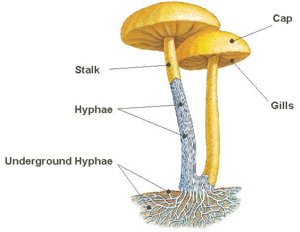
Slime moulds are eukaryotic organisms that have fungus-like features as well as some animal- or protists-like features. Slime moulds were previously classified as fungi because of some characteristics which both organisms share. Slime moulds like fungi produce spores and fruiting bodies; and these features warranted there earlier classification as fungi but this is no longer the case. They are motile organisms, and exhibit some amoebic-like features in locomotion.
Slime moulds are made up of two main divisions which are the Myxomycota (Myxomycetes) and the Acrasiomycota (Acrasiomycetes). Myxomycetes are the acellular slime moulds. They can also be called plasmodial slime moulds. Acellular (plasmodial) slime moulds are ubiquitously found in leaves, rotten woods and other moist environments as amoebic structures. The vegetative forms of plasmodial slime moulds in the environment exist as growing single mass of protoplasm.
The mass of protoplasm contain numerous diploid nuclei, and they are generally known as plasmodium (plural: plasmodia) because they lack cell walls. Following nutrient depletion or scarcity of water, the plasmodium form fruiting bodies which later develops into resistant spores that later re-germinate when environmental conditions becomes favourable again.
Plasmodial slime moulds move by cytoplasmic streaming, and their amoeboid movement helps them to absorb nutrient from the environment. Physarum species is a typical example of acellular or plasmodial slime moulds. Acrasiomycetes are cellular slime moulds. The vegetative forms of cellular slime moulds exist as single amoebae in the environment.
Cellular slime moulds have haploid nuclei, and they have independent cells unlike acellular slime moulds that have a mass of protoplasm. Cellular slime moulds unlike the acellular types feed on other microorganisms such as bacteria and yeast which they ingest via the process of phagocytosis. They form spores during unfavourable environmental conditions but re-germinate into their original amoebic form when normal conditions returns.
References
Anaissie E.J, McGinnis M.R, Pfaller M.A (2009). Clinical Mycology. 2nd ed. Philadelphia, PA: Churchill Livingstone Elsevier. London.
Brown G.D and Netea M.G (2007). Immunology of Fungal Infections. Springer Publishers, Netherlands.
Calderone R.A and Cihlar R.L (eds). Fungal Pathogenesis: Principles and Clinical Applications. New York: Marcel Dekker; 2002.
Chakrabarti A and Slavin M.A (2011). Endemic fungal infection in the Asia-Pacific region. Med Mycol, 9:337-344.
Chemotherapy of microbial diseases. In: Chabner B.A, Brunton L.L, Knollman B.C, eds.
Germain G. St. and Summerbell R (2010). Identifying Fungi. Second edition. Star Pub Co.
Ghannoum MA, Rice LB (1999). Antifungal agents: Mode of action, mechanisms of resistance, and correlation of these mechanisms with bacterial resistance. Clin Microbiol Rev, 12:501–517.
Gillespie S.H and Bamford K.B (2012). Medical Microbiology and Infection at a glance. 4th edition. Wiley-Blackwell Publishers, UK.
Larone D.H (2011). Medically Important Fungi: A Guide to Identification. Fifth edition. American Society of Microbiology Press, USA.
Madigan M.T., Martinko J.M., Dunlap P.V and Clark D.P (2009). Brock Biology of Microorganisms, 12th edition. Pearson Benjamin Cummings Inc, USA.
Mahon C. R, Lehman D.C and Manuselis G (2011). Textbook of Diagnostic Microbiology. Fourth edition. Saunders Publishers, USA.
Discover more from Microbiology Class
Subscribe to get the latest posts sent to your email.