Fungi unlike other groups of microorganisms possess or have unique structures which distinguish them from other microbial cells. Morphologically, fungi exist in any of the following structural forms:
Mycelium is the long branching structures of fungal hyphae (Figure 1). Mycelia are specialized structures used by fungal cells to absorb nutrients from their environment or supporting medium. They are known to penetrate the substrate on which they form to acquire nutrients by absorption. Two types of mycelia are exhibited by fungal organisms viz: vegetative mycelia and aerial mycelia. Vegetative mycelia act as the root of a fungal organism, and they are known to penetrate the substrate on which the fungus is growing. They help to absorb nutrient and water for the growing fungal cell. Another type of mycelia which do not penetrate nutrient substrate but grow above it is the aerial mycelia. Aerial mycelia are critical for asexual reproduction in imperfect fungi, and they usually bear or carry conidia or fungal spores. They serve as important reproductive structures in fungi.
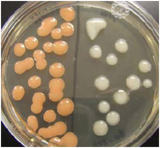
YEAST CELL
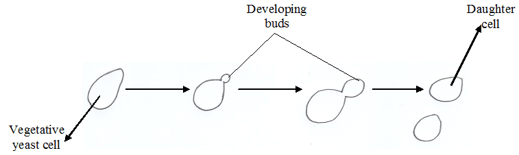
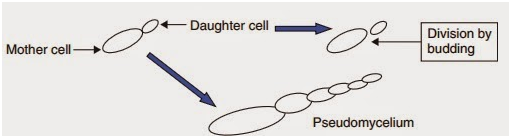
Yeasts are the unicellular forms of fungi. They usually reproduce by budding, an asexual reproduction pattern. Budding is a type of asexual reproduction in which the daughter cell emanates or develops from the parent cell. In budding, the daughter cell develops entirely from the mother or parent cell as a localized outgrowth. Structurally, yeast is oval or spherical in form (Figure 2).
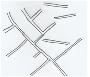
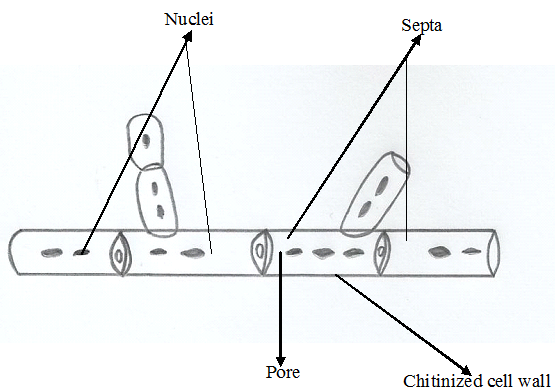
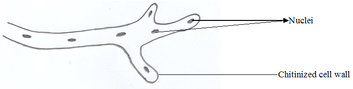
Hyphae is the tube-like extension of a fungal cell. They are usually rigid or thick in form. A mass of hyphae is required for the formation of mycelia. Hyphae formation by a fungal cell can be septate i.e. with cross-walls (Figure 3) or non-septate i.e. without cross-walls (Figure 4). Fungi produce two types of hyphae: aerial hyphae and vegetative hyphae.
Aerial hyphae are branching structures of fungi that do not penetrate the supporting medium but instead they project above the surface of the mycelia. They usually bear the reproductive structures of the filamentous fungi (i.e. moulds).
Vegetative hyphae can also be called the substrate hyphae. They are the hyphae that penetrate the supporting medium on which the fungus is growing. Substrate or vegetative hyphae absorb nutrients from the supporting medium necessary for fungal growth. Generally, mycelia are a mass of hyphae.
Pseudomycelium or Pseudohypha is the less-rigid hyphae formed by some fungi (Figure 5). They are different from the true-hyphae which are known to be very rigid.
References
Anaissie E.J, McGinnis M.R, Pfaller M.A (2009). Clinical Mycology. 2nd ed. Philadelphia, PA: Churchill Livingstone Elsevier. London.
Beck R.W (2000). A chronology of microbiology in historical context. Washington, D.C.: ASM Press.
Black, J.G. (2008). Microbiology: Principles and Explorations (7th ed.). Hoboken, NJ: J. Wiley & Sons.
Brooks G.F., Butel J.S and Morse S.A (2004). Medical Microbiology, 23rd edition. McGraw Hill Publishers. USA.
Brown G.D and Netea M.G (2007). Immunology of Fungal Infections. Springer Publishers, Netherlands.
Calderone R.A and Cihlar R.L (eds). Fungal Pathogenesis: Principles and Clinical Applications. New York: Marcel Dekker; 2002.
Chakrabarti A and Slavin M.A (2011). Endemic fungal infection in the Asia-Pacific region. Med Mycol, 9:337-344.
Champoux J.J, Neidhardt F.C, Drew W.L and Plorde J.J (2004). Sherris Medical Microbiology: An Introduction to Infectious Diseases. 4th edition. McGraw Hill Companies Inc, USA.
Chemotherapy of microbial diseases. In: Chabner B.A, Brunton L.L, Knollman B.C, eds. Goodman and Gilman’s The Pharmacological Basis of Therapeutics. 12th ed. New York, McGraw-Hill; 2011.
Chung K.T, Stevens Jr., S.E and Ferris D.H (1995). A chronology of events and pioneers of microbiology. SIM News, 45(1):3–13.
Germain G. St. and Summerbell R (2010). Identifying Fungi. Second edition. Star Pub Co.
Ghannoum MA, Rice LB (1999). Antifungal agents: Mode of action, mechanisms of resistance, and correlation of these mechanisms with bacterial resistance. Clin Microbiol Rev, 12:501–517.
Gillespie S.H and Bamford K.B (2012). Medical Microbiology and Infection at a glance. 4th edition. Wiley-Blackwell Publishers, UK.
Larone D.H (2011). Medically Important Fungi: A Guide to Identification. Fifth edition. American Society of Microbiology Press, USA.
Levinson W (2010). Review of Medical Microbiology and Immunology. Twelfth edition. The McGraw-Hill Companies, USA.
Madigan M.T., Martinko J.M., Dunlap P.V and Clark D.P (2009). Brock Biology of Microorganisms, 12th edition. Pearson Benjamin Cummings Inc, USA.
Mahon C. R, Lehman D.C and Manuselis G (2011). Textbook of Diagnostic Microbiology. Fourth edition. Saunders Publishers, USA.
Discover more from Microbiology Class
Subscribe to get the latest posts sent to your email.