SEMEN MICROSCOPY
AIM: To determine any abnormality in a seminal fluid (semen) as an aid in the diagnosis of male infertility.
MATERIAL/APPARATUS: Semen specimen, microscope, test tube, test tube rack, glass slide, cover slip, semen diluting fluid (sodium bicarbonate formalin), improved neubauer counting chamber, bulb pipette or Pasteur pipette, calibrated cylinder (5 ml).
The microscopy of a semen specimen involves the following procedures:
- Determination of the semen volume.
- Determination of the semen viscosity.
- Determination of the motility of the sperm cells in the semen specimen.
- Determination of the morphology of the sperm cells in the semen specimen.
- Sperm cell count.
In addition to the above parameters, the time of production, time of collection, time of examination, and the appearance of the semen specimen is also recorded. The appearance of a semen specimen can be grayish or white. These parameters are also reported together with other analysis carried out.
SEMEN VOLUME
The volume of a semen specimen can be determined by emptying or pouring the semen specimen into a calibrated cylinder that is about 5 ml. The volume of normal semen is between 2 – 5 ml and above.
SEMEN VISCOSITY
A semen specimen can be viscous, non-viscous, slightly viscous or highly viscous. Due to the presence of fibrinolysin in the semen, it usually becomes liquefied within 1 hour. You can determine how viscous a semen specimen is by touching it with a sterile inoculating loop or stick and raising it up. Normal semen is thick and viscous when ejaculated or produced.
SPERM CELL MOTILITY
The procedure for determining the motility of a sperm cell is as follows:
- Place one drop of well mixed semen on a clean glass slide.
- Cover with a cover slip.
- Focus the slide using ×10 and ×40 objective lens of a microscope. Ensure that the condenser iris of the microscope is sufficiently closed in order to give a good contrast while viewing.
- Examine several fields of the slide and report your result.
REPORTING THE RESULT: A sperm cell can be actively motile (rapid and progressive) or weakly motile (slow and non-progressive). Count a total of 100 spermatozoa/sperm cell, and note how many spermatozoa are motile out of the 100 cells counted. Record the percentage of sperm cells that is motile and non-motile. Normal semen should have more than 50 % motile spermatozoa within 60 minutes of ejaculation.
SPERM CELL MORPHOLOGY
The procedure for determining the morphology of a sperm cell is as follows:
- Place a drop of well mixed semen on a clean glass slide.
- Make a thin smear of the semen on the slide.
- Heat-fix the smear.
- Stain the smear with either Giemsa stain or methylene blue stain.
- Allow for 5 minutes.
- Wash off with water after 5 minutes and allow to dry.
- Add a drop of immersion oil on the slide.
- Focus with ×100 objective lens.
- Examine the different fields for any abnormality in the morphology of the spermatozoa.
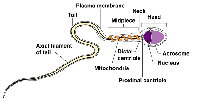
REPORTING OF THE RESULT: Estimate the number of spermatozoa showing normal morphology and abnormal morphology, and report accordingly. Normal spermatozoa measures about 50-70 µm in length, and each consists of an oval-shaped head (with acrosomal cap), a short middle piece, and a long tail (Figure 1). In normal semen sample, at least 50 % of spermatozoa should show normal morphology.
SEMINAL FLUID (SEMEN) CULTURE
AIM: To isolate pathogenic bacteria from semen specimen as an aid in the diagnosis of infertility in male.
MATERIAL/APPARATUS: Semen specimen, grease pencil, incubator, anaerobic jar, Bunsen burner, inoculating loop, blood agar (BA), chocolate agar (CA).
METHOD/PROCEDURE FOR SEMEN CULTURE
- Label the CA and BA plates with the patient’s name and laboratory number using the grease pencil.
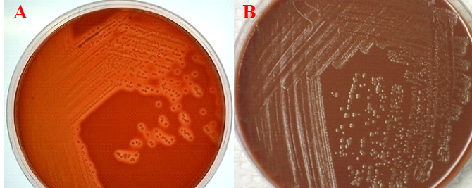
- Aseptically culture the semen specimen on both the CA and BA plates. This is usually done by using a sterilized inoculating wire loop to transfer the semen specimen onto the culture media plate. And the sample is spread or streaked across the culture plate.
- Incubate the BA plate in the incubator at 37oC overnight.
- Incubate the CA plate in the anaerobic jar at 37oC overnight.
- Examine culture media plates after incubation and look out for significant bacterial growth.
- Subculture the isolated organisms onto freshly prepared culture media plates (e.g. CA and BA) for the isolation of pure cultures.
- Perform biochemical test to identify the isolated bacteria.
- Perform antimicrobial sensitivity test only when a significant bacteria is isolated.
REPORTING OF THE RESULT: Significant bacteria growth should be reported. The culture of semen specimen is carried out first before any microscopical analysis in order to avoid contaminating the specimen.
SPERM CELL COUNT
The procedure for determining the amount of sperm cells in a semen sample is as follows:
- Make a 1:10 dilution of the semen specimen using a calibrated test tube and semen diluting fluid by placing 9 drops of the semen diluting fluid (sodium bicarbonate formalin) in a clean test tube standing on a test tube rack.
- Add 1 mL of well mixed semen specimen to the semen diluting fluid in the tube, and mix well. Avoid bubble production when mixing the semen and the diluent. In this way, a 1:10 dilution of a semen specimen is made.
- I in 10 dilutions is usually made for small semen specimen. For large semen specimen, 1:100 dilutions is made.
- The counting of spermatozoa in a semen specimen without dilution will be very difficult because the sperm cells are still alive and actively moving. This is why the semen diluting fluid is used so that they can inactivate or kill the spermatozoa, making the count easy. And this procedure is normally done after determining the motility and morphology of the sperm cells.
- Using a Pasteur’s pipette, fill an improved Neubauer counting chamber with the well mixed diluted semen specimen.
- Wait for about 4-5 minutes for the sperm cells to settle, before viewing.
- View the counting chamber with ×10 objective lens of a microscope. Make sure the condenser iris of the microscope is sufficiently reduced in or order to give a good contrast.
- Count the sperm cells in each of the 4 fields of the Neubauer counting chamber.
REPORTING THE RESULT: The formula for counting and reporting a sperm cell count is:
Number of sperm cells counted × 104 × dilution factor.
The unit of measurement is: Sperm cells/ml.
Normal sperm cell count is above 20 × 106 sperm cells/ml.
Abnormal sperm cell count is below 20 × 106 sperm cells/ml.
References
Basic laboratory procedures in clinical bacteriology. World Health Organization (WHO), 1991. Available from WHO publications, 1211 Geneva, 27-Switzerland.
Beers M.H., Porter R.S., Jones T.V., Kaplan J.L and Berkwits M (2006). The Merck Manual of Diagnosis and Therapy. Eighteenth edition. Merck & Co., Inc, USA.
Biosafety in Microbiological and Biomedical Laboratories. 5th edition. U.S Department of Health and Human Services. Public Health Service. Center for Disease Control and Prevention. National Institute of Health. HHS Publication No. (CDC) 21-1112.2009.
Cheesbrough M (2010). District Laboratory Practice in Tropical Countries. Part I. 2nd edition. Cambridge University Press, UK.
Cheesbrough M (2010). District Laboratory Practice in Tropical Countries. Part 2. 2nd edition. Cambridge University Press, UK.
Discover more from Microbiology Class
Subscribe to get the latest posts sent to your email.