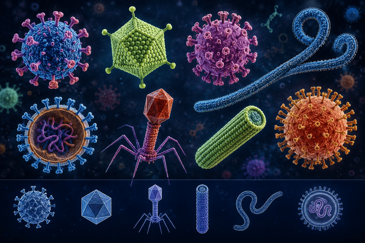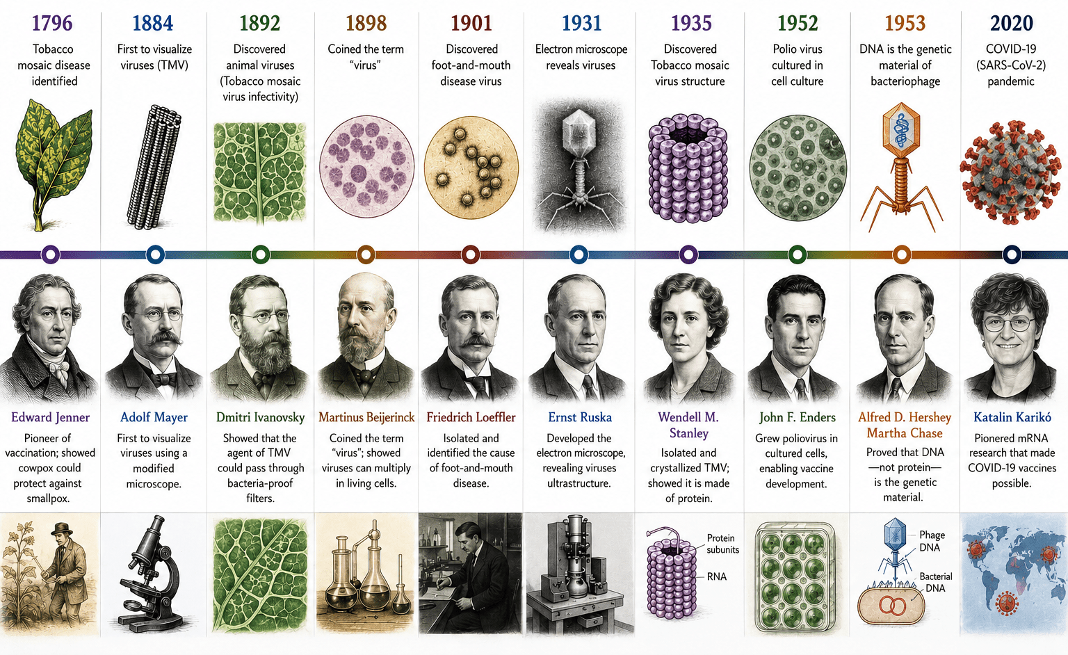

Viral replication refers to the biological process through which viruses generate new copies of themselves within a living host cell. Unlike cellular organisms that possess independent metabolic systems and reproduce through mechanisms such as cell division, viruses are obligate intracellular entities that lack the machinery required for autonomous growth and reproduction. Consequently, they depend entirely on the metabolic resources, biosynthetic pathways, and cellular structures of a susceptible host cell to complete their life cycle and produce progeny virions.
Replication in viruses differs fundamentally from reproduction in other microorganisms such as bacteria. Bacteria are living cells capable of independent multiplication, commonly through binary fission, in which one cell divides to produce two genetically identical daughter cells. Viruses, however, do not reproduce by cellular division because they are not cellular organisms. Instead, they replicate through a coordinated intracellular process that converts an infected host cell into a temporary viral production system. Once inside the host, the virus redirects cellular activities away from normal physiological functions and toward the synthesis of viral materials.
The replication process begins when a virus successfully infects a suitable host cell. Host specificity is an important feature of viral replication because viruses can only multiply in cells that possess compatible surface receptors and intracellular conditions capable of supporting viral development. Following entry into the host cell, the viral genome either deoxyribonucleic acid (DNA) or ribonucleic acid (RNA) is delivered into the cellular environment. At this stage, the virus initiates control over essential cellular processes to create conditions favorable for viral multiplication.
The viral genome serves as the central template for replication and directs the production of all molecular components required for generating new infectious particles. Using host-derived energy, ribosomes, enzymes, and structural resources, the virus orchestrates the synthesis of viral proteins and the replication of its genetic material. Structural proteins such as capsid proteins are produced alongside copies of viral nucleic acids to ensure that complete viral particles can be assembled. Some viruses also induce modifications in host cellular architecture to create specialized intracellular environments that enhance replication efficiency.
The interaction between viruses and host cells is highly dynamic and often leads to significant alterations in cellular metabolism. In many cases, viral infection suppresses normal host functions and prioritizes the manufacture of viral components. This redirection of cellular machinery enables rapid amplification of viral particles within a relatively short period. Depending on the type of virus and the host cell involved, replication may result in cellular damage, programmed cell death, or prolonged persistence of the virus within host tissues.
Although the details of replication vary among different viral families, the process generally follows a coordinated sequence of events that begins with recognition of a susceptible host cell and culminates in the formation and release of mature infectious virions. Throughout replication, viral nucleic acids are expressed and copied, viral proteins are synthesized, and newly formed viral components are organized into structurally complete particles. These mature virions are subsequently released from the host cell, enabling them to infect neighboring cells and sustain the cycle of infection. Viral replication is a highly specialized intracellular process that underpins viral survival, transmission, and pathogenesis. The dependence of viruses on host cellular machinery distinguishes them from other microorganisms and explains why successful viral multiplication can only occur within living cells.
General steps involved in viral replication
Although replication mechanisms differ among viruses depending on their genetic material, structural characteristics, and host range, the viral life cycle generally follows a sequence of coordinated events that allows successful multiplication within a susceptible host cell. These stages ensure that the virus enters the host, exploits cellular resources, synthesizes new viral components, and ultimately produces infectious progeny capable of continuing the infection cycle.
The replication process begins with attachment, a highly specific interaction between the virus and receptors located on the surface of the host cell (Figure 1). Viral surface proteins or attachment structures recognize and bind to complementary molecules on the host membrane, determining host specificity and tissue tropism. This selective interaction explains why certain viruses infect only particular cell types or species.
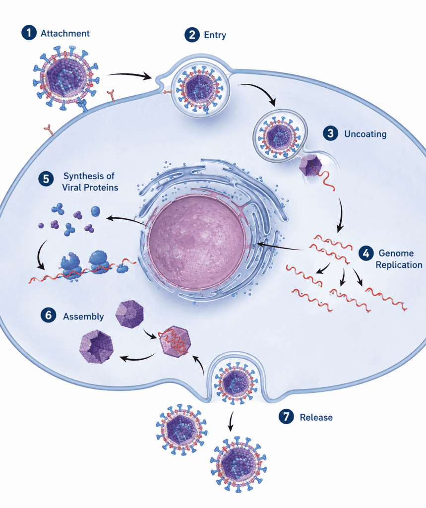
Following attachment is penetration, during which the virus or its genetic material gains entry into the host cell. Different viruses employ distinct mechanisms of entry depending on their structural properties. Enveloped viruses frequently enter through membrane fusion or receptor-mediated uptake, whereas many non-enveloped viruses gain access through endocytosis or direct penetration mechanisms. Successful entry positions the viral genome within the intracellular environment where replication can proceed.
Once inside the host cell, the virus undergoes uncoating, a process in which the protective protein coat surrounding the viral genome is removed. Uncoating releases the viral nucleic acid into the appropriate cellular compartment, making it accessible for expression and replication. The efficiency of this stage is essential because failure to expose the viral genome prevents subsequent stages of viral multiplication.
The next phase involves expression and replication of viral nucleic acid, during which viral genes are transcribed and translated using host cellular machinery. Viral genomes direct the synthesis of proteins required for replication, regulation, and structural formation. Simultaneously, multiple copies of the viral genome are generated to provide genetic material for new viral particles. The exact mechanisms differ among DNA viruses, RNA viruses, and reverse-transcribing viruses, but all aim to amplify viral genetic information.
This is followed by biosynthesis of viral components, where structural proteins such as capsid proteins and accessory molecules are produced in large quantities. These components accumulate within the host cell and prepare for the formation of complete viral particles. During assembly, newly synthesized viral genomes are packaged into structural protein shells to produce immature virions. Precise molecular interactions ensure correct organization and maturation of viral structures.
The final stage of viral replication is release, where mature virions exit the host cell to initiate new rounds of infection. Release may occur through cell rupture or controlled budding processes, depending on the viral type. This stage completes the replication cycle and enables viral dissemination within the host and transmission to new hosts.
Attachment Stage of Viral Replication
As aforementioned, viruses usually gain entry into their host cell through infection. Attachment or adsorption is the first step in viral replication; and this stage is critical because without it the infecting virus cannot gain entry into its target host cell. In adsorption (attachment), the infecting virus attaches to specific receptors on the cell membrane of its target host cell through its capsid or surface proteins and this interaction between the infecting virus and the target host cell is vital for viral entry.
Absence of a particular receptor site on the host cell membrane that the infecting virus can recognize means that the infecting virus will not attach and this ultimately prevent infection because the infecting virus cannot attach. One of the major aims of an infecting virus is to replicate its genome especially in a host cell, and in order to achieve this the virus must find a way to first of all enter the target host cell and then takeover its metabolic and cellular machinery to manufacture its own viral components so that new virions can be generated and released. But this cannot be possible if the infecting virus fails to attach itself first to specific protein molecules (inclusive of other lipoproteins, carbohydrates and glycoproteins) found on the cell or plasma membrane of the target host cell.
The first step involved in the replication of a virus is the attachment or adsorption of the infecting virus to the surface proteins found on the surface of its host cell. The protein molecules found on the capsid of the virus interact specifically with the surface receptors on the host cell; and this facilitates the entry or penetration of the infecting virus into the cell. After entry, the infecting virus uncoats and releases its nucleic acid genome (DNA or RNA) from its nucleocapsid or capsid. The released nucleic acid genome takes over the cellular machinery of the host cell and starts expressing its own genetic makeup. The expression of viral nucleic acid genome within the host cell leads to the biosynthesis of specific viral proteins and viral nucleic acids required for the assembling of new virions. Newly synthesized virions must be packaged into a complete virion or viral particle before it can be released from the host cell to infect new cells; and this is prerequisite for the infection of new cells within the organism. After proper assembling or packaging, the newly synthesized virions are released from the host cell through cell lysis (for naked viruses) or through budding (for enveloped viruses).
These receptors are unique and specific in nature, and viruses usually have several multiple protein sites that can bind to host cell receptors in a specific fashion in order to facilitate their entry into the host cell. Viral pathogenesis (i.e. the ability of a virus to cause infection in its host organism or cell) is largely dependent on the ability of the infecting virus to attach to receptors on the target host cell membrane; otherwise there will be no initiation of infection because viral entry will not be facilitated if the infecting virus fails to adsorb or attach to a specific receptor expressed the surface of a susceptible host cell.
It is noteworthy that susceptible host cells that fail to express specific receptors for viral attachment cannot be infected by the infecting virus; and the host cell can also become resistant to the infecting virus in cases where the target receptor on the surface of the host cell becomes mutated and altered in such a way that it cannot be recognized. The host cell is said to be resistant in such scenarios. However, some mutated viruses can still come up and attach to the mutated cell surface receptors. The cell surface receptors on the host cell generally determine whether the host cell will be infected or not by a particular infecting virus.
Penetration (Entry) Stage of Viral Replication
The penetration or entry stage is the phase of viral replication that follows successful attachment of the virus to the surface of a susceptible host cell. After recognizing and binding to specific cellular receptors, the virus must gain access to the intracellular environment because viral replication cannot occur outside a living host cell. Entry therefore represents a critical step in the viral life cycle, as failure to penetrate the host cell prevents subsequent replication, synthesis of viral components, and production of new virions.
Penetration refers to the process through which a virus, or more specifically its genetic material, crosses the host cell membrane and enters the cell. The primary objective of this stage is to deliver the viral genome into the appropriate intracellular compartment where replication and gene expression can occur. Since viruses lack independent metabolic systems, successful entry enables them to exploit host cellular machinery for multiplication.
One of the most common mechanisms of viral entry is endocytosis, a naturally occurring cellular process by which cells internalize external materials enclosed within membrane-bound vesicles. During viral infection, endocytosis allows the host cell membrane to surround and engulf the attached virus, forming an intracellular compartment known as an endosome. This mechanism is advantageous because many viruses exploit existing cellular transport pathways to gain access into host cells without immediately disrupting membrane integrity. The mode of penetration varies depending largely on whether the infecting virus is non-enveloped (naked) or enveloped, although receptor-mediated endocytosis remains a common pathway for both groups.
For non-enveloped viruses, entry usually occurs through endocytosis. Following receptor binding, the host cell membrane invaginates around the virus and encloses it within an endosomal vesicle. Once internalized, environmental changes within the endosome, such as alterations in pH or enzymatic activity, trigger structural modifications in the virus that facilitate escape from the vesicle and release of the viral genome into the cytoplasm. Because non-enveloped viruses lack a lipid membrane, they often rely on specialized capsid proteins to penetrate or destabilize the endosomal membrane and allow genome delivery.
In contrast, enveloped viruses possess an outer lipid envelope surrounding the viral capsid. This envelope plays an important role in facilitating entry into host cells. Many enveloped viruses enter through membrane fusion, a process in which the viral lipid envelope merges with the plasma membrane of the host cell. Fusion creates a passage through which viral contents are released directly into the cytoplasm. In other cases, enveloped viruses may first enter through endocytosis and subsequently fuse with the membrane of the endosome to release their nucleocapsid into the host cell interior. Following penetration, the virus proceeds to the next stage of replication, where uncoating occurs and the viral nucleic acid becomes available for expression and replication. Overall, the penetration stage is a highly regulated and essential event that determines the success of infection and establishes the foundation for productive viral replication within susceptible host cells.
Uncoating Stage of Viral Replication
Uncoating is a critical stage in the viral replication cycle during which the viral genetic material is released from its protective protein covering after successful entry into the host cell. This process marks the transition from viral entry to active intracellular replication because it exposes the viral genome either deoxyribonucleic acid (DNA) or ribonucleic acid (RNA) to the internal environment of the host cell where viral gene expression and genome replication can begin. Without successful uncoating, viral infection cannot progress beyond the initial stages, making this step essential for productive viral replication.
Following attachment and penetration into a susceptible host cell, viruses remain enclosed within a protein shell known as the capsid, or in some viruses, a more complex nucleocapsid composed of nucleic acid associated with viral proteins. The viral genome must be liberated from these structural components before it can access host cellular machinery. Uncoating therefore involves the physical or biochemical disassembly of the viral particle to expose the genome and permit subsequent replication events.
The mechanisms of uncoating vary among different viral groups and are influenced by both viral characteristics and host cell factors. In many viruses, uncoating is initiated by environmental changes encountered after entry into the host cell, such as alterations in pH, enzymatic activity, or interactions with intracellular compartments. Cellular enzymes and physiological conditions facilitate the breakdown of viral structural proteins, allowing the viral nucleic acid to become accessible. Some viruses complete uncoating within endocytic vesicles, while others transport their genetic material to specific intracellular sites such as the cytoplasm or nucleus before complete disassembly occurs.
Once released, the viral genome becomes functionally active and directs the host cell to support viral multiplication. At this point, viral nucleic acids begin controlling cellular biosynthetic processes by redirecting the host’s transcriptional and translational machinery toward the synthesis of viral molecules. Viral proteins required for replication, regulation, and structural assembly are produced, while copies of the viral genome are generated to support the formation of progeny virions.
Uncoating also represents an important checkpoint in host defense against viral infection. Host cells possess innate protective mechanisms that may interfere with this stage and limit viral propagation. Intracellular enzymes and antiviral responses can destabilize viral structures, degrade viral components, or prevent successful genome release. Such defenses reduce the ability of viruses to establish productive infections. Although enzymatic mechanisms contribute to antiviral protection, host immune responses generally involve multiple coordinated pathways that act before and after genome exposure.
An important consequence of uncoating is that the infecting parental virion loses its original infective identity. Once the capsid or nucleocapsid is disassembled and separated from the genome, the intact extracellular viral particle no longer exists. Infectivity is subsequently maintained through the replication and assembly of newly synthesized viral genomes into complete progeny virions. Uncoating is a decisive stage in viral replication because it enables the transition from a structurally protected viral particle to an active genetic program within the host cell. Successful genome release ultimately determines whether infection progresses to the production of new infectious viral particles.
Expression of Viral Nucleic Acid and Biosynthesis of Viral Components
After uncoating as aforementioned, the viral nucleic acid genome (DNA or RNA) becomes expressed within the host cell and this stage usually follows the central dogma of molecular biology in which DNA is transcribed to mRNA that is later translated to specific proteins. Specific mRNA must be transcribed from the viral genome (e.g. DNA) for the successful expression and duplication of the genetic information of the infecting virus. DNA replication mainly occurs in the nucleus of the infected host cell for DNA viruses; while for RNA viruses (whose genome is mainly mRNA in form), the viral RNA is mainly replicated in the cytoplasm of the cell. However, some DNA viruses (e.g. poxviruses) can still replicate their genome in the cytoplasm; and this variation is observable in some RNA viruses (e.g. retroviruses) that replicate their genome in the nucleus of the infected host cell.
Following infection, it is critical to produce new copies of the viral genome inside the host cell and this is usually accompanied by the synthesis of viral proteins as shall be highlighted later. An understanding of the replication mechanisms of infecting viruses (DNA and RNA viruses inclusive) is vital to the development of novel drugs that can interfere with and inhibit the pathogenic or virulent capabilities of pathogenic viruses especially in viral disease conditions. The synthesis of virus nucleic acid and proteins is vital to viral replication, and this is carried out by the metabolism of the host cell as directed by the infecting virus. New copies of viral genome and specific viral proteins must be synthesized in the infected host cell prior to the formal replication of the said virus. The replication of a particular virus is usually directed by the orientation or structure of its genome. Generally, the messenger ribonucleic acid (mRNA) of a virus is defined as “positive sense” (designated with the symbol ‘+’) while its complementary strand is defined as “negative sense” (designated with the symbol ‘—’).
This implies that a nucleic acid strand that has the same sequence as mRNA is designated (+) strand while the one with a complementary strand is designated (—) strand. Andsince all viruses (both DNA and RNA viruses) must express their genes or genetic makeup as functional mRNA molecules early enough in the course of their infection or disease development in vivo (i.e. within a living host cell), the mRNA (which is unique in carrying and decoding the genetic information for protein synthesis from the gene) of the infecting virus must always be in preference to the host’s mRNAs potential viral proteins must be synthesized for the coupling of new virions. Viruses as aforementioned are microbes that are composed mainly of nucleic acids and protein molecules and envelopes in the case of enveloped virus. The mRNA is responsible for the translation of genetic information it encodes (especially for the synthesis of specific viral proteins). It converts the genetic information it is carrying into a sequence of polypeptide in order to form a particular protein molecule; and this phenomenon is of utmost importance to virus because if the viral capsid (which is proteinous in nature) is not well synthesized, the viral genome will not be well packaged or assembled; and this generally exposes the virus to destruction by the host immune mechanisms and by other antiviral agents.
Thus, in order to direct the host cell’s translational machinery to make viral proteins; it is critical that the correct viral mRNAs which are designated as positive sense or negative sense depending on the genomic orientation of the virion are always available and well aligned to carry out this important genetic transformation. The phrases ‘sense’ and ‘strand’ are used synonymously in describing the genomic orientation of viruses. It is noteworthy that some viruses can have both the negative and positive strand, and for such viruses; their genomic orientation is said to be ambisense in nature. Different viral families inclusive of DNA and RNA viruses have different genomic orientation that can either be single-stranded(ss) or double stranded (ds); and the polarity of their mRNA can either be positive sense, negative sense or ambisense as the case may be. These variations or features are critical in understanding the replication pattern of each of these viruses that make up the different viral families; and because of the differences in the transcription of the genomes of DNA viruses and RNA viruses during viral replication, it is important to use these designations as aforementioned in the discussions of viral replication for clarity.
The replication pattern of a particular virus is usually determined by the structural orientation and makeup of its genome. For example, viruses with single-stranded positive sense RNA (ss+RNA) genome make use of their positive RNA (+RNA) as mRNA during transcription. These viruses uses the ribosomes and enzymes of the host cell to decode the information encoded in the +RNA genome of the virus during translation to produce protein molecules (e.g. RNA-dependent RNA polymerase). RNA-dependent RNA polymerase is responsible for transcribing the RNA genome of RNA viruses into RNA genomes so that such viruses can be further replicated. Eukaryotic cells do not possess enzymes for the replication of RNA genomes; and thus the infecting virus must provide replicase enzymes such as the RNA-dependent RNA polymerase which initiate the replication of RNA genome of viruses in the host cell(s). For viruses containing single-stranded negative sense RNA (ss-RNA) genome, their ss-RNA genome must first be converted to a +RNA strand.
The +RNA strand is used as an mRNA template for transcription to the genomic –RNA genome. And this is enzymatically catalyzed by RNA-dependent RNA polymerase proteins. In the replication of double stranded RNA (dsRNA) genome of dsRNA viruses, the –RNA strand of the dsRNA genome must first be converted to a complementary +RNA strand that will serve as a template for the replication of the viral genome. RNA-dependent RNA polymerase also plays a role in the replication of the viral genome. Retroviruses (e.g. human immunodeficiency virus, HIV) are RNA viruses that differ markedly from other RNA viruses in that the retroviruses synthesize mRNA and replicate their genome by means of an RNA-dependent DNA polymerase (also known as reverse transcriptase, RT). Hepadnaviruses are another group of animal viruses that utilizes RT in the replication of their genome apart from the viruses in the Retroviridae family (e.g. Retroviruses).
The Retroviruses have an ss+RNA. They do not use their ss+RNA genome as an mRNA template in the replication of their genome because it is not used as a messenger RNA or message. Rather, the RNA genome of Retroviruses is first transcribed into complementary DNA strands by RT in a genetic process known as reverse transcription (which is quite different from the central dogma of molecular biology – in which DNA is transcribed to RNA and then translated to protein). Double stranded DNA (dsDNA) viruses depend solely on the cellular DNA replication of their host cell since the mRNA of dsDNA viruses is transcribed in the same manner akin to DNA replication of host cells. For single stranded DNA (ssDNA) viruses, their ssDNA is first converted to a dsDNA molecule that is transcribed to form mRNA. The host RNA polymerase plays a role in the transcription of the genome of DNA viruses into mRNA; and the viral mRNAs are later translated to specific proteins in the ribosomes located within the cytoplasm of the host cell.
The newly synthesized viral proteins are transported back to the nucleus of the host cell where the progeny viral particles are assembled and released from the infected host cell(s). It is however, noteworthy that new viral proteins are required for the replication of a viral genome (whether DNA or RNA genome) within an infected host cell; and the synthesis of these viral proteins are encoded by mRNAs transcribed from the genome of the infecting virus. And as aforementioned, the viral genomic RNA can also serve as the mRNA (especially in RNA viruses), and in others, the viral genome can serve as a template for the synthesis of the mRNA – which is to be translated for viral protein synthesis. And some viruses such as the Retroviruses contain reverse transcriptase or RNA-dependent DNA polymerase that helps to carry out the transcriptional process in which the RNA genome of the virus is converted to ds DNA that act as the template for the synthesis of mRNA by normal cellular enzymes of the infected cells.
With the exception of Poxviruses and Hepadnaviruses, all animal DNA viruses replicate in the nucleus of their host cells; and the replication of viral genome varies from one virus to another depending on the genome the virus possess and its configuration i.e. whether it is double-stranded (ds) or single-stranded (ss) and whether it is positive sense (+) or negative sense (—). All RNA viruses replicate in the cytoplasm of the infected host cells with the exception of Orthomyxoviruses and Retroviruses (which can replicate in both the nucleus and cytoplasm).
Assembly Stage of Viral Replication
The assembly of synthesized viral proteins and other associated molecules or particles is critical for viral pathogenesis in living cells. The viral proteins and viral genome must be packaged into a complete virion or viral particle before it can be released from the host cell. Virions carry out a self-assembling mechanism of the viral proteins and viral genomes within the infected host cell. The release of naked viruses form the host cell after packaging is quite different from the release of enveloped viral particles. Viral assembly is the last stage in the life cycle of viral infection; and it is accompanied by the release of the complete viral particles from the infected host cell.
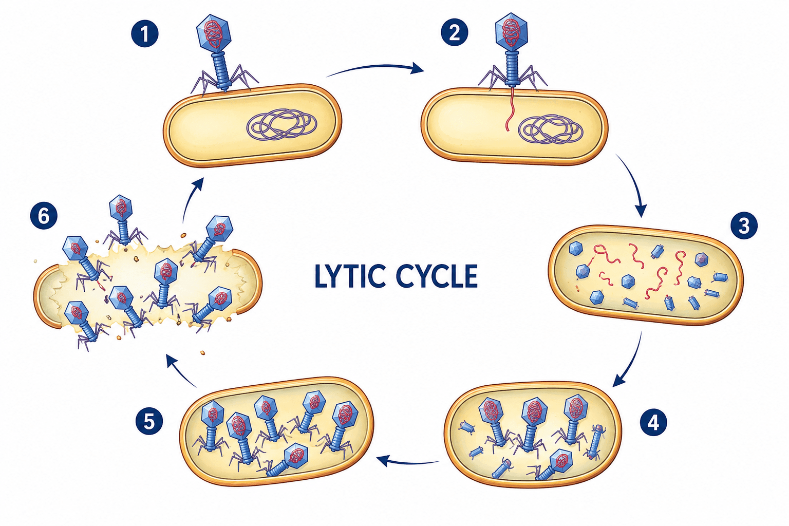
Viral replication can occur in any of two ways: lytic cycle of viral replication and lysogenic cycle of viral replication. The lytic cycle is the normal process of viral reproduction involving penetration of the cell membrane, nucleic acid synthesis, and lysis of the host cell. It involves the reproduction of viruses using a host cell to manufacture more viruses; the viruses then burst out of the cell. Viruses overtake a living host cell and use the cellular machinery of the host cell to reproduce and form its own molecules – since viruses are not capable of independent or self-replication.
The lytic cycle is a viral replication pathway in which a bacteriophage actively hijacks the host bacterium to produce new viral particles (Figure 2). It begins with attachment to the bacterial surface followed by injection of viral genetic material into the host cell. The host machinery is then redirected to replicate viral genomes and synthesize viral proteins. Newly assembled virions accumulate inside the cell until the host membrane is enzymatically broken down. This results in cell lysis and the release of progeny viruses that can infect neighboring cells.
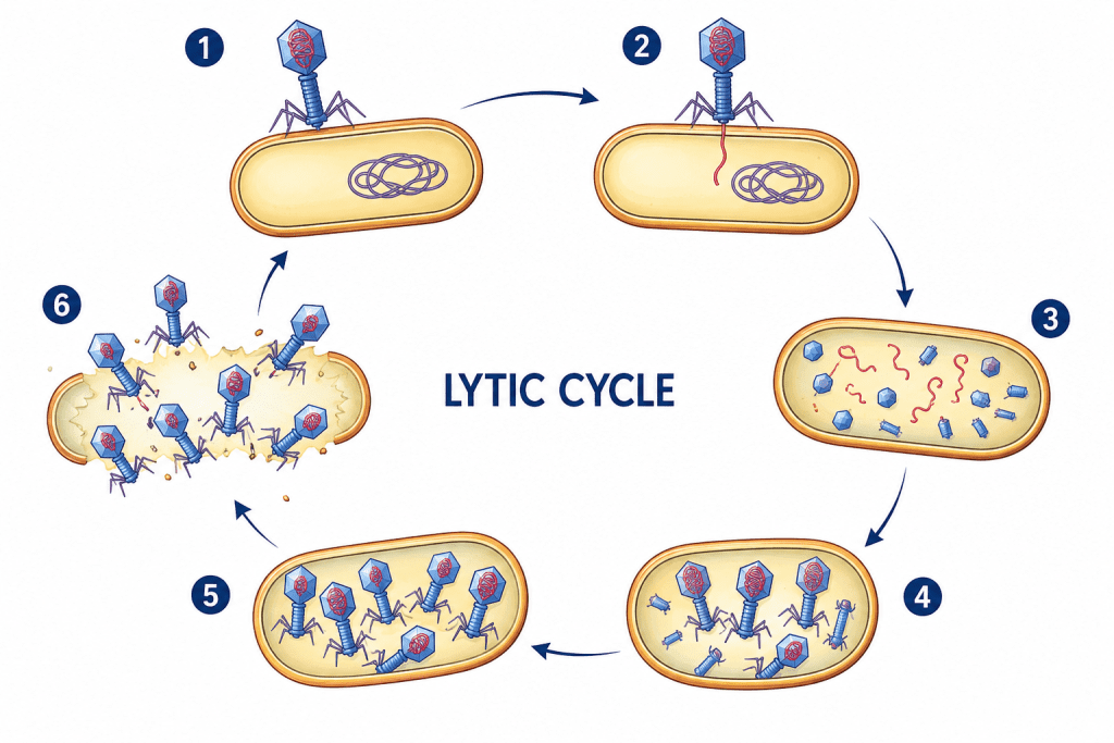
And one of the ways the infecting virus may choose to leave the affected host cell is by destroying the host cell. Viruses usually leave their host cell by cutting (lysing) their way out of the host cell. This is called the lytic cycle of a virus replication. In a lytic cycle, the virus reproduces thousands to millions of times in just a few hours and they produce many viral progeny during this time or process. This result to the weakening of the host cell wall enough that the cell will lyse, or burst open, setting the army of new viruses free. The lytic cycle results in the destruction of the infected cell and its membrane unlike the lysogenic cycle which does not lead to the destruction of the host cell and its membrane.
The lysogenic cycle involves the incorporation of the viral genome into the host cell genome, this infecting the host cell from within. It is a form of viral reproduction involving the fusion of the nucleic acid of a bacteriophage with that of a host, followed by proliferation of the resulting prophage. In the lysogenic cycle, the phage DNA first integrates into the bacterial chromosome to produce the prophage. When the bacterium reproduces, the prophage is also copied and is present in each of the daughter cells. The daughter cells can continue to replicate with the prophage present or the prophage can exit the bacterial chromosome to initiate the lytic cycle. Prophage is the latent form of the virus genome that remains within the host cell without destroying it.
The lysogenic cycle is a viral replication strategy in which the viral genome integrates into the host bacterium’s chromosome instead of immediately producing new virions (Figure 3). In this dormant state, the integrated viral DNA called a prophage replicates passively as the host cell divides, allowing the virus to persist without killing the host. Environmental triggers such as UV radiation or chemical stress can activate the prophage, causing it to exit the genome and enter the lytic cycle. Once induced, the virus rapidly produces new particles, leading to host cell lysis and release of progeny phages. This cycle enables long-term viral survival while maintaining a latent infection within bacterial populations.
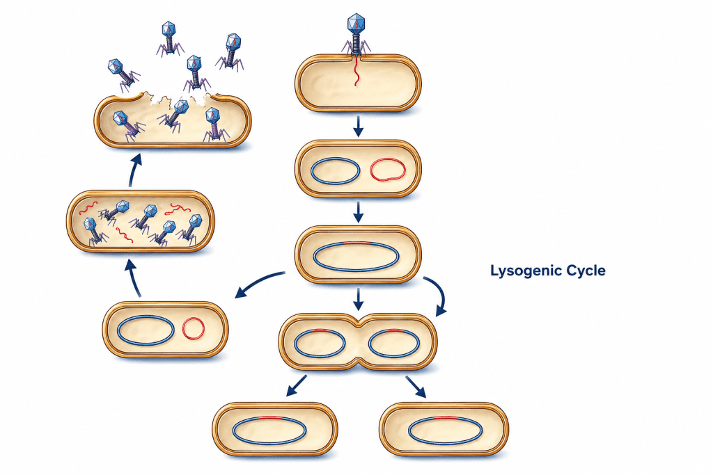
The difference between the lytic and lysogenic cycles of viral replication is that in the lytic phage, the viral DNA exists as a separate molecule within the bacterial (host) cell, and replicates separately from the host (bacterial) DNA while the location of viral DNA in the lysogenic cycle is within the host (bacterial) DNA. And while the lytic cycle of viral replication leads to the destruction of the host cell and membrane, the lysogenic cycle of viral replication does not lead to the destruction or lysis of the host cell and its membrane.
Lytic cycle is a type of viral life cycle that ends with host cell lysis and the release of numerous and newly synthesized viral progenies. This type of viral replication (i.e. lytic cycle) is usually carried out by virulent viruses that naturally lyse their host cells during the reproductive cycle. However, in lysogenic cycles, viruses engage in a different type of relationship with their host. And in this type of viral replication (i.e. lysogenic cycle), the viral genome does not take total control of the host and destroy it while it is still synthesizing new virions or phages as the case may be.
In lysogenic cycle, the viral genome remains within the host cell and replicates its own genome alongside the host (bacterial) genome to produce new cells that continue to grow and divide over a long period of time. The infected host cell looks normal; and each of the infected host cells can go on to produce new virions or phages which will lyse under normal conditions. The infecting virus establishes a type of relationship known as lysogeny – in which both the virus and the host cell coexist without destroying each other.
Lysogeny is defined as a relationship in which a virus or phage genome remains within its host cell (e.g. a bacterial cell) after infection and reproduces alongside the host genome instead of taking total control of the host cell and destroying it in the process of replication. The viruses that enter into this type of relationship with their host cell are known as temperate phages while the bacterial host cell that are able to produce new viral or phage particles under these conditions are known as lysogens; and they are said to be lysogenic.
Release of Mature and Complete Virions
The release of mature and complete virions represents the final stage of the viral replication cycle and marks the transition from intracellular viral multiplication to the spread of infection within the host. At this stage, newly synthesized viral genomes and structural proteins have been successfully assembled into complete infectious particles capable of initiating subsequent rounds of infection. The mechanism through which viruses leave infected cells varies depending largely on whether the virus possesses an outer lipid envelope. Once assembly and packaging of viral nucleic acid are completed, the newly formed virions must exit the host cell to infect neighboring cells or disseminate to distant tissues. This release process is essential for viral survival and transmission because virions confined within host cells cannot establish productive infections elsewhere.
For naked (non-enveloped) viruses, release commonly occurs through cell lysis or programmed cell death pathways. During lytic release, the accumulation of viral particles disrupts normal cellular integrity, resulting in rupture of the plasma membrane and destruction of the infected cell. This process liberates large numbers of mature virions simultaneously into the extracellular environment. Some naked viruses may also induce apoptosis, a regulated form of cell death, which facilitates viral dissemination while limiting certain inflammatory responses. Because naked viruses do not possess a lipid membrane, they do not require membrane acquisition during release and are typically discharged immediately once virion maturation and packaging are completed.
In contrast, enveloped viruses employ a more controlled release mechanism known as budding. During this process, mature nucleocapsids migrate toward regions of the host cell membrane that contain viral envelope proteins previously inserted into the lipid bilayer. As the nucleocapsid pushes outward through the membrane, it becomes enclosed within a portion of the host-derived lipid membrane, thereby acquiring its characteristic viral envelope. This envelope contains embedded viral glycoproteins that play essential roles in attachment and entry into future host cells.
The maturation of enveloped viruses frequently occurs concurrently with the budding process. In many cases, virions become fully infectious only after undergoing structural and biochemical modifications during or immediately following release. Unlike lytic release, budding may allow prolonged survival of the infected host cell and continuous production of viral particles over an extended period. Consequently, enveloped viruses can establish persistent infections while minimizing immediate cellular destruction.
Following release, mature virions encounter and infect new susceptible host cells. Viral infection begins with attachment to specific cellular receptors, followed by entry and disassembly of the viral particle, which releases the viral genome into the intracellular environment to initiate another replication cycle. The outcome of viral release and subsequent disease development depends on multiple interacting factors. These include the quantity of virions released, the virulence and replication efficiency of the infecting virus, the host immune status, tissue and organ involvement, environmental influences, and the duration of viral persistence within the host. Therefore, viral release not only completes the replication cycle but also plays a central role in determining transmission dynamics, pathogenicity, and the overall progression of viral disease.
HOST IMMUNITY TO VIRAL INFECTION
Most viral infections do not result to disease development with the exception of AIDS and some haemorrhagic viral infections such as Ebola and Lassa fever that leads to serious disease in the affected host and even death. The effectiveness of the immune system of the viral-infected host is one singular factor there is that can significantly affect the outcome of a particular viral infection in an individual. Even though the immune system may not eliminate totally the infecting virus, a strong immune system helps to contain the pathogenicity or virulence of a given pathogenic virus.
The administration of potent vaccines and other antiviral agents to affected hosts and other susceptible host can help to treat the disease or infection and even prevent the contamination of a particular viral infection respectively. Vaccine development (which is mainly based on the isolation or cultivation, attenuation or inactivation and the direct or indirect injection of a susceptible host with killed, live or purified subunits of causative pathogenic microorganism) has saved mankind from the onslaught of some life-threatening infectious diseases such as measles and smallpox through the control and the complete eradication of these diseases in some cases.
The timely vaccination of a large population of susceptible host against a particular viral infection helped to minimize the spread of the pathogen in the immunized populace via herd immunity. And vaccination/immunization has helped to increase the life expectancy of mankind through the protection of the population from life-threatening diseases especially those caused by pathogenic viruses. Viral infection unlike other microbial infections stimulate an immune response in the host; and this serves to protect the host from immediate attack and even from futuristic viral infection through specific immune responses.
Pathogenic viruses as obligate intracellular microorganisms engage in uniquely intimate host-parasite relationships with the living organisms (plant, animal or man) that they infect, and this is due in part to the fact that viruses only exist or replicate in living cells. In the course of their pathogenicity or virulence in a particular host, pathogenic viruses express gene products that act to circumvent one or more of the several antiviral defense mechanisms (e.g. production of interferons) developed by the host organisms.
Nevertheless, the host resistance to viral infections involves both the humoral immunity and cell-mediated immunity. While some viral infections (e.g. influenza and common colds) can be contained by the host’s immune system; some others such as the causative agent of AIDS (i.e. HIV) overpowers the host’s immune system and makes it incapable to fight against the invading viral agent. Infections with some pathogenic viruses may lead to apoptosis; and this programmed cell death is a host defense mechanism that can be inhibited by some viruses.
Antibodies produced by humoral immunity can neutralize pathogenic viruses by interfering with their attachment to host cells; and the production of antibodies also enhances the destruction of viral particles via phagocytosis. However, antibodies cannot completely eliminate the infecting virus once the virion has incorporated its genome into that of the host. The cell-mediated immunity is one of the major important arms of the immune system that interfere with viral replication in vivo.
Activated lymphocytes including cytotoxic T lymphocytes (CTL), helper T cells (CD+4) and cytotoxic T cells (CD+8) can recognize and destroy viral infected cells especially when these organisms bud off from their host cells. The production of interferons (which are protein substances produced by cells during viral infection) helps to reduce the spread of virus especially in some benign viral infections such as influenzae and cold.
They stimulate the production of natural killer (NK) cells and T cells; and interferons also accelerate the immune response of a host to viral infection. And by acting on other effector cells of the immune system, interferons (which are antiviral cytokines) generally reduce the susceptibility of other uninfected cells of the host to the invading virus, and they do so by localizing the pathogenic virus so that they do not easily spread in the host’s body.
References
Acheson N.H (2011). Fundamentals of Molecular Virology. Second edition. John Wiley and Sons Limited, West Sussex, United Kingdom.
Alan J. Cann (2005). Principles of Molecular Virology. 4th edition. Elsevier Academic Press, Burlington, MA, USA.
Alberts B, Bray D, Johnson A, Lewis J, Raff M, Roberts K and Walter P (1998). Essential Cell Biology: An Introduction to the Molecular Biology of the Cell. Third edition. Garland Publishing Inc., New York.
Barrett J.T (1998). Microbiology and Immunology Concepts. Philadelphia, PA: Lippincott-Raven Publishers. USA.
Black, J.G. (2008). Microbiology: Principles and Explorations (7th ed.). Hoboken, NJ: J. Wiley & Sons.
Brian W.J Mahy and Mark H.C van Regenmortel (2010). Desk Encyclopedia of Human and Medical Virology. Elsevier Academic Press, San Diego, USA.
Brooks G.F., Butel J.S and Morse S.A (2004). Medical Microbiology, 23rd edition. McGraw Hill Publishers. USA.
Cann A.J (2011). Principles of Molecular Virology. Fifth edition. Academic Press, San Diego, United States.
Carter J and Saunders V (2013). Virology: Principles and Applications. Second edition. Wiley-Blackwell, New Jersey, United States.
Champoux J.J, Neidhardt F.C, Drew W.L and Plorde J.J (2004). Sherris Medical Microbiology: An Introduction to Infectious Diseases. 4th edition. McGraw Hill Companies Inc, USA.
Dimmock N (2015). Introduction to Modern Virology. Seventh edition. Wiley-Blackwell, New Jersey, United States.
Dimmock N.J, Easton A.J and Leppard K.N (2001). Introduction to modern virology. 5th edition. Blackwell Science publishers. Oxford, UK.
Discover more from Microbiology Class
Subscribe to get the latest posts sent to your email.