AIM: To detect the presence of pus cells and predominant bacteria in sputum specimens as an aid in the diagnosis of lower respiratory tract infections like pneumonia or bronchopneumonia.
MATERIAL/APPARATUS: Sputum specimen, glass slide, microscope, immersion oil, inoculating loop, Bunsen burner, piece of stick.
METHOD/PROCEDURE FOR SPUTUM SAMPLE MICROSCOPY
- Transfer a purulent part of the sputum specimen to a clean glass slide using a sterilized inoculating loop or a piece of clean stick.
- Make a thin smear of the purulent sputum on the glass slide.
- Place the slide in a safe place away from dust for it to dry.
- Heat fix the smear on the slide by passing it thrice over a Bunsen burner flame.
- Perform Gram staining technique.
- Allow the Gram stained slide to dry. Add a drop of immersion oil on the slide.
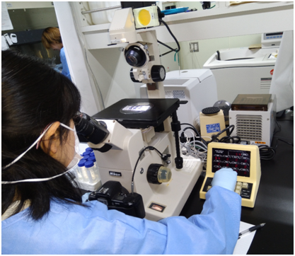
- Examine/observe the stained slide under the microscope using the ×100/oil immersion objective lens; and look out for pus cells and predominant bacteria in the smear.
REPORTING OF THE RESULT: Describe and report the appearance of the sputum specimen. The appearance of a sputum specimen can be described with any of the following parameters:
- Purulent sputum: Green – looking, mostly pus.
- Mucopurulent sputum: Green – looking with pus and mucus.
- Mucoid sputum: Mostly mucous.
- Muco-salivary sputum: mucous with a small amount of saliva.
- Bloody sputum: Sputum containing some amount of blood.
NOTE: The presence of blood in a sputum specimen can be due to Paragonimiasis caused by the parasite, Paragonimus westermani. P. westermani is a human lung fluke which affects the lungs.
Examine the slide carefully under the microscope and look especially for:
- Capsulated Gram positive diplococcic (e.g. Streptococcus pneumoniae).
- Gram positive cocci in groups (e.g. Staphylococcus aureus).
- Capsulated Gram negative rods (e.g. Klebsiella pneumoniae).
- Gram negative rods and coccobacilli (e.g. Haemophilus influenzae).
- Gram negative diplococci that are usually found in and between pus cells (e.g. Moraxella catarrhalis).
The result of a Gram stained smear of sputum specimen should be reported with utmost care and caution, as cocci, diplococcic, rods, and streptococci can also be seen in a normal sputum sample as normal flora. This is because these organisms form part of the normal microbial flora of the human upper respiratory tract. The clinical presentation or diagnosis of the patient as given by the physician can also help in taking care of this.
References
Basic laboratory procedures in clinical bacteriology. World Health Organization (WHO), 1991. Available from WHO publications, 1211 Geneva, 27-Switzerland.
Beers M.H., Porter R.S., Jones T.V., Kaplan J.L and Berkwits M (2006). The Merck Manual of Diagnosis and Therapy. Eighteenth edition. Merck & Co., Inc, USA.
Biosafety in Microbiological and Biomedical Laboratories. 5th edition. U.S Department of Health and Human Services. Public Health Service. Center for Disease Control and Prevention. National Institute of Health. HHS Publication No. (CDC) 21-1112.2009.
Cheesbrough M (2010). District Laboratory Practice in Tropical Countries. Part I. 2nd edition. Cambridge University Press, UK.
Cheesbrough M (2010). District Laboratory Practice in Tropical Countries. Part 2. 2nd edition. Cambridge University Press, UK.
Collins C.H, Lyne P.M, Grange J.M and Falkinham J.O (2004). Collins and Lyne’s Microbiological Methods. Eight edition. Arnold publishers, New York, USA.
Disinfection and Sterilization. (1993). Laboratory Biosafety Manual (2nd ed., pp. 60-70). Geneva: WHO.
Garcia L.S (2010). Clinical Microbiology Procedures Handbook. Third edition. American Society of Microbiology Press, USA.
Garcia L.S (2014). Clinical Laboratory Management. First edition. American Society of Microbiology Press, USA.
Fleming, D. O., Richardson, J. H., Tulis, J. I. and Vesley, D. (eds) (1995). Laboratory Safety: Principles and practice. Washington DC: ASM press.
Dubey, R. C. and Maheshwari, D. K. (2004). Practical Microbiology. S.Chand and Company LTD, New Delhi, India.
Gillespie S.H and Bamford K.B (2012). Medical Microbiology and Infection at a glance. 4th edition. Wiley-Blackwell Publishers, UK.
Discover more from Microbiology Class
Subscribe to get the latest posts sent to your email.