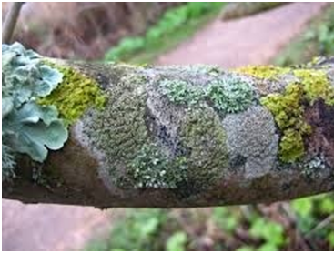
Lichen is simply a symbiotic association of slow-growing microorganisms that is composed mainly of a fungus and cyanobacteria or green algae. They are a composition of twin or double organisms, and are very rich in chemical compounds as expressed by the varying colours that they produce on the surfaces where they form. Lichens grow on a wide variety of surfaces including tree trunks, roof tops of houses, bare soils and rock surfaces where they form colourful growths that may include yellow, red, black, white, oranges, green and brown [Figure 1].
Though an association between cyanobacteria (a prokaryote) and fungi are often the most versatile lichen association known; green algae (a eukaryote) can also go into association with a fungus (a eukaryote) to form a lichen relationship. Lichens are widespread and can be found in both terrestrial and aquatic environments. They can reproduce sexually and asexually, and the morphology of lichens is usually made up of a lichen thallus that comprises of fungal hyphae (used for attachment to surfaces) and photobiont or phycobiont cells (used for photosynthetic activity).
The fungal hyphae of the lichen are the main determinants of lichen structure or morphology, and it is generally referred to as mycobiont. Cyanobacteria are assemblages of bacteria that have the innate ability to carry out oxygenic photosynthesis. The two partners in a lichen association are usually a fungus and a cyanobacteria or green algae. The fungal partner is called a mycobiont while the cyanobacteria partner is known as a phycobiont.
Notably, it is not all species or genera of fungi and cyanobacteria that can form a mutualistic lichen association. Ascomycetes (commonly known as sac fungi) are the class of fungi that go into symbiotic association with a cyanobacterium to form lichens. Both organisms in a lichen association interact amongst each other on only a mutual basis. They both derive a reciprocated type of benefit from each other such as nutrient sharing and protection from untoward effects in their surrounding environment.
While the fungus mainly provides structure, absorb water and provide protection for the cyanobacterium, the cyanobacteria on the other hand is highly photosynthetic and thus acts as the primary food producer by providing carbohydrates for both itself and its fungal partner in the lichenized association.
Their ability tocolonize any surface is based on this mutual relationship between the two organisms in the lichen association. Most lichen association use cyanobacteria as their photosynthetic partner. Due to their ability to tolerate extreme conditions in their environment, cyanobacteria are widespread and ubiquitous in the soil, water and on rock surfaces.
On the surfaces of lakes, streams, rivers and ponds, cyanobacteria (e.g. Nostoc spp. and Anabaena spp.) are notorious in forming blooms (community of microorganisms comprising mainly of algae and cyanobacteria) that cover water surfaces and render it almost unhygienic and dirty-looking.
CATEGORY OF LICHENS
Lichens are amongst one of the least forms of life known to mankind. Their very nature which comprises of a symbiosis of two different organisms (a fungus and a cyanobacterium) is unique and this gives them the expertise to inhabit environmental areas which are usually harsh to other microbial forms of life.
Lichens unlike other microbial forms of life come in various colours and structures; and they can inhabit a wide variety of surfaces such as tree barks, rocks, streams, leave surfaces and so on. They provide microhabitats for other forms of life such as insects and other small animals. Lichens exist in three different forms:
- Crustose lichens: Crustose lichens are crust-like lichens which usually inhabit the bark of trees, soils and rock surfaces. They have a flat structure (thallus) that is securely adhered to the lower surfaces of its substrate. They form a variety of colours that includes yellow, green and red. Examples of crustose lichens include Acarospora species and Lecanora species.
- Fruticose lichens: Fruticose lichens are the most highly developed forms of lichens. They can also be referred to as stalked lichens due to their structure and ability to form fruiting bodies. Usnea species, the source of usnic acids used as potential lead compounds for antiviral drug development belong to this category of lichens. Fruticose lichens form on trees and on soils.
- Foliose lichens: They are leaf-like lichens which also from on tree trunks and rock surfaces. They have a characteristic puffed body with a black undersurface. Examples of foliose lichens include Pseudocyphellaria species and Hypogymnia species.
References
Anaissie E.J, McGinnis M.R, Pfaller M.A (2009). Clinical Mycology. 2nd ed. Philadelphia, PA: Churchill Livingstone Elsevier. London.
Beck R.W (2000). A chronology of microbiology in historical context. Washington, D.C.: ASM Press.
Black, J.G. (2008). Microbiology: Principles and Explorations (7th ed.). Hoboken, NJ: J. Wiley & Sons.
Brooks G.F., Butel J.S and Morse S.A (2004). Medical Microbiology, 23rd edition. McGraw Hill Publishers. USA.
Brown G.D and Netea M.G (2007). Immunology of Fungal Infections. Springer Publishers, Netherlands.
Calderone R.A and Cihlar R.L (eds). Fungal Pathogenesis: Principles and Clinical Applications. New York: Marcel Dekker; 2002.
Chakrabarti A and Slavin M.A (2011). Endemic fungal infection in the Asia-Pacific region. Med Mycol, 9:337-344.
Champoux J.J, Neidhardt F.C, Drew W.L and Plorde J.J (2004). Sherris Medical Microbiology: An Introduction to Infectious Diseases. 4th edition. McGraw Hill Companies Inc, USA.
Chemotherapy of microbial diseases. In: Chabner B.A, Brunton L.L, Knollman B.C, eds. Goodman and Gilman’s The Pharmacological Basis of Therapeutics. 12th ed. New York, McGraw-Hill; 2011.
Chung K.T, Stevens Jr., S.E and Ferris D.H (1995). A chronology of events and pioneers of microbiology. SIM News, 45(1):3–13.
Germain G. St. and Summerbell R (2010). Identifying Fungi. Second edition. Star Pub Co.
Ghannoum MA, Rice LB (1999). Antifungal agents: Mode of action, mechanisms of resistance, and correlation of these mechanisms with bacterial resistance. Clin Microbiol Rev, 12:501–517.
Gillespie S.H and Bamford K.B (2012). Medical Microbiology and Infection at a glance. 4th edition. Wiley-Blackwell Publishers, UK.
Larone D.H (2011). Medically Important Fungi: A Guide to Identification. Fifth edition. American Society of Microbiology Press, USA.
Levinson W (2010). Review of Medical Microbiology and Immunology. Twelfth edition. The McGraw-Hill Companies, USA.
Madigan M.T., Martinko J.M., Dunlap P.V and Clark D.P (2009). Brock Biology of Microorganisms, 12th edition. Pearson Benjamin Cummings Inc, USA.
Mahon C. R, Lehman D.C and Manuselis G (2011). Textbook of Diagnostic Microbiology. Fourth edition. Saunders Publishers, USA.
Discover more from Microbiology Class
Subscribe to get the latest posts sent to your email.