
The increasing prevalence of microbial infections and the continuous emergence of microorganisms with reduced susceptibility to existing antimicrobial agents have intensified the need for reliable methods to assess antimicrobial performance. In research, healthcare, pharmaceutical development, food safety, environmental monitoring, and biotechnology, determining whether an antimicrobial substance can effectively inhibit or eliminate microorganisms is an essential component of evaluating its potential application. The measurement of antimicrobial efficacy or activity therefore provides an important link between the development of an antimicrobial intervention and the evidence required to demonstrate its biological performance.
Antimicrobial substances encompass a broad range of naturally occurring, synthetic, and semi-synthetic agents designed to act against microorganisms. Their effectiveness, however, cannot be assumed solely from their chemical composition or intended use. Antimicrobial performance may vary substantially according to the target microorganism, concentration of the active substance, exposure conditions, environmental characteristics, and experimental design. Systematic measurement is necessary to establish whether an antimicrobial agent produces a meaningful response under defined conditions and to allow its performance to be compared across different formulations, organisms, and applications.
The assessment of antimicrobial activity has developed into an important area of microbiological investigation, supported by a range of laboratory approaches. These approaches generate measurable evidence of microbial responses following exposure to an antimicrobial agent and can provide information about the extent and nature of its effect. Depending on the purpose of the investigation, measurements may focus on inhibition of microbial growth, reduction in viable microorganisms, changes in microbial population over time, or the effectiveness of an intervention under conditions that resemble its intended application. The choice of measurement approach consequently has a direct influence on how antimicrobial performance is interpreted.
Reliable assessment is particularly important because antimicrobial activity is influenced by the interaction between the antimicrobial agent and its biological and environmental context. An agent that demonstrates substantial activity under controlled laboratory conditions may behave differently when tested in a more complex system. Variations in microbial species or strains, inoculum characteristics, contact conditions, medium composition, temperature, exposure duration, and the physical properties of the antimicrobial formulation can all affect observed outcomes. Measurement therefore extends beyond obtaining a numerical result; it involves generating reproducible evidence that can support meaningful interpretation of antimicrobial performance.
The measurement of antimicrobial efficacy/activity also contributes to the standardisation and validation of antimicrobial technologies. Consistent assessment enables researchers to evaluate newly developed compounds, formulations, materials, and treatment strategies against established performance expectations. It further supports comparisons between experimental findings and helps identify differences in antimicrobial performance that may otherwise remain difficult to distinguish.
Against this background, the measurement of antimicrobial efficacy/activity represents a fundamental aspect of antimicrobial research. It provides the experimental basis for determining how effectively an antimicrobial intervention performs against selected microorganisms and under specified conditions.
Major methods for antimicrobial susceptibility testing (AST)
The antimicrobial susceptibility of a particular pathogen to a selected antimicrobial agent can be determined through laboratory procedures that evaluate the organism’s response to controlled exposure. These methods are central to microbiological investigations because they provide an experimental basis for distinguishing susceptible organisms from those that exhibit reduced or limited responsiveness to an antimicrobial compound. Although susceptibility testing can be performed using several specialised techniques, the principal approaches are broadly grouped into diffusion methods and dilution methods. Each category incorporates different procedures and generates information that can be interpreted according to established microbiological criteria.
1. Disk diffusion method
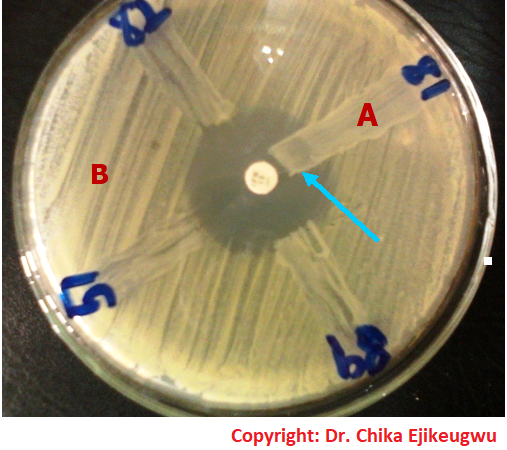
The disk diffusion method, commonly associated with the Kirby-Bauer technique, is one of the most widely used qualitative or semi-quantitative approaches for AST. The Kirby-Bauer technique (disk diffusion test) is a standardized laboratory method used to measure how well specific bacteria respond to various antibiotics. It is a standardized technique that is used for determining the susceptibility of bacteria to antimicrobial agents. In performing the Kirby-Bauer technique or disk diffusion test, a standardised microbial inoculum is distributed across the surface of an appropriate solid growth medium, after which paper disks containing defined amounts of antimicrobial agents are positioned on the inoculated surface. During incubation, the antimicrobial compound diffuses outward from the disk and interacts with the growing microorganism.
Where the organism is susceptible, microbial growth is inhibited around the antimicrobial disk, producing a visible zone of inhibition (Figure 1). The diameter of this zone is measured and interpreted using appropriate susceptibility criteria. Larger or smaller zones do not simply represent antimicrobial potency in isolation because diffusion characteristics, growth conditions, inoculum density, and the properties of the test organism can influence the observed diameter.
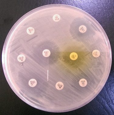
The disk diffusion approach may be applied to a wide variety of clinically relevant organisms and antimicrobial agents. For example, the susceptibility of Staphylococcus aureus to antibiotics such as oxacillin or erythromycin, or that of Escherichia coli to agents such as ciprofloxacin or gentamicin, may be assessed using appropriately selected disks. Variations of diffusion-based testing include well diffusion, in which antimicrobial solutions are introduced into wells cut into an inoculated agar surface, and agar plug or agar diffusion techniques, which can be useful for investigating antimicrobial compounds produced by microorganisms or plant-derived materials.
The Clinical and Laboratory Standards Institute (CLSI) provides standardised criteria for interpreting disk diffusion results, with zone-diameter breakpoints used to classify bacterial isolates according to their expected antimicrobial response. In CLSI M100, these breakpoints allow measured inhibition zones to be categorized as susceptible (S), intermediate (I), or resistant (R) categories, thereby translating a measured zone diameter into a clinically meaningful susceptibility result. The appropriate breakpoint is organism- and antimicrobial-specific and should be based on the current CLSI standards.
2. Dilution method
The dilution method determines antimicrobial susceptibility by exposing microorganisms to progressively varying concentrations of an antimicrobial agent. Unlike disk diffusion, this approach directly examines microbial growth across a concentration gradient and is particularly valuable when a quantitative estimate of antimicrobial activity is required.
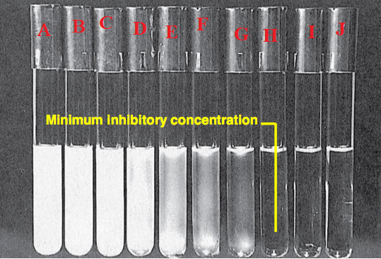
Two principal forms are broth dilution and agar dilution. Broth dilution may be performed as macrodilution or microdilution, depending on the scale and format of the test. In broth microdilution, serially decreasing concentrations of an antimicrobial agent are prepared in wells of a microtitre plate and inoculated with the test organism. Following incubation, microbial growth is assessed, commonly by observing turbidity or another measurable indicator. The lowest concentration that prevents visible growth is designated the minimum inhibitory concentration (MIC). Further testing of organisms from inhibitory concentrations can be used to determine the minimum bactericidal concentration (MBC) or corresponding minimum fungicidal concentration where appropriate.
The MIC is the lowest concentration of an antimicrobial agent that prevents visible growth of a microorganism under defined laboratory conditions (Figure 2). It is commonly determined using broth or agar dilution techniques, in which microorganisms are exposed to progressively decreasing concentrations of the test agent. Following incubation, the concentration at which no detectable microbial growth occurs is identified as the MIC. This measurement provides a quantitative indication of antimicrobial activity and is useful for comparing the susceptibility of different microorganisms or evaluating the relative effectiveness of antimicrobial agents.
The MBC is the lowest concentration of an antimicrobial agent capable of killing a microorganism rather than merely suppressing its growth. It is generally determined after MIC testing by transferring samples from concentrations showing no visible growth onto antimicrobial-free growth media. Following incubation, the lowest concentration that produces no subsequent microbial growth is considered the MBC. This measurement complements the MIC by distinguishing inhibitory effects from lethal activity and can therefore provide additional information about the antimicrobial agent’s killing capacity.
Agar dilution follows a related principle, with defined antimicrobial concentrations incorporated into solid media before inoculation. Diffusion and dilution approaches provide complementary means of characterizing antimicrobial susceptibility, ranging from comparative inhibition patterns to concentration-based measurements of microbial response.
The CLSI provides standardized criteria for interpreting antimicrobial dilution tests, particularly MIC results. Following the CLSI M07 methodology, the measured MIC is compared with the organism- and antimicrobial-specific clinical breakpoints published in CLSI M100. These breakpoints allow the MIC to be assigned an interpretive category, such as susceptible, intermediate, susceptible-dose dependent, or resistant, thereby translating a quantitative laboratory result into clinically meaningful susceptibility information.
Agar disk diffusion method
The agar disk diffusion method is a standardized AST technique in which antimicrobial-impregnated disks are placed on the surface of an agar medium inoculated with a test microorganism, allowing the agent to diffuse through the medium and produce a measurable zone of growth inhibition. The resulting inhibition pattern provides a qualitative basis for categorizing the microorganism as susceptible, intermediate, or resistant to the antimicrobial agent according to established interpretive standards.
The method is among the most extensively adopted approaches for routine antimicrobial susceptibility testing because it combines procedural simplicity, relatively low cost, reproducibility, and the capacity to evaluate several antimicrobial agents simultaneously. It is particularly suitable for many rapidly growing aerobic and facultatively anaerobic bacteria, including clinically important organisms such as Staphylococcus aureus, Escherichia coli, and Pseudomonas aeruginosa. Its broad applicability has made it an established component of diagnostic microbiology and antimicrobial surveillance in laboratories worldwide.
The procedure begins with the preparation of a suitable agar plate and a standardized suspension of the microorganism under investigation. The bacterial suspension is distributed evenly across the agar surface to produce a confluent or near-confluent lawn of growth after incubation. Paper disks containing predetermined quantities of selected antimicrobial agents are then positioned on the inoculated surface. Each disk serves as a localized source of the antimicrobial compound, allowing the substance to migrate outward through the hydrated agar.
As diffusion proceeds, the antimicrobial agent establishes a concentration gradient around the disk. Concentrations are highest close to the disk and progressively decrease with increasing distance from its centre. During incubation, microorganisms that are sufficiently susceptible to the agent fail to grow within an area where the antimicrobial concentration is inhibitory. This produces a distinct, usually circular zone of inhibition, while organisms capable of tolerating the antimicrobial concentration continue to grow closer to the disk.
Following incubation under the specified conditions, the diameter of each inhibition zone is measured in millimetres. The observed measurement is then interpreted using the appropriate breakpoint criteria established by recognized standards, such as those provided by the CLSI or other applicable susceptibility-testing frameworks. Interpretation does not depend solely on whether a zone appears large or small; rather, the measured diameter is evaluated against organism-drug-specific interpretive thresholds. This permits the isolate to be assigned to an appropriate susceptibility category.
The Kirby-Bauer disk diffusion method represents the best-known standardized form of this approach. Its value extends beyond determining the response of an individual isolate because multiple antimicrobial agents can be tested concurrently on the same agar surface, provided that appropriate spacing and test conditions are maintained. This makes the technique particularly practical when an organism must be examined against a panel of therapeutically relevant agents.
An important consideration is that the diameter of an inhibition zone cannot be interpreted as a direct measure of antimicrobial potency or used to rank different antimicrobial agents simply according to zone size. Diffusion characteristics, molecular properties of the antimicrobial, agar composition, inoculum density, incubation conditions, and organism-specific factors can all influence the resulting zone. Valid interpretation requires standardized methodology and the appropriate susceptibility breakpoints rather than visual comparison alone.
In terms of its clinical relevance, the agar disk diffusion provides clinically useful susceptibility information that can assist in selecting appropriate antimicrobial therapy for bacterial infections, recognizing resistant isolates, supporting antimicrobial stewardship, and monitoring changing resistance patterns. When performed and interpreted according to standardized procedures, it remains a practical and valuable tool for translating laboratory observations into clinically relevant antimicrobial susceptibility profiles.
Standardized procedure for the Kirby-Bauer disk diffusion method
The Kirby-Bauer disk diffusion method is performed under controlled conditions to ensure that differences in antimicrobial inhibition reflect the response of the test organism rather than inconsistencies in inoculum density, medium preparation, antimicrobial concentration, or incubation. A standardized bacterial suspension is therefore prepared before inoculation of the test medium. The following sequence describes the principal stages involved in carrying out the procedure in the laboratory:
1. Preparation of the bacterial inoculum: Using a sterile inoculating loop, select approximately 2-3 well-isolated colonies of the test bacterium from a fresh agar culture. The selected colonies should represent the organism being investigated and should be free from obvious contamination. Transfer the colonies aseptically into a sterile tube containing an appropriate volume of nutrient broth and mix thoroughly to produce a uniform bacterial suspension.
2. Incubation and standardization of turbidity: Incubate the inoculated nutrient broth at approximately 37°C for 3-4 hours, or until a light, visible turbidity develops. The purpose of this stage is to obtain sufficient bacterial growth for preparation of a standardized inoculum. After incubation, compare the turbidity of the bacterial suspension with a 0.5 McFarland turbidity standard, which provides a reference for achieving a consistent inoculum density.
If the suspension appears more concentrated or less turbid than the selected standard, its density should be appropriately adjusted. Sterile physiological saline may be used to dilute an overly dense suspension, while additional bacterial growth may be incorporated when the suspension is insufficiently turbid. Careful standardization at this stage is essential because variations in inoculum density can substantially influence the size of antimicrobial inhibition zones.
Alternative preparation using saline: The bacterial inoculum may also be prepared directly in sterile normal saline rather than nutrient broth. In this approach, approximately 2-3 colonies are suspended in saline and mixed thoroughly. The resulting suspension is visually compared with the 0.5 McFarland standard and adjusted with sterile saline or additional bacterial material until the desired turbidity is achieved. This alternative provides a direct route to inoculum standardization and can be incorporated into routine susceptibility testing where appropriate.
3. Inoculation of Mueller-Hinton (MH) agar: Dip a sterile cotton swab into the standardized bacterial suspension, ensuring that the swab absorbs an adequate amount of inoculum. Press and rotate the swab against the inner wall of the tube above the fluid level to remove excess suspension. This helps prevent excessive inoculation of the agar surface.
Using the charged swab, inoculate the entire surface of a MH agar plate systematically. The plate should be streaked in multiple directions to obtain an even bacterial lawn, with attention given to the margins and central areas of the agar surface. Uniform inoculation is necessary for the development of interpretable inhibition zones.
4. Application of antimicrobial disks: After inoculation, allow the agar surface to stand briefly with the lid in place, typically for approximately 10 minutes, to permit excess surface moisture to dissipate. Using sterile forceps or an appropriate disk dispenser, aseptically position the selected antimicrobial disks on the inoculated MH agar surface. The disks should make firm contact with the agar and be sufficiently separated to prevent overlapping inhibition zones.
5. Incubation: Invert the inoculated plates and incubate them under the specified conditions, commonly at approximately 35-37°C for the required incubation period. The precise temperature and duration should follow the applicable standardized protocol for the organism and antimicrobial agents being tested.
6. Examination, measurement and data interpretation: Following incubation, examine the agar plates for distinct zones of inhibition surrounding the antimicrobial disks. Measure the diameter of each zone, usually in millimetres, using an appropriate measuring device. The recorded measurements are then interpreted according to the relevant susceptibility breakpoints and standardized criteria to classify the organism appropriately. The CLSI provides standardised criteria for interpreting disk diffusion results, with zone-diameter breakpoints used to classify bacterial isolates according to their expected antimicrobial response.
Note: Careful attention to inoculum preparation, turbidity standardization, agar inoculation, disk placement, incubation, and zone measurement is fundamental to obtaining reproducible and clinically interpretable Kirby-Bauer susceptibility results.
Dilution susceptibility method
The dilution susceptibility method is a quantitative antimicrobial susceptibility testing approach in which a microorganism is exposed to a series of defined concentrations of an antimicrobial agent, and its growth response is subsequently assessed. Unlike diffusion-based procedures, which primarily rely on the dimensions of an inhibition zone, dilution testing establishes antimicrobial activity in relation to specific drug concentrations. It therefore provides a concentration-based framework for characterizing the response of a bacterial or fungal isolate to an antimicrobial substance.
In this method, standardized microbial inocula are introduced into culture media containing progressively varying concentrations of the antimicrobial agent. The test may be conducted in liquid broth or within solid agar, depending on the particular dilution technique employed. Following incubation under appropriate conditions, microbial growth is examined across the concentration series. The resulting pattern allows the concentration at which observable growth is inhibited to be identified and recorded.
A principal outcome of dilution susceptibility testing is the MIC. The MIC represents the lowest concentration of an antimicrobial agent that prevents visible growth of the test microorganism under the specified test conditions. It provides a quantitative measure of antimicrobial inhibition and is particularly useful when susceptibility needs to be expressed in concentration units rather than by comparative inhibition zones.
Dilution procedures can also be extended to determine the MBC. The MBC is the lowest concentration of an antimicrobial agent that produces a bactericidal effect, demonstrated by the absence of viable bacterial growth following appropriate subculture from concentrations showing inhibition. The term minimum lethal concentration (MLC) may also be encountered in antimicrobial literature, although terminology can vary according to the organism and experimental context. Whereas the MIC reflects inhibition of detectable growth, the MBC concerns the concentration associated with loss of bacterial viability.
An important feature of dilution susceptibility testing is the use of a serial concentration gradient. Rather than exposing microorganisms to a single antimicrobial concentration, the organism encounters a systematically arranged range of concentrations. This arrangement permits the response of the microorganism to be traced across increasing or decreasing antimicrobial exposure and provides a more precise basis for quantitative susceptibility assessment.
Several established techniques can be used to determine antimicrobial susceptibility through dilution or concentration-based procedures. The principal approaches include:
- Broth dilution method: The antimicrobial agent is incorporated into liquid growth medium at a series of predetermined concentrations before exposure to the test microorganism. Broth dilution may be conducted as macrodilution or microdilution, with microdilution commonly performed in multi-well plates.
- Agar dilution method: Defined concentrations of the antimicrobial agent are incorporated into solid agar media. Standardized microbial inocula are then applied to the prepared media, allowing growth responses at different antimicrobial concentrations to be compared.
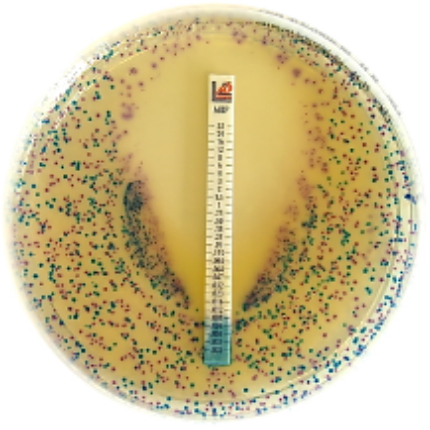
- E-test method: Also known as the gradient diffusion method, this technique uses a plastic strip containing a continuous gradient of antimicrobial concentrations. Following incubation on an inoculated agar surface, an elliptical inhibition zone develops, and the MIC is estimated at the point where the growth boundary intersects the concentration scale on the strip.
These approaches provide complementary quantitative means of assessing antimicrobial susceptibility. The resulting MIC and, where required, MBC values can subsequently be interpreted using appropriate susceptibility criteria, providing a structured basis for comparing antimicrobial activity among microbial isolates and therapeutic agents.
Broth dilution tests
Broth dilution testing is a quantitative antimicrobial susceptibility technique in which a standardized suspension of a test microorganism is exposed to a series of predetermined concentrations of an antimicrobial agent incorporated into a liquid growth medium. The method is designed to establish the concentration-dependent response of the microorganism and is particularly useful for determining the MIC of the antimicrobial agent.
In a conventional broth dilution procedure, the antimicrobial compound is prepared in a sequence of progressively decreasing concentrations, most commonly using two-fold serial dilutions. Each concentration is introduced into a separately labelled tube containing an appropriate broth medium capable of supporting the growth of the organism under investigation. The standardized bacterial inoculum is then added to each tube, ensuring that the microbial population is sufficiently uniform across the concentration series. A growth control containing the organism without the antimicrobial agent is also incorporated to confirm the viability of the test inoculum.
Following incubation under appropriate conditions, the tubes are examined for evidence of microbial growth. In conventional broth systems, growth is commonly recognized by the development of turbidity, resulting from the multiplication of bacterial cells within the liquid medium. A visibly turbid tube indicates that the organism has continued to grow at the corresponding antimicrobial concentration, whereas a clear tube indicates inhibition of detectable growth. The lowest antimicrobial concentration that prevents visible growth is recorded as the MIC.
The arrangement of antimicrobial concentrations in a dilution series provides a quantitative representation of microbial susceptibility. For example, if bacterial growth is observed at several lower concentrations but is absent at a higher concentration, the first concentration at which visible growth is suppressed represents the inhibitory threshold under the conditions of the assay. This concentration-based outcome distinguishes broth dilution from methods in which antimicrobial activity is inferred primarily from the physical dimensions of an inhibition zone.
Broth dilution testing may be performed as macrodilution or microdilution. Macrodilution traditionally involves a comparatively larger volume of broth contained in individual test tubes and may require considerable quantities of reagents and laboratory materials. The procedure can therefore become laborious when multiple antimicrobial concentrations or several test organisms are examined simultaneously. Microdilution addresses many of these practical limitations by transferring the dilution series into the individual wells of sterile microtitre plates. This miniaturized format permits numerous antimicrobial concentrations and isolates to be examined concurrently while substantially reducing reagent consumption and laboratory space.
Prepared antimicrobial panels have further streamlined broth microdilution by providing standardized concentration ranges for selected agents. These systems facilitate systematic susceptibility testing and can generate MIC values that are subsequently interpreted using established susceptibility breakpoints. Where determination of bactericidal activity is required, material from inhibitory concentrations can be subcultured onto antimicrobial-free media to assess whether viable organisms remain, thereby allowing estimation of the MBC.
Despite its quantitative value, broth dilution is not always the first-line method for routine susceptibility testing because it requires careful inoculum standardization, accurate preparation or use of antimicrobial concentration ranges, controlled incubation, and reliable interpretation of growth. Nevertheless, broth microdilution has become an important laboratory approach because it combines quantitative resolution with efficient testing of multiple antimicrobial agents and microbial isolates. Its ability to generate defined MIC values makes it particularly valuable for susceptibility characterization, resistance investigations, antimicrobial research, and situations requiring precise concentration-based assessment.
Procedure for the broth dilution susceptibility method
1. Prepare the test organism: Select a fresh, pure culture of the bacterial isolate and prepare a standardized microbial suspension using an appropriate procedure. The inoculum should be standardized to ensure comparable bacterial density throughout the test.
2. Prepare the antimicrobial solution: Prepare the antimicrobial agent at a suitable starting concentration using an appropriate sterile diluent and solvent, where required.
3. Prepare the dilution series: Make a series of two-fold serial dilutions of the antimicrobial agent in sterile broth. Each successive tube or well should contain a progressively lower concentration of the test antimicrobial.
4. Label the test vessels: Clearly identify each tube or microtitre-plate well according to its antimicrobial concentration. Include appropriate growth and sterility controls to facilitate interpretation of the results.
5. Add the broth medium: Dispense the appropriate volume of sterile broth into the designated test tubes or microtitre-plate wells.
6. Introduce the antimicrobial dilutions: Add the prepared antimicrobial concentrations to the corresponding broth vessels while maintaining the intended concentration gradient.
7. Inoculate the test organism: Add the standardized bacterial suspension to each antimicrobial-containing vessel. The same inoculum should be applied consistently throughout the test series.
8. Prepare the controls: Include a growth control containing the organism without antimicrobial agent to demonstrate that the organism is viable and capable of growth. A sterility control containing uninoculated broth may also be included to confirm that the medium is free from contamination.
9. Mix the preparations: Carefully mix the contents of the tubes or wells to distribute the microorganism and antimicrobial agent uniformly without introducing unnecessary contamination or excessive aeration.
10. Incubate the test: Incubate the inoculated tubes or microtitre plate under the standardized conditions appropriate for the organism and susceptibility-testing procedure.
11. Examine for microbial growth: After incubation, inspect each tube or well for evidence of growth. In conventional broth systems, growth is generally indicated by visible turbidity, whereas a clear preparation indicates the absence of detectable growth.
12. Determine the MIC: Arrange the antimicrobial concentrations from lowest to highest and identify the lowest concentration at which visible microbial growth is inhibited. This concentration is recorded as the MIC.
13. Determine the MBC when required: Where bactericidal activity is being assessed, material from selected tubes or wells showing inhibition may be subcultured onto antimicrobial-free growth medium. After appropriate incubation, the lowest antimicrobial concentration associated with the absence of viable growth is used to determine the MBC.
14. Record and interpret the findings: Document the observed growth pattern and corresponding antimicrobial concentrations. The resulting MIC, and MBC where determined, should be interpreted using the applicable standardized susceptibility criteria for the specific organism-antimicrobial combination. The CLSI provides standardized criteria for interpreting antimicrobial dilution tests, particularly MIC results.
15. Report the result: Present the final susceptibility result clearly, including the antimicrobial tested, the measured MIC where applicable, and the corresponding interpretive category according to the relevant laboratory standard.
Agar dilution susceptibility tests
Agar dilution susceptibility testing is a quantitative antimicrobial susceptibility method in which predetermined concentrations of an antimicrobial agent are incorporated into solid agar media and subsequently inoculated with a standardized microbial suspension. The technique provides a concentration-dependent assessment of microbial growth and is principally used to determine the MIC of an antimicrobial agent against a selected microorganism. Unlike broth dilution, in which the organism is exposed to antimicrobial concentrations in a liquid environment, agar dilution establishes the concentration gradient within a series of solid culture media.
The procedure begins with the preparation of an appropriate agar medium according to the manufacturer’s specifications and the requirements of the organism under investigation. A standardized antimicrobial stock solution is prepared from a suitable reference-grade or pharmaceutical-quality antimicrobial substance. From this stock, a sequence of predetermined concentrations is produced, commonly through two-fold serial dilution. Each concentration represents a separate test condition and is incorporated into an individual agar plate so that every plate contains a known and uniform concentration of the antimicrobial agent.
The antimicrobial-containing agar is prepared under controlled conditions to ensure adequate distribution of the drug throughout the medium. Once the plates have been prepared and solidified, standardized inocula of the test microorganisms are applied to their respective surfaces. Multiple isolates may be examined on the same concentration plate when the experimental design permits, allowing several organisms to be assessed across an identical antimicrobial concentration range. Appropriate growth and sterility controls are also incorporated to support reliable interpretation of the test.
Following inoculation, the plates are incubated under conditions appropriate for the microorganism being investigated. After incubation, each plate is examined for the presence or absence of visible microbial growth. Growth across the concentration series is compared sequentially, beginning with the lowest antimicrobial concentration. The MIC is identified as the lowest concentration of the antimicrobial agent at which visible growth is inhibited under the defined test conditions. Where additional viability assessment is required, material from selected concentrations may be subjected to further subculture to investigate bactericidal activity and determine an appropriate MBC.
Agar dilution requires several carefully coordinated stages, including selection and preparation of the antimicrobial substance, preparation of a stock solution, establishment of the desired concentration range, incorporation of the antimicrobial into the agar, preparation of standardized microbial inocula, inoculation of the test plates, incubation, and systematic examination of growth. Because each antimicrobial concentration generally requires a separately prepared agar plate, the method can be relatively labor-intensive and resource-demanding compared with some other susceptibility-testing approaches.
Despite these practical demands, agar dilution has considerable methodological value because it permits direct examination of microbial growth at precisely defined antimicrobial concentrations. It is particularly useful in reference laboratories, specialized susceptibility investigations, antimicrobial research, and experimental studies where accurate concentration-based comparisons are required. Its quantitative nature also makes it valuable for investigating unusual susceptibility patterns, evaluating novel antimicrobial compounds, and generating reproducible MIC data under controlled experimental conditions.
In terms of its clinical relevance, agar dilution provides a structured solid-medium platform for examining the relationship between antimicrobial concentration and microbial growth, thereby producing quantitative susceptibility information that can support laboratory investigation, antimicrobial research, and specialized microbiological assessment.
Procedure for agar dilution susceptibility testing
The agar dilution method is carried out systematically to expose a standardized microbial inoculum to a series of known antimicrobial concentrations incorporated into solid agar. The procedure can be organized into the following stages:
1. Preparation of the antimicrobial stock solution: Obtain a suitable reference antimicrobial substance and prepare a concentrated stock solution using the recommended solvent and concentration. The stock solution should be accurately prepared and appropriately labelled to ensure that the subsequent dilution series can be traced to a known starting concentration.
2. Selection of the antimicrobial concentration range: Determine the concentration range required for the investigation based on the expected susceptibility of the test organism. A series of concentrations is then prepared, commonly using two-fold serial dilutions. For example, each successive concentration may represent half of the preceding concentration, producing a systematic range for susceptibility assessment.
3. Preparation of the agar medium: Prepare the appropriate agar medium according to the manufacturer’s instructions and the requirements of the organism being tested. The medium should be adequately sterilized and maintained under suitable conditions before incorporation of the antimicrobial agent.
4. Incorporation of antimicrobial concentrations into agar: The predetermined antimicrobial concentrations are incorporated individually into portions of molten agar to produce separate plates containing known drug concentrations. Thorough mixing is required to promote an even distribution of the antimicrobial throughout each plate. A drug-free agar plate should also be prepared as a growth control.
5. Preparation of the test microorganism: Obtain a pure culture of the microorganism under investigation and prepare a standardized inoculum. The inoculum should have a consistent microbial density so that differences in growth between antimicrobial concentrations can be attributed primarily to the effect of the antimicrobial rather than variations in the number of organisms introduced.
6. Inoculation of the agar plates: Apply the standardized inoculum to the surface of each agar plate containing the different antimicrobial concentrations. The inoculation pattern should be consistent across the concentration series. Where several isolates are being investigated, each isolate should be clearly identified and tested according to the experimental design.
7. Inclusion of appropriate controls: Include suitable controls alongside the test plates. A growth control confirms that the organism is capable of growing under the test conditions, while sterility controls help demonstrate that the media and reagents are free from unintended microbial contamination.
8. Incubation of the inoculated plates: Incubate the plates under conditions appropriate for the microorganism and the standardized susceptibility-testing protocol being followed. Temperature, atmospheric conditions, and incubation duration should be maintained consistently for all plates because changes in these variables may influence microbial growth.
9. Examination of microbial growth: Following incubation, examine each plate for visible microbial growth. Record whether growth is present or absent at each antimicrobial concentration. The results should be assessed systematically from the lowest concentration through the highest concentration tested.
10. Determination of the MIC: Identify the lowest antimicrobial concentration at which no visible microbial growth occurs. This concentration is recorded as the MIC for the organism under the specified test conditions.
11. Further assessment of bactericidal activity: Where determination of the MBC is required, material from selected inhibitory concentrations can be subjected to an appropriate viability assessment through subculture onto antimicrobial-free medium. The concentration associated with the required loss of viable growth is then recorded according to the applicable testing criteria.
12. Recording and interpretation of results: Document the antimicrobial concentrations tested, the corresponding growth observations, and the resulting MIC and, where applicable, MBC. The findings should be interpreted using the relevant standardized criteria and reported with the organism, antimicrobial agent, concentration units, and test conditions clearly identified.
E-Test: gradient diffusion approach for MIC determination
The E-test (epsilometer test) is a quantitative antimicrobial susceptibility testing method used to determine the MIC of a selected antimicrobial agent against a particular microorganism. It combines the principles of agar diffusion and antimicrobial concentration gradients, providing a convenient bridge between conventional disk diffusion testing and dilution-based MIC determination. The technique is especially useful when a precise MIC value is required for an individual antimicrobial-organism combination.
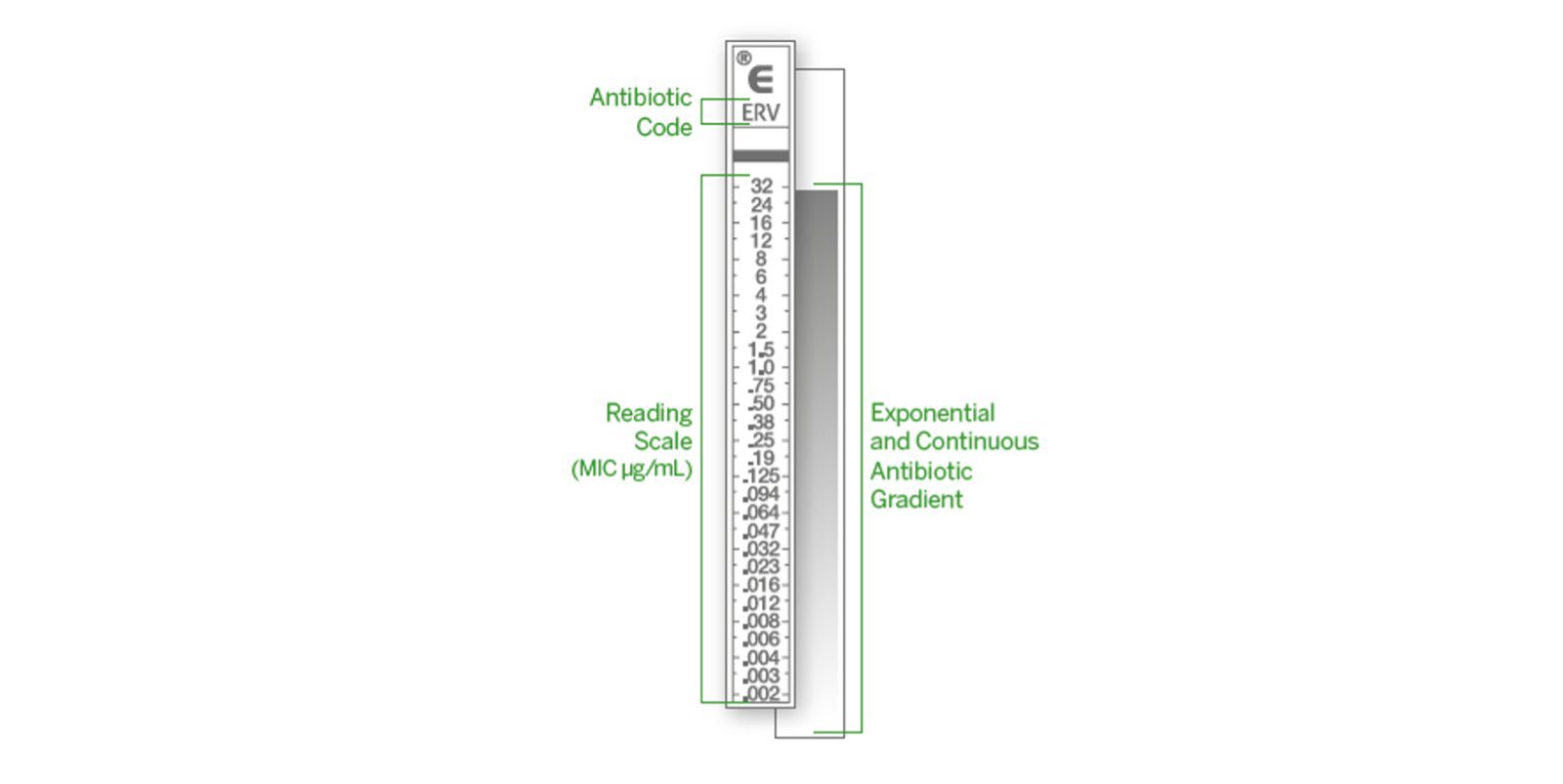
The E-test employs a narrow, non-porous plastic strip impregnated with a continuous, predefined gradient of antimicrobial concentrations. The strip is marked with a numerical concentration scale, allowing the MIC to be read directly from the test after incubation (Figure 3). Unlike a conventional antibiotic disk, which contains a relatively fixed quantity of an antimicrobial agent, the E-test strip provides a progressive concentration range along its length. This gradient permits the organism’s inhibitory response to be assessed at different antimicrobial concentrations within a single test.
Source: www.biomerieux.com/corp/en/our-offer/clinical-products/etest.html
To perform the test, a standardized suspension of the microorganism is evenly inoculated across the surface of an appropriate agar medium, commonly Mueller-Hinton agar for susceptible bacterial isolates. The antimicrobial strip is then placed aseptically on the freshly inoculated agar surface, ensuring complete contact between the underside of the strip and the medium. Once positioned, the antimicrobial agent begins to diffuse from the strip into the surrounding agar, establishing a concentration gradient that corresponds to the values printed along the strip.
Following incubation under the conditions appropriate for the organism being tested, microbial growth occurs on the agar surface except where the antimicrobial concentration is sufficient to inhibit growth. This produces a characteristic elliptical zone of inhibition surrounding the strip (Figure 4). The elongated shape of the inhibition zone reflects the continuous concentration gradient generated by the E-test strip rather than the single-point drug loading characteristic of a conventional disk.
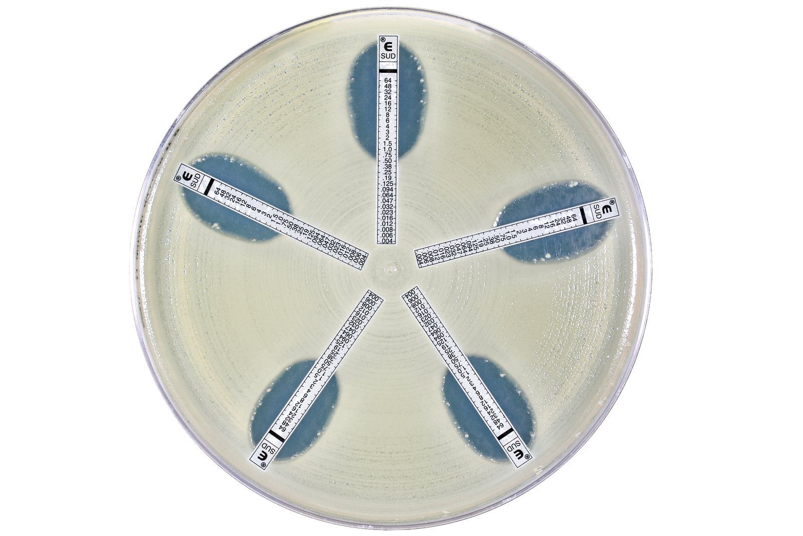
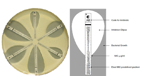
The MIC is determined at the point where the margin of the elliptical inhibition zone intersects the concentration scale printed on the E-test strip. This reading provides the estimated lowest antimicrobial concentration capable of preventing visible growth under the defined test conditions. If the growth boundary falls between marked values, the result is interpreted according to the manufacturer’s reading instructions and applicable susceptibility-testing standards.
Test reading scale: The E-test strip’s concentration scale should be read directly at the intersection between the edge of the inhibition ellipse and the printed MIC scale; the corresponding value represents the MIC result as shown in Figure 5.
Source: www.biomerieux-diagnostics.com/etest
Several features make the E-test attractive for laboratory susceptibility testing. It requires only one antimicrobial strip per organism-drug combination, produces a readily interpretable visual endpoint, and can provide MIC values for antimicrobial agents for which conventional disk diffusion may offer limited quantitative information. It may also be particularly useful when susceptibility falls near a clinically important breakpoint and a concentration-specific result is desirable.
Accurate E-test interpretation depends on appropriate inoculum preparation, agar quality, strip placement, incubation conditions, and careful recognition of the growth boundary. The technique therefore provides a practical and informative means of generating quantitative susceptibility data while retaining the operational simplicity of an agar-based diffusion procedure.
Procedure for performing the E-Test for MIC determination
The E-test is carried out using a standardized microbial inoculum, an appropriate agar medium, and a commercially prepared antimicrobial gradient strip. Consistency at each stage is important because the final MIC value depends on the uniformity of the inoculum, the condition of the agar surface, and the correct positioning and interpretation of the strip.
1. Preparation of the test inoculum: Begin with a fresh, pure culture of the microorganism under investigation. Select well-isolated colonies using a sterile inoculating loop and prepare a suspension in sterile physiological saline or another appropriate diluent. Adjust the suspension to the required turbidity, commonly equivalent to a 0.5 McFarland standard, to provide a suitably standardized inoculum.
2. Inoculation of the agar plate: Dip a sterile cotton swab into the standardized suspension and remove excess fluid by pressing the swab against the inner wall of the tube. Evenly inoculate the entire surface of a suitable agar plate, commonly MH agar for routine bacterial susceptibility testing. Streak the plate in several directions to establish a uniform bacterial lawn, ensuring that the inoculum reaches the entire agar surface.
3. Preparation of the inoculated plate: After inoculation, allow the plate to stand briefly with the lid in place so that excess surface moisture can be absorbed or evaporate. The surface should be sufficiently dry to prevent displacement of the E-test strip or excessive spreading of the inoculum. Excessive moisture may distort the developing inhibition ellipse and consequently affect MIC interpretation.
4. Application of the E-test strip: Using sterile forceps or an appropriate strip applicator, remove the E-test strip from its packaging without contaminating the antimicrobial surface. Place the strip onto the inoculated agar with the MIC scale facing upward. Ensure that the strip lies completely flat and makes firm, uninterrupted contact with the agar surface. Avoid moving the strip once it has contacted the medium.
5. Incubation: Invert the plate and incubate it under the conditions recommended for the organism and antimicrobial combination being examined. Temperature, atmospheric conditions, and incubation duration should follow the applicable standardized procedure or the manufacturer’s instructions. During incubation, the antimicrobial agent diffuses continuously from the strip into the agar, producing a concentration gradient along its length.
6. Examination of the inhibition pattern: Following incubation, examine the plate for the characteristic elliptical zone of inhibition surrounding the E-test strip. The microorganism should demonstrate visible growth outside the inhibitory region, while growth is suppressed within the area where the antimicrobial concentration is sufficient to inhibit the organism.
7. Determination and recording of the MIC: Read the MIC directly from the numerical scale printed on the strip. The endpoint is located at the intersection between the edge of the inhibition ellipse and the concentration scale. Record the corresponding antimicrobial concentration as the MIC. If the ellipse intersects the scale between two values or an atypical growth pattern is observed, interpretation should follow the specific manufacturer’s instructions and applicable susceptibility-testing guidelines rather than relying on visual estimation alone.
8. Documentation and interpretation: Record the organism tested, antimicrobial agent, MIC value, incubation conditions, and any unusual features observed during reading. The MIC should subsequently be interpreted using the appropriate organism-specific susceptibility breakpoints where such criteria are available. This final interpretation allows the quantitative E-test result to be translated into a clinically meaningful susceptibility category.
Note: The reliability of the E-test depends on maintaining a standardized inoculum, uniform agar inoculation, proper strip placement, controlled incubation, and accurate identification of the inhibition-ellipse endpoint. These elements collectively ensure that the MIC obtained represents the antimicrobial concentration associated with inhibition under the defined test conditions.
References
Balouiri, M., Sadiki, M., & Ibnsouda, S. K. (2016). Methods for in vitro evaluating antimicrobial activity: A review. Journal of Pharmaceutical Analysis, 6(2), 71–79.
Chika, E., Ifeanyichukwu, I., Michael, A., & Charles, E. (2013). Susceptibility and detection of extended spectrum β-lactamase enzymes from otitis media pathogens. American Journal of Infectious Diseases, 9(1), 24–29.
CLSI. Performance Standards for Antimicrobial Susceptibility Testing. 32nd ed. CLSI supplement M100. Clinical and Laboratory Standards Institute; 2022.
Chung, E., Ren, G., Johnston, I., Matharu, R. K., Ciric, L., Walecka, A., & Cheong, Y.-K. (2023). Applied methods to assess the antimicrobial activity of metallic-based nanoparticles. Bioengineering, 10(11), 1259.
Cunliffe, A. J., Askew, P. D., Stephan, I., Iredale, G., Cosemans, P., Simmons, L. M., Verran, J., & Redfern, J. (2021). How do we determine the efficacy of an antibacterial surface? A review of standardised antibacterial material testing methods. Antibiotics, 10(9), 1069.
Curtin, J., & Cormican, M. (2003). Measuring antimicrobial activity against biofilm bacteria. Reviews in Environmental Science and Bio/Technology, 2, 285–291.
Ejikeugwu, C., Obi, C., Oluyemisi, O. E., Ujam, N. T., Blessing, U., Edeh, C., Nwezeagu, F., & Iroha, I. (2020). Susceptibility profile and multiple antibiotics resistance indexes (MARI) of clinical isolates of Staphylococcus aureusfrom urinary tract infections (UTIs). Journal of Bacteriology and Mycology, 7(3), 1134.
Gajic, I., Kabic, J., Kekic, D., Jovicevic, M., Milenkovic, M., Mitic Culafic, D., Trudic, A., Ranin, L., & Opavski, N. (2022). Antimicrobial susceptibility testing: A comprehensive review of currently used methods. Antibiotics, 11(4), 427.
Gonzalez-Pastor, R., Carrera-Pacheco, S. E., Zúñiga-Miranda, J., Rodríguez-Pólit, C., Mayorga-Ramos, A., Guamán, L. P., & Barba-Ostria, C. (2023). Current landscape of methods to evaluate antimicrobial activity of natural extracts. Molecules, 28(3), 1068.
Hossain, T. J. (2024). Methods for screening and evaluation of antimicrobial activity: A review of protocols, advantages, and limitations. European Journal of Microbiology and Immunology, 14(2), 97–115.
Kavela, S., Kakkerla, S., & Thupurani, M. K. (2025). Broad-spectrum antimicrobial activity and in vivo efficacy of SK1260 against bacterial pathogens. Frontiers in Microbiology, 16, 1553693.
Perez-Gavilan, A., de Castro, J. V., Arana, A., et al. (2021). Antibacterial activity testing methods for hydrophobic patterned surfaces. Scientific Reports, 11, 6675.
Doern G.V (1995). Susceptibility tests of fastidious bacteria. Manual of Clinical Microbiology, 6th edition, Murray P.R, Baron E.J, Pfaller M.A, Tenover F.C, Yolken R, American Society for Microbiology, Washington DC, Pp. 1342-1349.
Livermore D.M, Winstanley T.B, Shannon K.P (2001). Interpretative reading: recognizing the unusual and inferring resistance mechanisms from resistance phenotypes. J Antimicrob Chemother, 48 Suppl 1:87-102.
Discover more from Microbiology Class
Subscribe to get the latest posts sent to your email.