The genotypic detection and characterization of antibiotic resistant genes in pathogenic bacteria is more specific in characterizing resistance genes than the phenotypic detection methods. The term genotypic is derived from genotype – which is the complete genetic makeup of an organism. Genotype describes the overall composition of the genetic information found in an organism. Thus, genotypic detection methods used for antimicrobial susceptibility studies detects specific nucleic acid component that is responsible for drug resistance in pathogenic bacteria.
Genotypic detection and characterization techniques are DNA-based unlike the phenotypic methods that are merely antibiotic-based. They are generally used to validate the susceptibility test results obtained with a phenotypic testing method. Phenotypic techniques used for antimicrobial susceptibility testing are not sufficient enough to characterize the genes that are largely responsible for a particular resistance problem, and because of this the genotypic detection and characterization methods are included to detect and establish the genes responsible for the resistance in the pathogenic bacteria.
The genotypic techniques generally involves gene amplification in the test bacteria, and the polymerase chain reaction (PCR) is employed to achieve this. Restriction fragment length polymorphism (RFLP), PCR-single-stranded conformation polymorphism (SSCP), Random Amplified Polymorphic DNA (RAPD), Fluorescent in situ hybridization (FISH),DNA microarray technique, ligase – chain reaction (LCR) and nucleotide sequencing are examples of the different DNA-based techniques employed in for the detection and characterization of antibiotic resistance genes in pathogenic bacteria.
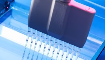
In summary, the PCR amplification of resistance genes in pathogenic bacteria is carried out with the aid of a thermal cycler in addition to other instruments/equipments that may involve some of the following processes:
- Denaturing at 94°C for 1 min.
- Annealing at 54°C for 45 sec.
- Elongation at 72°C for 1-2 min.
- Visualization and scanning of the electrophoresed PCR products. The separated DNA bands are visualized by staining with ethidium bromide, and viewed under ultraviolet (UV) light. Note: It is best practice to include a molecular marker or ladder alongside the running of the PCR products in the gel electrophoresis system.
Molecular markers are mixtures of standard DNA fragments with known sizes, and they are used to estimate, compare and identify the size of the individual separated DNA bands. The presence of a PCR product of the appropriate size after gel electrophoresis experiment (as informed by the molecular markers or DNA ladders) indicates the presence in the sample of a particular gene of interest or piece of DNA.
- Single Strand Conformational Polymorphism (SSCP): SSCP is a rapid method for mutational screening. Single point mutations can cause major differences in the folded form of single stranded DNA. These differences can be detected as differences in electrophoretic mobility using the SSCP technique. Single stranded DNA can adopt multiple conformations under non-denaturing conditions. In the absence of a complementary strand, DNA will anneal short internal complementary sequences, forming a complex “knot“.
A DNA molecule follows a complex path of folding to reach its final form, influenced by the solution environment and temperature. The path consists of a series of annealing steps, each stabilizing and to some extent directing the next. Minor alterations in the sequence of the DNA will disrupt the annealing process and result in a different final shape. The compactness of these structures will determine how fast the single stranded DNA migrates through a non-denaturing gel. Such differences in electrophoretic mobility between nearly identical strands serve as the basis for the technique known as single strand conformation polymorphism (SSCP).
Samples are denatured with heat and then rapidly cooled. Rapid cooling favors self-annealing, because insufficient time is allowed for complementary strands to collide and orient for duplex formation. The re-natured samples are analyzed on a gel opposite the control DNA. All DNA fragments must be of the same length. Mutant samples will show mobility different from the control DNA. The gel matrix used must be optimized for the resolution of DNA conformers of the same length. Various combinations of Acrylamide and Bis-Acrylamide are usually used to run the gel electrophoresis technique.
- Random Amplified Polymorphic DNA (RAPD): RAPD technique is mainly used to examine phylogenetic distance among different microbial species. It is a type of PCR reaction, but the segments of DNA that are amplified are random. RAPD is a modified PCR technique that provides useful data for the comparison of different microbial types; and it helps researchers to amplify a whole DNA segment extracted from microbial cell. The scientist performing RAPD creates several arbitrary, short primers (8–12 nucleotides), then proceeds with the PCR using a large template of genomic DNA, hoping that fragments will amplify.
By resolving the resulting patterns, a semi-unique profile can be gleaned from a RAPD reaction. RAPD has been used to characterize, and trace, the phylogeny of diverse plant and animal species. In the RAPD technique, multiple 10 base pair (bp) oligonucleotide primers are added each to an individual sample of DNA which is then subjected to PCR. The resulting amplified DNA markers are random polymorphic segments with band sizes from 100 to 3000 bp depending upon the genomic DNA and the primer. The RAPD technique is sensitive, fast, and it requires the use of no radioactive probes, and is easily performed.
RAPD markers are limited in their usefulness, however, in that they are dominant alleles, so it is necessary to prepare many closely linked markers to insure reliable comparisons among microbial or plant populations. RAPD produces a large number of DNA bands of various sizes from each of the different samples which are prepared. These bands migrate according to size during electrophoresis. To analyze RAPD data, one must first count the total number of unique bands (thus if several lanes share a band, that band is only counted once toward the total) for each primer used.
- Restriction fragment length polymorphism (RFLP): RFLP is a technique that exploits variations in homologous DNA sequences. It refers to a difference between samples of homologous DNA molecules from differing locations of restriction enzyme sites, and to a related laboratory technique by which these segments can be illustrated. In RFLP analysis, the DNA sample is broken into pieces (and digested) by restriction enzymes and the resulting restriction fragments are separated according to their lengths by gel electrophoresis technique.
RFLP analysis was the first DNA profiling technique despite the fact that it is now becoming obsolete as a result of novel and inexpensive DNA sequencing technologies. RFLP was an important tool in genome mapping, localization of genes for genetic disorders, determination of risk for disease, and paternity testing. The basic technique for detecting restriction fragment length polymorphisms (RFLPs) involves fragmenting a sample of DNA by a restriction enzyme, which can recognize and cut DNA wherever a specific short sequence occurs, in a process known as a restriction digest.
The resulting DNA fragments are then separated by length through a process known as agarose gel electrophoresis, and transferred to a membrane via the Southern blot procedure. Hybridization of the membrane to a labeled DNA probe then determines the length of the fragments which are complementary to the probe. A RFLP occurs when the length of a detected fragment varies between individuals. Each fragment length is considered an allele, and can be used in genetic analysis.
RFLP analysis was the basis for early methods of genetic fingerprinting, and it is useful in the identification of samples retrieved from crime scenes, in the determination of paternity, and in the characterization of genetic diversity or breeding patterns in animal populations. However, the RFLP technique is slow and cumbersome to perform. It usually requires a large amount of sample DNA; and the combined process of probe labeling, DNA fragmentation, electrophoresis, blotting, hybridization, washing, and autoradiography could take up to a month to complete.
- Fluorescent in situ hybridization (FISH): FISH is a cytogenetic technique that uses fluorescent probes that bind to only those parts of the chromosome with a high degree of sequence complementarity. It is used to detect and localize the presence or absence of specific DNA sequences on chromosomes. FISH is often used for finding specific features in DNA for use in genetic counseling, medicine, and species identification. FISH can also be used to detect and localize specific RNA targets (mRNA) in cells, circulating tumor cells, and tissue samples.
- Ligase chain reaction (LCR): The ligase chain reaction (LCR) is a method of DNA amplification. While the better-known PCR carries out the amplification by polymerizing nucleotides, LCR instead amplifies the nucleic acid used as the probe. For each of the two DNA strands, two partial probes are ligated to form the actual one; thus, LCR uses two enzymes: a DNA polymerase and a DNA ligase. Each cycle results in a doubling of the target nucleic acid molecule.
A key advantage of LCR is greater specificity as compared to PCR. It has been widely used for the detection of single base mutations, as in genetic diseases. LCR and PCR may be used to detect gonorrhea and chlamydia, and may be performed on first-catch urine samples, providing easy collection and a large yield of organisms. Endogenous inhibitors limit the sensitivity, but if this effect could be eliminated, LCR and PCR would have clinical advantages over any other methods of diagnosing gonorrhea and chlamydia.
- DNA microarray: DNAmicroarrayis simply a high-throughput molecular biology technique that is used to determine how genes work at any given point in time in a cell i.e. how genes are turned off or on in a cell. Similar to the traditional southern blotting technique (which is mainly used to detect the presence of a piece of DNA molecule in a sample), DNA microarray allows scientists to array several thousands of DNA on a microarray or microscopic slide at a very short time and in a precise manner.
The technology of DNA microarray has become an indispensable research tool within the life sciences; and this high-throughput technique is also applied in medicine, pharmaceutical companies and even in biotechnology to enhance productivity and understanding of the genetic bases of living organisms inclusive of microbes. With DNA microarray, it is possible for molecular biologists to monitor the expression of thousands of the genes of an organism in real-time i.e. at a simultaneous fashion.
DNA microarray is a gene-expression profiling-based technique that is computer-enabled and is generally used to identify multiple and similar genes that are usually co-expressed in a cell at a short span of time.
References
Arora D.R (2004). Quality assurance in microbiology. Indian J Med Microbiol, 22:81-86.
Ashutosh Kar (2008). Pharmaceutical Microbiology, 1st edition. New Age International Publishers: New Delhi, India.
Barenfanger J, Drakel C and Kacich (1999). Clinical and Financial Benefits of Rapid Bacterial Identification and Antimicrobial Susceptibility Testing. Journal of Clinical Microbiology, 37(5):1415-1418.
Denyer S.P., Hodges N.A and Gorman S.P (2004). Pharmaceutical Microbiology. 7th ed. Blackwell Publishing Company, USA.
Doern G, Brueggemannn A.B, Perla R, Daly D, Halkias D, Jones R.N, Saubolle M.A (1997). Multicenter laboratory evaluation of the bioMerieux Vitek antimicrobial susceptibility testing system with 11 antimicrobial agents versus members of the family Enterobacteriaceae and Pseudomonas aeruginosa. J Clin Microbiol, 35:2115–2119.
Doern G, Vautour R, Gaudet M, Levy B (1994). Clinical impact of rapid in vitro susceptibility testing and bacterial identification. J Clin Microbiol, 32:1757–1762.
Doern G.V (1995). Susceptibility tests of fastidious bacteria. Manual of Clinical Microbiology, 6th edition, Murray P.R, Baron E.J, Pfaller M.A, Tenover F.C, Yolken R, American Society for Microbiology, Washington DC, Pp. 1342-1349.
Funke G, Monnet D, deBernardis C, von Graevenitz A, Freney J (1998). Evaluation of the VITEK 2 system for rapid identification of medically relevant Gram-negative rods. J Clin Microbiol, 36:1948–1952.
Garcia L.S (2010). Clinical Microbiology Procedures Handbook. Third edition. American Society of Microbiology Press, USA.
Hart C.A (1998). Antibiotic Resistance: an increasing problem? BMJ, 316:1255-1256.
Livermore D.M, Winstanley T.B, Shannon K.P (2001). Interpretative reading: recognizing the unusual and inferring resistance mechanisms from resistance phenotypes.
J Antimicrob Chemother, 48 Suppl 1:87-102.
Madigan M.T., Martinko J.M., Dunlap P.V and Clark D.P (2009). Brock Biology of Microorganisms, 12th edition. Pearson Benjamin Cummings Inc, USA.
Mahon C. R, Lehman D.C and Manuselis G (2011). Textbook of Diagnostic Microbiology. Fourth edition. Saunders Publishers, USA.
National Committee for Clinical Laboratory Standards. Performance Standards for antimicrobial susceptibility testing. 8th Informational Supplement. M100 S12. National Committee for Clinical Laboratory Standards, 2002. Villanova, Pa.
Washington J.A (1993). Rapid antimicrobial susceptibility testing: technical and clinical considerations. Clin Microbiol Newsl, 15:153–155.
Discover more from Microbiology Class
Subscribe to get the latest posts sent to your email.


