The term “electrophoresis” refers to the movement of a solid particle (e.g. nucleic acids) through a polymer matrix or gel under the influence of electric field. Electrophoresis is a molecular biology technique that is used to separate nucleic acid molecules and other macromolecules mainly on the basis of their charge to mass ratio as they migrate through a gel in an electric field.
Gel electrophoresis technique is a molecular biology technique that is used to separate nucleic acid molecules (DNA and RNA) according to their sizes and conformation or charges. It is generally used in the molecular biology laboratory for the separation and purification of nucleic acid fragments. The process occurs in an electrophoretic tank or chamber laden with specialized type of gel (e.g. agarose) through which controlled electric charge is allowed to pass through (Figure 1).
A dye (e.g. ethidium bromide) is added to the gel so that the nucleic acid fragments can be visualized under ultraviolet (UV) light. The size range of nucleic acid fragments that can be separated using agarose gel is usually in the range of 0.2 kb to 20 kb. Gel electrophoresis is an important technique that is used to analyze the products of a PCR reaction, and it allows separated fragments of nucleic acid molecules or protein molecule from a given organism or cell to form characteristic pattern of bands known as fingerprints when they are passed through a gel under the influence of electric charges.
Gel electrophoresis is the technique of separating charged molecules such as DNA in an electric field. Fragments of separated nucleic acid molecules move through gel in electric fields according to their different sizes, and this serves as the basis for the utilization of electrophoresis to identify the individual fragments of a particular DNA. It is worthy of note that after isolating a piece of DNA from an organism, and cutting same into different fragments using restriction enzymes; there is need to study the individual fragments, and this can only be made possible through electrophoresis which gives a detailed analysis of each fragment of the nucleic acid. Deoxyribonucleic acid (DNA) is a negatively charged nucleic acid molecule because of its phosphate groups.
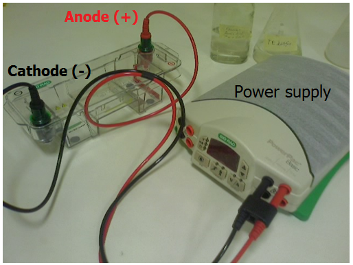
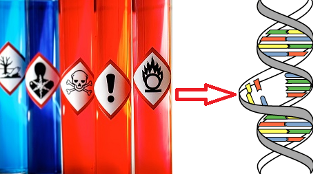
When the separated DNA fragments is placed in a gel and allowed to move through an electric field (with positive and negative ends), the DNA molecule tends to move towards the positive terminus (i.e. the anode) of the electric charge than the negative terminus (or cathode) because of its notable negative charge. Smaller molecules of DNA migrate through the gel faster than the larger molecules because of the sieving nature of the gel used for gel electrophoresis technique. The separated DNA fragment is allowed to run for a specific amount of time, and the DNA fragments are visualized under UV light after the addition of ethidium bromide (EtBr) which makes the bands visible. DNA is a colourless macromolecule, and EtBr is used in gel electrophoresis to make the different bands of the nucleic acid (DNA) visible. EtBr is mutagenic and can cause cancer; thus it should always be handled with utmost care.
The EtBr intercalate between the nitrogenous bases of the double stranded DNA molecule, and this causes the DNA molecule to fluoresce or produce an orange colour when the gel carrying the DNA fragments is photographed or illuminated with UV light. Electrophoretic technique is the most versatile method of analyzing, identifying and purifying the fragments of nucleic acid molecules (DNA and RNA) and proteins; and it is unique because it separates macromolecules according to their sizes and charges.
Agarose and polyacrylamide are the two notable gels used in electrophoresis experiment. While agarose gel is used in most simple electrophoresis techniques (e.g. separation of nucleic acid molecules), polyacrylamide gel is mainly used in advanced electrophoresis such as those that has to do with protein separation. Several electrophoresis techniques are available and they include agarose gel electrophoresis, pulse field gel electrophoresis (PFGE) and polyacrylamide gel electrophoresis (PAGE) amongst others.
References
Alberts B, Bray D, Lewis J, Raff M, Roberts K and Watson J.D (2002). The molecular Biology of the Cell. Fourth edition. New York, Garland, USA.
Alcamo I.E (2000). DNA Technology: The Awesome Skill. Elsevier Science and Technology Books. Philadelphia, PA, USA.
Ausubel, F.M., Brent, R., Kingston, R.E., Moore, D.D., Seidman, J.G., Smith, J.A., Struhl, K., eds (2002). Short Protocols in Molecular Biology, 5th edn. John Wiley & Sons, New York.
Bourgaize D, Jewell T.R and Buiser R.G (1999). Biotechnology: Demystifying the Concepts. Pearson Education, San Francisco, CA.
Chen I and Dubnau D (2004). DNA uptake during bacterial transformation. Nat. Rev. Microbiol. 2 (3): 241–249.
Clark D.P and Pazdernik N (2010). Biotechnology. First edition. Elsevier Science and Technology Books, Amsterdam, Netherlands.
Cooper G.M and Hausman R.E (2004). The cell: A Molecular Approach. Third edition. ASM Press.
Dale J (2003). Molecular genetics of bacteria. Jeremy W. Dale and Simon Park (4th eds.). John Wiley & Sons Ltd, West Sussex, UK. Pp. 312-313.
Das H.K (2010). Textbook of Biotechnology. Fourth edition. Wiley edition. Wiley India Pvt, Ltd, New Delhi, India.
Hames B.D and Rickwood D (1998). Gel Electrophoresis of Proteins: A Practical Approach 3rd Edition. The Practical Approach Series, Oxford University Press.
Lewis R (2004). Human Genetics: Concepts and Applications. Sixth edition. McGraw Hill Publishers, USA.
Lodish H, Berk A, Matsudaira P, Kaiser C.A, Kreiger M, Scott M.P, Zipursky S.L and Darnell J (2004). Molecular Cell Biology. Fifth edition. Scientific American Books, Freeman, New York, USA.
Madigan M.T., Martinko J.M., Dunlap P.V and Clark D.P (2009). Brock Biology of Microorganisms, 12th edition. Pearson Benjamin Cummings Inc, USA.
McPherson M and Moller S (2002). PCR: The Basics. 2nd edition. Taylor and Francis Group. New York, USA.
Sambrook, J., Russell, D.W. (2001). Molecular Cloning: a Laboratory Manual, 3rd edn. Cold Spring Harbor Laboratory Press, New York.
Singleton P and Sainsbury D (1995). Dictionary of microbiology and molecular biology, 3d ed. New York: John Wiley and Sons.
Southern, E.M. (2000). Blotting at 25. Trends in Biochemical Science, 25, 585–588.
Synder L, Peters J.E, Henkin T.M and Champness W (2013). Molecular Genetics of Bacteria. Fourth edition. American Society of Microbiology Press, USA.
Tamarin Robert H (2002). Principles of Genetics. Seventh edition. Tata McGraw-Hill Publishing Co Ltd, Delhi.
Discover more from Microbiology Class
Subscribe to get the latest posts sent to your email.