Voges-Proskauer (VP) test is used to identify Gram negative bacteria that produce 2,3-butanediol (acetoin) from the fermentation of glucose. The Voges–Proskauer test determines the capability of some organisms to produce non acidic or neutral end products, such as acetyl methyl carbinol from glucose or sugar fermentation.
VP is a test used to detect acetoin in a bacterial broth culture. The test is performed by adding alpha-naphthol and potassium hydroxide to the Voges-Proskauer broth which has been inoculated with bacteria. In the presence of atmospheric oxygen and 40 % potassium hydroxide, acetoin is converted to diacetyl.
Alpha–naphthol serves as a catalyst to bring out a red complex. Some bacteria utilize glucose producing acetyl methyl carbinol which is oxidized in the presence of alkali to diacetyl. The presence of diacetyl is confirmed by the formation of red colour with arginine, creatine or creatinine.
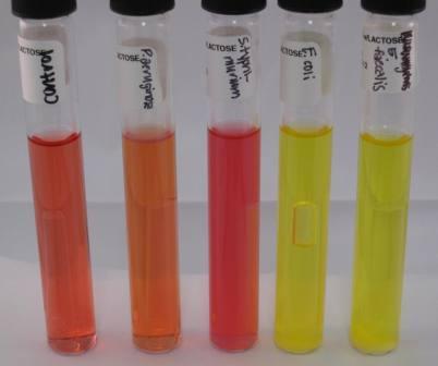
VP positive bacteria turn the medium in which they are inoculated to brownish-red to pink because they produce acetoin. Bacteria that are VP negative turn the inoculated medium to brownish-green to yellow. Klebsiella pneumoniae and Enterobacter aerogenes are VP positive bacteria.
PROCEDURE FOR VOGES PROSKAUER TEST
- Prepare and sterilize glucose-phosphate as the growth medium and cool to room temperature.
- Inoculate the glucose-phosphate medium with the test bacterium.
- Incubate medium at 37oC overnight.
- Add 5 mg of creatine followed by 5 ml of 40 % NaOH and shake the tube very well.
- Observe tube(s) for a change in colour. Change of the colour of the medium to brownish-red to pink signifies a positive VP test (Figure 1).
References
Basic laboratory procedures in clinical bacteriology. World Health Organization (WHO), 1991. Available from WHO publications, 1211 Geneva, 27-Switzerland.
Beers M.H., Porter R.S., Jones T.V., Kaplan J.L and Berkwits M (2006). The Merck Manual of Diagnosis and Therapy. Eighteenth edition. Merck & Co., Inc, USA.
Biosafety in Microbiological and Biomedical Laboratories. 5th edition. U.S Department of Health and Human Services. Public Health Service. Center for Disease Control and Prevention. National Institute of Health. HHS Publication No. (CDC) 21-1112.2009.
Cheesbrough M (2010). District Laboratory Practice in Tropical Countries. Part I. 2nd edition. Cambridge University Press, UK.
Cheesbrough M (2010). District Laboratory Practice in Tropical Countries. Part 2. 2nd edition. Cambridge University Press, UK.
Collins C.H, Lyne P.M, Grange J.M and Falkinham J.O (2004). Collins and Lyne’s Microbiological Methods. Eight edition. Arnold publishers, New York, USA.
Disinfection and Sterilization. (1993). Laboratory Biosafety Manual (2nd ed., pp. 60-70). Geneva: WHO.
Garcia L.S (2010). Clinical Microbiology Procedures Handbook. Third edition. American Society of Microbiology Press, USA.
Garcia L.S (2014). Clinical Laboratory Management. First edition. American Society of Microbiology Press, USA.
Fleming, D. O., Richardson, J. H., Tulis, J. I. and Vesley, D. (eds) (1995). Laboratory Safety: Principles and practice. Washington DC: ASM press.
Dubey, R. C. and Maheshwari, D. K. (2004). Practical Microbiology. S.Chand and Company LTD, New Delhi, India.
Gillespie S.H and Bamford K.B (2012). Medical Microbiology and Infection at a glance. 4th edition. Wiley-Blackwell Publishers, UK.
Discover more from Microbiology Class
Subscribe to get the latest posts sent to your email.