Triple sugar iron agar (TSIA) is a differential agar medium used to different lactose-fermenting Enterobacteriaceae from non-lactose fermenting enterobacteria. The medium usually contains three sugars viz: sucrose, glucose, and lactose, and phenol red as a pH indicator. TSIA is a synonymous biochemical test to Kliger’s iron agar (KIA) test.
Both TSIA and KIA are used to test the ability of an Enterobacteriaceae to ferment glucose and lactose. These agar medium also test the ability of an enteric organism to reduce sulphur and produce gas (e.g. hydrogen sulphide, H2S). TSIA/KIA is commonly used to identify E. coli, Salmonella species, Proteus species and Shigella species.
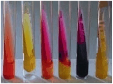
A yellow butt and pink-red slope shows glucose fermentation and acid production only. Lactose fermentation (inclusive of glucose fermentation) also turns the medium yellow. Salmonella and Shigella shows this type of reaction. Production of gas (which often cause cracks in the medium) during the fermentation of any of these sugars also leads to the lifting up of the agar off the bottom of the tube.
Salmonella paratyphi and some feacal commensals are known to cause cracks in TSIA/KIA media. A yellow slope and a yellow butt indicate lactose fermentation, and this show the presence of a lactose fermenter e.g. E. coli. A pink-red slope and butt indicates that the organism is a non-fermenter, and thus cannot ferment either glucose or lactose.
Pseudomonas aeruginosa shows this type of reaction. Production of a black precipitate or colour that covers the butt indicates the production of H2S gas. Salmonella species especially S. typhimurium is notorious in the production of H2S gas in TSIA/KIA media. Shigella species are not known to produce H2S gas in TSIA/KIA media.
PROCEDURE FOR TSIA
- Prepare TSIA/KIA media according to manufacturer’s instructions.
- Use a pure culture of the test isolate to perform this test.
- Pick a speck or colony of the test isolate using a straight inoculating loop.
- Stab the TSIA/KIA medium with the collected test isolate (making sure the stab reaches the butt of the tube) and streak the slope upon removal.
- Incubate inoculated tube(s) at 37oC for 18-24 hrs.
- Observe the tube(s) for glucose and lactose fermentation. Also examine the tube(s) for H2S production.
References
Basic laboratory procedures in clinical bacteriology. World Health Organization (WHO), 1991. Available from WHO publications, 1211 Geneva, 27-Switzerland.
Beers M.H., Porter R.S., Jones T.V., Kaplan J.L and Berkwits M (2006). The Merck Manual of Diagnosis and Therapy. Eighteenth edition. Merck & Co., Inc, USA.
Biosafety in Microbiological and Biomedical Laboratories. 5th edition. U.S Department of Health and Human Services. Public Health Service. Center for Disease Control and Prevention. National Institute of Health. HHS Publication No. (CDC) 21-1112.2009.
Cheesbrough M (2010). District Laboratory Practice in Tropical Countries. Part I. 2nd edition. Cambridge University Press, UK.
Cheesbrough M (2010). District Laboratory Practice in Tropical Countries. Part 2. 2nd edition. Cambridge University Press, UK.
Collins C.H, Lyne P.M, Grange J.M and Falkinham J.O (2004). Collins and Lyne’s Microbiological Methods. Eight edition. Arnold publishers, New York, USA.
Disinfection and Sterilization. (1993). Laboratory Biosafety Manual (2nd ed., pp. 60-70). Geneva: WHO.
Garcia L.S (2010). Clinical Microbiology Procedures Handbook. Third edition. American Society of Microbiology Press, USA.
Garcia L.S (2014). Clinical Laboratory Management. First edition. American Society of Microbiology Press, USA.
Fleming, D. O., Richardson, J. H., Tulis, J. I. and Vesley, D. (eds) (1995). Laboratory Safety: Principles and practice. Washington DC: ASM press.
Dubey, R. C. and Maheshwari, D. K. (2004). Practical Microbiology. S.Chand and Company LTD, New Delhi, India.
Gillespie S.H and Bamford K.B (2012). Medical Microbiology and Infection at a glance. 4th edition. Wiley-Blackwell Publishers, UK.
Discover more from Microbiology Class
Subscribe to get the latest posts sent to your email.