Sugar (glucose) utilization test or carbohydrate fermentation test is used to detect bacteria that ferment various sugars (e.g. glucose) as well as convert pyruvate (the end product of glycolysis) into gaseous by-products (e.g. hydrogen and CO2). Bacteria in an effort to generate energy can ferment various simple sugars including glucose, sucrose, mannitol, and lactose, and this serves as basis for their identification in the laboratory.
Thus the glucose test, lactose test or sucrose test can be performed to determine the type of carbohydrate or sugar utilized by a particular organism. Sugars are fermented or metabolized by microbes through different metabolic pathways depending on the types of microbial species present and the environment in which the organism is located.
It is noteworthy that when carbohydrates or sugars are fermented by bacteria, they produce acidic products in the process; and a change in the pH of the medium due to carbohydrate or sugar fermentation can be detected when the fermentation of a given carbohydrate molecule has occurred. Acids are also produced in the process; and acids lower the pH of the medium.
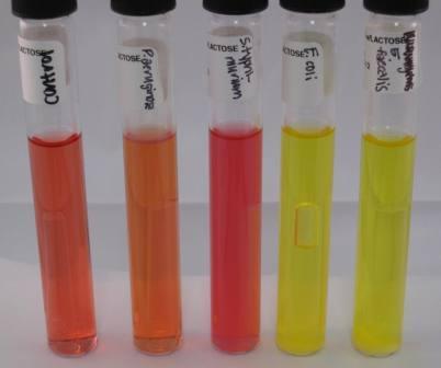
This will cause the pH indicator (phenol Red) in the medium to turn yellow, thus showing a positive test result. When a bacterium does not ferment the carbohydrate, the media remains red. Sometimes during fermentation, gas is produced. The Durham tube will then have a gas bubble trapped within it.
Sugar utilization test is performed in broth medium containing different fermentable carbohydrate or sugar, and also very special tubes known as Durham tubes – which helps the researcher to detect gas production. The sugars are added to a peptone medium to which a pH indicator is also added. The Durham tubes are known to collect the gases produced from the sugar fermentation. Gases produced during fermentation process can be detected by Durham’s tubes (small inverted tubes placed in the broth medium), within the liquid culture medium.
If gas is produced as a result of the fermentation process, the liquid medium inside the Durham’s tube will be replaced by the gas in the form of bubbles. Sugar utilization test is a differential test which is specifically used to distinguish Gram negative enterobacteria based on their ability to ferment glucose and produce gas at the same time. All Gram negative enteric bacteria are glucose fermenters but not all of them produce gas.
Gram negative enterobacteria that show gas production are able to convert pyruvic acid to formic acid which is further broken-down to produce H2 and CO2. The gases produced (i.e. H2 and CO2) are trapped in the Durham tube(s), and they appear as bubbles at the top of the tube. Proteus mirabilis and Escherichia coli are both glucose fermenters and gas producers.
PROCEDURE FOR SUGAR UTILIZATION TEST
- Prepare the baseline medium such as peptone water according to the manufacturer’s instructions.
- Dispense 9 ml portion of the basal medium into clean test tubes or bijou bottles.
- Add about 2-3 ml drops of 1 % Andrade’s indicator or any other pH indicator (e.g. bromothymol blue and phenol red) into the basal medium.
- Carefully and aseptically insert clean Durham tube(s) into all the aliquoted portions of the basal medium, taking care to avoid the inclusion of air bubbles into the Durham tube(s).
- Sterilize the tubes in the autoclave at 121oC for 15 mins and cool to normal room temperature.
- Prepare 1 % aqueous solution of the test sugar.
- Sterilize test sugar at 115oC for 15 mins in the autoclave. Membrane filtration technique can also be used to sterilize the test sugar.
- Aseptically introduce 1 ml of the 1 % sugar into each 9 ml portion of the basal medium containing the Durham tubes.
- Inoculate all the portions with the test bacteria while leaving one tube uninoculated. This last tube without the test bacteria will serve as the negative control tube.
- Incubate all inoculated tubes overnight at 37oC.
- Examine tubes for sugar utilization and gas production after incubation (Figure 1). A change in the colour of the indicator used signifies substrate (sugar) utilization with acid production: Andrade’s indicator to pink; phenol red to yellow; and bromothymol blue to yellow. Correspondingly, the presence of gas bubbles in the Durham tube(s) indicates gas production.
References
Basic laboratory procedures in clinical bacteriology. World Health Organization (WHO), 1991. Available from WHO publications, 1211 Geneva, 27-Switzerland.
Beers M.H., Porter R.S., Jones T.V., Kaplan J.L and Berkwits M (2006). The Merck Manual of Diagnosis and Therapy. Eighteenth edition. Merck & Co., Inc, USA.
Biosafety in Microbiological and Biomedical Laboratories. 5th edition. U.S Department of Health and Human Services. Public Health Service. Center for Disease Control and Prevention. National Institute of Health. HHS Publication No. (CDC) 21-1112.2009.
Cheesbrough M (2010). District Laboratory Practice in Tropical Countries. Part I. 2nd edition. Cambridge University Press, UK.
Cheesbrough M (2010). District Laboratory Practice in Tropical Countries. Part 2. 2nd edition. Cambridge University Press, UK.
Collins C.H, Lyne P.M, Grange J.M and Falkinham J.O (2004). Collins and Lyne’s Microbiological Methods. Eight edition. Arnold publishers, New York, USA.
Disinfection and Sterilization. (1993). Laboratory Biosafety Manual (2nd ed., pp. 60-70). Geneva: WHO.
Garcia L.S (2010). Clinical Microbiology Procedures Handbook. Third edition. American Society of Microbiology Press, USA.
Garcia L.S (2014). Clinical Laboratory Management. First edition. American Society of Microbiology Press, USA.
Fleming, D. O., Richardson, J. H., Tulis, J. I. and Vesley, D. (eds) (1995). Laboratory Safety: Principles and practice. Washington DC: ASM press.
Dubey, R. C. and Maheshwari, D. K. (2004). Practical Microbiology. S.Chand and Company LTD, New Delhi, India.
Gillespie S.H and Bamford K.B (2012). Medical Microbiology and Infection at a glance. 4th edition. Wiley-Blackwell Publishers, UK.
Discover more from Microbiology Class
Subscribe to get the latest posts sent to your email.