There abound several numbers of microscopes that can be used by a microscopist to view specimens, samples, and microorganisms in the laboratory. The choice of the microscope to be used is usually dependent on the task to be performed by the user and on the type of specimen or microorganism to be investigated. In scientific laboratories, research institutes, hospitals, pharmaceutical industries, and academic environments, microscopes play a significant role in the observation and study of organisms and materials that cannot be seen clearly with the unaided human eye. Their importance in microbiology, medicine, biology, pathology, and other scientific disciplines cannot be overemphasized because they provide a means for detailed examination and accurate analysis of minute structures.
Normally, all the different types of microscopes are geared towards serving the same purpose, which is to magnify and make clear small forms of life, especially microorganisms, which the normal human eyes cannot easily see. Through magnification and illumination, microscopes enable scientists and laboratory personnel to observe the shape, size, arrangement, structure, and movement of microorganisms and other tiny particles. This has greatly contributed to scientific discoveries and advancements in medicine and technology over the years. Without the use of microscopes, many microorganisms responsible for diseases, fermentation, environmental changes, and industrial processes would remain unknown.
Nevertheless, microscopes still vary in their technicality, choice, and function. Experience on the part of the user of a microscope is very important to obtaining better results from their usage. Proper handling, maintenance, and understanding of the operational principles of a microscope are essential in order to achieve accurate observations and prevent damage to the equipment. A skilled microscopist must understand how to prepare specimens correctly, adjust illumination, focus accurately, and interpret observations effectively. Inadequate knowledge or poor handling of the microscope may result in inaccurate findings, blurred images, or destruction of delicate specimens.
Microscopes have become indispensable tools in modern science because they assist in diagnosis, teaching, research, and quality control. In medical laboratories, they are used for the identification of microorganisms associated with infections and diseases. Laboratory scientists rely heavily on microscopes to examine blood cells, tissues, parasites, fungi, and bacteria. In educational institutions, microscopes help students gain practical understanding of biological structures and processes through direct observation. Research scientists also use microscopes extensively in carrying out investigations aimed at improving human health, agriculture, food production, and environmental management.
The effectiveness of a microscope largely depends on its magnifying power and resolving ability. Magnification refers to the enlargement of the image of a specimen, while resolution refers to the ability to distinguish two closely related objects as separate entities. A good microscope should therefore provide clear, sharp, and detailed images that allow accurate interpretation of observations. Proper lighting and focusing are equally important in achieving high-quality images during microscopic examination. The preparation of specimens also contributes significantly to the quality of observation obtained under the microscope.
Microscopy as a scientific technique has undergone tremendous development over the years. Early microscopes were relatively simple and provided limited magnification, but technological advancement has led to the production of highly sophisticated instruments capable of producing detailed and precise images. These improvements have enhanced scientific investigations and expanded the scope of microbiological and biomedical research. Modern microscopes are designed to improve visibility, reduce errors, and enable the study of structures that were previously impossible to observe.
In addition to their scientific importance, microscopes also contribute significantly to industrial and environmental studies. They are used in food industries to examine contaminants and ensure quality control, while environmental scientists use them to study microorganisms present in water, soil, and air samples. In forensic science, microscopes aid in the examination of evidence such as hair, fibers, and other minute materials used in criminal investigations. Their usefulness therefore extends beyond microbiology and medicine into several other professional and scientific fields.
The successful use of microscopes requires regular care and maintenance. Microscopes should always be cleaned properly after use and stored in safe, dust-free environments to preserve their efficiency and durability. Lenses must be handled carefully to avoid scratches, while mechanical parts should be protected from damage. Proper maintenance not only prolongs the lifespan of the microscope but also ensures accurate and reliable observations during laboratory work.
Microscopes remain one of the most important instruments in science and laboratory practice. They have greatly improved the ability of scientists and researchers to study microorganisms and other minute structures that are invisible to the naked eye. Although microscopes differ in design, complexity, and application, they all serve the fundamental purpose of magnifying and clarifying tiny objects for detailed examination. Adequate knowledge, experience, and proper handling techniques are essential for obtaining the best results from their use. Through continuous technological advancement, microscopes continue to support scientific discoveries, medical diagnosis, education, and research across various fields of study.
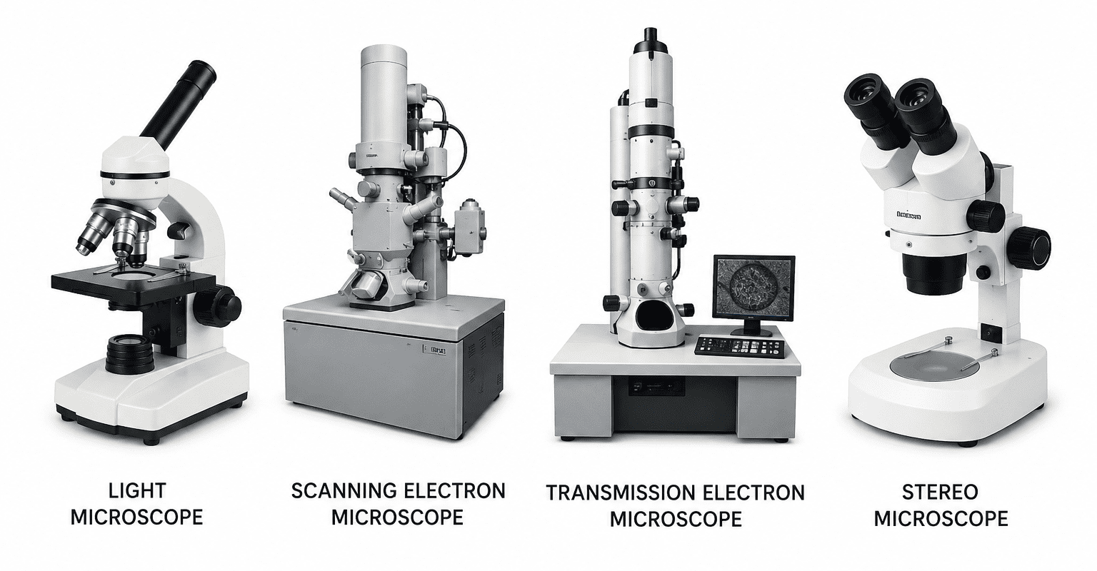
Compound light microscope
The compound light microscope is one of the most commonly used instruments in biological and medical laboratories for the observation of microorganisms, cells, tissues, and other minute specimens. It operates by making use of visible light to illuminate the structure of specimens placed on the stage of the microscope. The light passes through or reflects from the specimen and is then magnified through a system of lenses to produce a clear and enlarged image for observation. Because of its simplicity, effectiveness, and accessibility, the compound light microscope remains one of the most versatile and widely available magnifying instruments found in academic institutions, research laboratories, hospitals, and diagnostic centers throughout the world.
The term “compound” refers to the presence of two lens systems that work together to achieve magnification. These include the objective lens and the ocular lens, also known as the eyepiece lens. The objective lens is positioned close to the specimen and forms the primary magnified image, while the ocular lens further enlarges the image so that it can be viewed clearly by the observer. This dual magnification system enables the compound light microscope to provide higher magnification and better resolution than simple microscopes.
Compound light microscopes are usually fitted with several objective lenses of varying magnifying powers mounted on a revolving nosepiece. Common objective lenses include low-power, medium-power, high-power, and oil immersion lenses. These lenses allow the user to observe specimens at different levels of magnification depending on the nature of the sample and the detail required. The microscope also contains important components such as the condenser, diaphragm, illuminator, coarse adjustment knob, and fine adjustment knob, all of which contribute to proper focusing and illumination of the specimen.
The compound light microscope is particularly important in microbiology because it allows scientists and laboratory personnel to study the morphology, arrangement, and characteristics of microorganisms such as bacteria, fungi, algae, and protozoa. It is also extensively used in histology, pathology, hematology, and other branches of biological science for the examination of tissues and cells. Proper staining techniques are often employed to improve the visibility and contrast of specimens viewed under the microscope.
There are about four different types of compound light microscopes widely available today, each designed to meet specific laboratory and research needs. Despite differences in design and application, all compound light microscopes function primarily to magnify and provide clear visualization of microscopic specimens for scientific study and analysis.
The four commonly recognized types of compound light microscopes are:
- Bright-field microscope: This is the most widely used type of microscope, where light passes directly through the specimen to produce a dark image against a bright background.
- Dark-field microscope: This microscope produces bright images of specimens against a dark background and is useful for observing living, unstained microorganisms.
- Phase-contrast microscope: Phase-contrast microscope enhances the contrast of transparent and colorless specimens, making it suitable for viewing living cells without staining.
- Fluorescence microscope: This type of microscope uses ultraviolet or fluorescent light to observe specimens that have been stained with fluorescent dyes or naturally fluoresce.
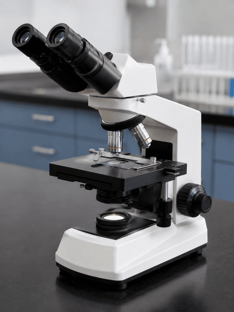
The Bright Field Microscope
The bright field microscope is the most commonly used type of microscope in microbiology laboratories, medical institutions, research centers, and educational establishments. It is widely employed for teaching purposes, routine laboratory investigations, and the observation of microorganisms and stained specimens that do not require highly specialized microscopic techniques. Due to its simplicity, affordability, and effectiveness, the bright field microscope remains one of the most important and accessible instruments used in biological and medical sciences.
The bright field microscope operates by allowing visible light to pass directly through a specimen, thereby producing a magnified image that can be observed through the eyepiece lens. It is usually fitted with several objective lenses of varying magnification powers and one or two ocular lenses that work together to produce a clear, enlarged, and detailed image of the specimen under examination. Most modern bright field microscopes are binocular, meaning they possess two eyepieces to provide greater viewing comfort and reduce eye strain during prolonged use (Figure 1). The combined action of the objective and ocular lenses enables the microscope to achieve high magnification and improved resolution.
One important characteristic of the bright field microscope is that it forms a dark image of the specimen against a bright illuminated background, which explains the origin of the term “bright field.” This contrast allows the observer to visualize the structure, arrangement, and morphology of microorganisms and cells more clearly. However, many microorganisms are naturally transparent and colorless, making them difficult to observe directly under ordinary light. To overcome this limitation, specimens are usually stained and fixed before examination under the bright field microscope.
Staining is an essential laboratory procedure that improves contrast between the specimen and its surrounding environment. Different staining techniques are used to introduce color variations into the cellular structures of microorganisms, thereby making them more visible and distinguishable. Common stains such as methylene blue, crystal violet, safranin, and Gram stains are frequently employed in microbiology laboratories for this purpose. Fixation, which often involves heat or chemical treatment, helps preserve the structure of the specimen and firmly attaches it to the microscope slide during examination.
The bright field microscope is generally limited in its ability to observe living cells because living organisms are often transparent and lack sufficient contrast for clear visualization. For this reason, it is mainly used for viewing dead cells, stained microorganisms, and preserved specimens. Despite this limitation, the microscope remains highly valuable in microbiology, pathology, histology, and hematology for routine examinations and diagnostic procedures.
The bright field microscope continues to serve as a fundamental tool in scientific laboratories because of its reliability, ease of use, and effectiveness in the observation of stained specimens and microorganisms. Its role in teaching, research, and medical diagnosis remains indispensable in modern biological science.
Phase Contrast Microscope
The phase contrast microscope is a specialized type of compound light microscope designed to observe living, unstained microorganisms in their natural state. Unlike the bright-field microscope, which typically requires staining to enhance visibility, the phase contrast microscope eliminates the need for staining, thereby preserving the integrity and viability of the cells under observation. This is particularly important in microbiological and biomedical studies where staining procedures can distort cellular structures, alter physiological conditions, or eventually kill the organism. By allowing direct visualization of live cells, the phase contrast microscope provides a more accurate representation of natural cellular processes.
This microscope is widely used in the study of both prokaryotic and eukaryotic cells. In eukaryotic cells, it is particularly useful for observing dynamic processes such as cytoplasmic streaming, cell division, and organelle movement. In prokaryotic organisms, it is commonly applied in examining bacterial cell shape, arrangement, motility, and the presence of internal structures such as spores and inclusion bodies. Because many bacteria are transparent and lack pigmentation, they are difficult to observe under standard bright-field illumination without staining. The phase contrast microscope overcomes this limitation, making it an essential tool in microbiology, cell biology, and medical research.
The working principle of the phase contrast microscope (Figure 2) is based on the differences in refractive index within various components of a microbial cell. When light passes through a specimen, different parts of the cell such as the cytoplasm, nucleus, and cell wall bend (refract) light to varying degrees due to differences in density and composition. These subtle differences are normally invisible to the human eye under standard light microscopy. However, the phase contrast microscope converts these small differences in phase shifts of light waves into variations in brightness and contrast, allowing internal structures to become visible without staining.
In phase contrast microscopy, the resulting image typically appears as a dark or grayish specimen against a bright background, providing enhanced contrast that highlights structural details within the cell. This optical enhancement is achieved through specialized components in the microscope, including a phase plate and an annular diaphragm, which manipulate the light waves as they pass through and around the specimen. The interaction between direct and diffracted light rays produces the final image with improved clarity and contrast.
Phase contrast microscopy is widely used in biomedical and clinical research due to its ability to observe living cells in wet mounts and natural conditions. It is particularly valuable in studying microbial behavior, cell morphology, and physiological responses to environmental changes, antibiotics, and other chemical agents. Researchers also rely on it to monitor cell growth and division in real time, making it an important tool in experimental microbiology and pharmacological studies.
A closely related technique is the differential interference contrast (DIC) microscope, which also enhances contrast in unstained specimens. Although it operates on a slightly different optical principle, it produces images with improved clarity and a pseudo three-dimensional appearance. The DIC microscope is useful for examining fine cellular details such as endospores, cell walls, vacuoles, and nuclei in both prokaryotic and eukaryotic cells. It provides sharper images with greater depth perception compared to phase contrast microscopy in some applications.
In addition to DIC microscopy, modern advancements in optical imaging have led to the development of highly sophisticated instruments such as the confocal scanning laser microscope and the atomic force microscope. These advanced systems are capable of producing high-resolution, three-dimensional images of living cells and tissues. They have greatly expanded the ability of scientists to study biological structures at the microscopic and even nanoscopic levels, contributing significantly to modern cell biology, medical diagnostics, and molecular research. The phase contrast microscope remains a fundamental and indispensable tool in microbiology and biomedical sciences due to its unique ability to visualize living, unstained microorganisms with enhanced contrast and clarity.
The Dark Field Microscope
The dark field microscope is a specialized form of light microscope used primarily for the observation of living, unstained cells and microorganisms. It is particularly valuable in microbiology and medical diagnostics where it is important to examine organisms in their natural, living state without the use of staining techniques that might kill or alter them. This method provides enhanced contrast, making it easier to visualize organisms that would otherwise appear transparent or nearly invisible under conventional light microscopy.
The dark field microscope is equipped with a modified condenser known as a dark field condenser. This condenser is designed in such a way that it prevents direct light from entering the objective lens. Instead of allowing light to pass straight through the specimen, it directs light at an angle so that only oblique rays reach the specimen on the stage. As a result, the specimen itself is not directly illuminated. Only light that is scattered or diffracted by structures within the specimen is captured by the objective lens.
This unique illumination technique produces a characteristic visual effect in which the specimen appears bright or luminous against a dark or black background. The background remains dark because no direct light enters the objective lens, while the light that is scattered by the specimen creates a glowing outline or detailed image of the organism. This contrast greatly enhances the visibility of small and delicate structures that might otherwise be difficult to detect. In dark field microscopy, the image formation depends primarily on refracted and reflected light rather than transmitted light. This makes it especially useful for observing thin, transparent organisms such as spirochetes, protozoa, and certain bacteria that are not easily stained or visualized using a bright field microscope. It is also widely used in clinical laboratories for the detection of pathogens in body fluids such as blood or cerebrospinal fluid.
One of the major advantages of dark field microscopy is its ability to allow the observation of living cells in motion. Cell structures, shapes, and especially motility can be clearly observed, providing valuable information about the behavior and characteristics of microorganisms. This is particularly important in the diagnosis of certain infectious diseases where motility is a key identifying feature. Although it provides excellent contrast and is highly useful for specific applications, the dark field microscope requires careful alignment and proper handling to produce accurate results. Any misalignment of the condenser or light source can significantly affect image quality. Despite this, it remains an essential tool in microbiology due to its ability to reveal fine structural details and dynamic behavior in living, unstained specimens.
The Fluorescence Microscope
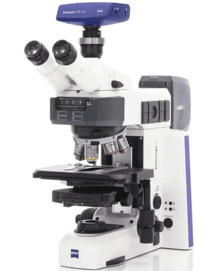
The fluorescence microscope is a specialized type of light microscope used to observe microorganisms, cells, and tissues that exhibit fluorescence, meaning they are able to absorb light at one wavelength and emit it at a different, usually longer, wavelength (Figure 3). This property produces bright, vividly colored images that stand out clearly against a dark background, making it especially useful for detecting and studying specific structures within biological samples. It is widely applied in microbiology, medical diagnostics, immunology, and molecular biology for its high sensitivity and specificity in identifying target organisms or cellular components.
Fluorescence microscopy is particularly useful for observing living cells or microorganisms that naturally fluoresce or have been stained with fluorescent dyes. Some microorganisms naturally contain compounds that emit light when exposed to certain wavelengths. However, in most laboratory applications, fluorescence is achieved through the use of fluorescent stains or fluorochromes. These dyes bind selectively to specific cellular structures or molecules, allowing scientists to visualize organisms that would otherwise be difficult to detect. For example, pathogens such as Mycobacterium tuberculosis and Escherichia coli can be labeled with fluorescent dyes or antibodies that bind to their surface structures, making them visible under fluorescence microscopy.
Unlike traditional light microscopes such as the bright-field, dark-field, or phase-contrast microscope, which form images by passing visible light directly through a specimen, fluorescence microscopy relies primarily on the light emitted by the specimen itself after excitation. In other words, the image is not formed from transmitted light but from emitted fluorescence. This fundamental difference allows fluorescence microscopes to produce highly specific and high-contrast images, even when the target structures are present in very small quantities within complex biological samples.
In fluorescence microscopy, the specimen is first exposed to high-energy light, typically ultraviolet (UV) light or other specific wavelengths such as blue or violet light. This excitation light is directed onto the sample through a system of filters and mirrors designed to isolate the appropriate wavelength. When the fluorescent molecules within the specimen absorb this energy, they become excited and subsequently release light at a lower energy level. This emitted light is then collected and passed through additional filters before being detected and magnified to form a visible image.
The ability to selectively highlight specific structures makes fluorescence microscopy an essential tool in modern biological research. It is widely used in the detection of antigen-antibody reactions, particularly in immunofluorescence techniques where antibodies tagged with fluorescent markers bind to specific antigens in tissue or cell samples. This application is highly valuable in clinical diagnostics for identifying infectious agents, autoimmune disorders, and cancer-related markers. It also plays a critical role in microbial ecology, where it is used to study microbial diversity, population dynamics, and interactions within environmental samples such as soil and water.
In addition, fluorescence microscopy is extensively used in genetic and cellular research, including the study of DNA, RNA, and protein localization within cells. It allows scientists to track biological processes in real time, providing insight into cell division, gene expression, and intracellular transport mechanisms. Its high sensitivity and ability to detect minute quantities of biological material make it one of the most powerful tools in modern laboratory science. The fluorescence microscope represents a significant advancement in microscopy techniques due to its ability to provide detailed, specific, and high-contrast images of biological specimens. Its wide range of applications in research, diagnostics, and environmental studies continues to make it an indispensable instrument in scientific investigation.
Electron Microscope
The electron microscope is a high-resolution imaging instrument that uses a beam of electrons, rather than visible light, to generate magnified images of biological and non-biological specimens. Unlike the light microscope, which relies on glass lenses to bend and focus photons, the electron microscope uses electromagnetic lenses to control and direct electron beams. Because electrons have a much shorter wavelength than visible light, this technique achieves far superior resolving power, enabling visualization of ultrastructural details that are not resolvable using conventional optical microscopy.
In electron microscopy, the specimen is placed in a highly controlled environment, typically a vacuum chamber, to prevent scattering of electrons by air molecules. This requirement makes electron microscopy fundamentally incompatible with the observation of living organisms in their natural, hydrated state. Instead, specimens must be fixed, dehydrated, and often embedded in resin or coated with conductive materials prior to observation. As a result, electron microscopy is primarily used for detailed structural analysis of cells, tissues, microorganisms, and macromolecular complexes rather than for real-time imaging of living cells.
The principle of image formation in an electron microscope depends on the interaction between electrons and the specimen. When an electron beam is generated and accelerated at high voltage, it is focused onto a very thin section of the specimen using electromagnetic lenses. As electrons pass through or interact with the specimen, they are scattered to varying degrees depending on the density and composition of the material. These interactions are then converted into a visible image, either on a fluorescent screen, photographic film, or more commonly today, a digital imaging system integrated into the microscope.
There are two major categories of electron microscopes: the transmission electron microscope (TEM) and the scanning electron microscope (SEM). The transmission electron microscope produces images by transmitting electrons through ultra-thin sections of a specimen. This allows for the visualization of internal structures such as organelles within eukaryotic cells, bacterial cell architecture, and even viral particles. TEM is especially valuable in cell biology and virology because it reveals internal ultrastructural details at the nanometer scale.
In contrast, the scanning electron microscope (SEM) generates images by scanning a focused electron beam across the surface of a specimen. The electrons reflected or emitted from the surface are detected and used to construct a three-dimensional image of the specimen’s exterior. SEM is particularly useful for studying surface morphology, including the texture of microbial colonies, the arrangement of cells in tissues, and the fine structural features of viruses or inorganic materials. Together, TEM and SEM provide complementary perspectives: internal ultrastructure versus surface topology.
One of the most important advantages of electron microscopy is its exceptionally high resolving power. While light microscopes are typically limited to a resolution of about 200 nanometers due to the diffraction limit of visible light, electron microscopes can achieve resolutions in the range of 0.1 to 1 nanometer, depending on the instrument and technique used. This represents an improvement of roughly 1,000-fold over light microscopy. This extraordinary resolution is made possible by the extremely short wavelength of electrons when accelerated to high energies.
Because of this high resolution, electron microscopy allows scientists to study biological structures at the molecular and subcellular level. Features such as ribosomes, membrane bilayers, cytoskeletal filaments, and viral capsid structures can be clearly resolved. This capability has made electron microscopy indispensable in fields such as microbiology, structural biology, pathology, and materials science.
However, electron microscopes have several limitations. They are large, complex, and extremely expensive instruments that require specialized facilities. Their operation demands highly trained personnel, as specimen preparation and imaging procedures are technically demanding and time-consuming. In addition, because electron microscopy requires a vacuum environment and extensive sample processing, it cannot be used to observe living cells in real time, which limits its application in dynamic biological studies.
Electron microscopes are typically installed in dedicated laboratory rooms designed to maintain environmental stability, including vibration isolation, electromagnetic shielding, and controlled temperature conditions. Many modern systems are also equipped with integrated digital cameras and computer-based imaging software, allowing for precise capture, processing, and analysis of micrographs. The electron microscope is a powerful analytical tool that has revolutionized modern biology and materials science. By replacing light with electrons and glass lenses with electromagnetic lenses, it enables visualization of structures at an unprecedented level of detail. Despite its limitations in cost, complexity, and inability to image living specimens, its contribution to understanding the ultrastructure of cells, microorganisms, and nanomaterials remains fundamental to scientific research.
Transmission Electron Microscope (TEM)
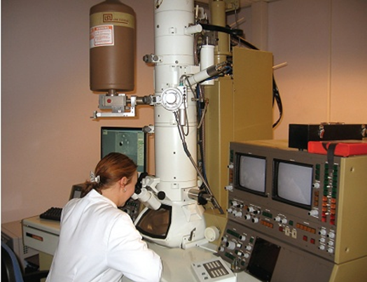
The transmission electron microscope (TEM) is a high-resolution imaging instrument used to visualize the ultrastructural organization of microorganisms, viruses, and subcellular components at nanometer-scale resolution (Figure 4). Unlike light microscopy, which is limited by the diffraction of visible light, TEM uses a beam of electrons accelerated at high voltage and transmitted through an ultrathin specimen. Because electrons have much shorter wavelengths than photons, TEM can achieve resolution sufficient to reveal fine structural details such as membranes, ribosomes, nucleic acid organization, and intracellular inclusions.
For biological applications, specimens must be extensively prepared to withstand the vacuum environment and electron beam. Microorganisms are typically fixed using chemical fixatives such as glutaraldehyde and osmium tetroxide to preserve structural integrity and stabilize lipids and proteins. Following fixation, the sample is dehydrated and embedded in a resin before being cut into ultrathin sections (approximately 50-100 nm thick) using an ultramicrotome equipped with a diamond or glass knife. These thin sections are necessary because electrons have limited penetration depth, and excessive thickness would scatter electrons and reduce image clarity.
Staining is a critical step in TEM preparation because biological materials are inherently electron-lucent (poor at scattering electrons). Heavy metal stains such as uranyl acetate and lead citrate are commonly used to enhance contrast by increasing electron density in specific cellular structures. This allows detailed visualization of internal features such as the cytoplasmic membrane, cell wall architecture, nucleoid region, and organelles in eukaryotic microorganisms.
In addition to thin-section TEM, negative staining is widely used for examining surface structures and viral morphology. In this technique, the background is stained with electron-dense compounds (e.g., phosphotungstic acid or uranyl acetate), while the specimen itself remains relatively unstained. This creates a high-contrast silhouette that highlights external features such as viral capsids, envelopes, bacterial capsules, pili, and flagella. Negative staining is particularly valuable in virology because it allows rapid visualization of intact viral particles without requiring extensive sectioning. TEM is primarily used to study internal ultrastructural organization and fine morphological details of cells and viruses. It provides critical insights into microbial architecture, pathogenesis, and cellular organization that cannot be resolved using light microscopy.
Scanning Electron Microscopy (SEM)
Scanning Electron Microscopy (SEM) is an advanced imaging technique widely used for the high-resolution, three-dimensional visualization of microorganisms and other biological specimens. Unlike Transmission Electron Microscopy (TEM), which is primarily used to examine internal ultrastructural details of cells through ultra-thin sections, SEM is specifically designed to investigate the external morphology of specimens. It provides detailed information about surface architecture, making it particularly valuable for studying microbial cell shape, surface textures, appendages such as pili or flagella, and structural changes associated with environmental or experimental conditions.
In SEM analysis, the specimen is typically fixed and dehydrated to preserve structural integrity and prevent distortion under vacuum conditions. Since biological samples are generally non-conductive, they are coated with an ultra-thin conductive layer of heavy metals such as gold, platinum, or silver. This coating enhances electron conductivity and improves signal generation by facilitating the emission and scattering of electrons when exposed to the electron beam.
During operation, a focused beam of high-energy electrons is systematically scanned across the surface of the specimen in a raster pattern. As these electrons interact with the metal-coated surface, they generate secondary electrons and backscattered electrons. These emitted electrons are collected by specialized detectors within the microscope. The signals are then converted into electrical impulses and processed to construct a detailed image displayed on a viewing screen. The resulting image provides a striking three-dimensional appearance due to variations in electron emission intensity from different surface topographies.
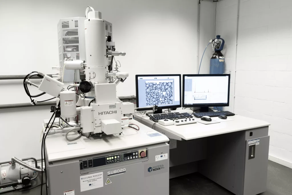
Additionally, SEM systems are equipped with digital imaging and storage capabilities, allowing high-resolution micrographs to be captured and archived for further analysis (Figure 5). These images can also be enhanced or analyzed quantitatively using specialized software. SEM is extensively used in microbiology, materials science, and biomedical research due to its ability to reveal fine surface details at nanometer-scale resolution. It is particularly useful in studying microbial adhesion, biofilm formation, pathogen-host interactions, and structural responses to antimicrobial agents. Together with TEM, SEM forms a complementary suite of electron microscopy techniques that has significantly advanced modern biological and medical research by enabling comprehensive visualization of both internal and external cellular structures.
REFERENCES
Beck R.W (2000). A chronology of microbiology in historical context. Washington, D.C.: ASM Press.
Cheesbrough, M (2006). District Laboratory Practice in Tropical countries Part I Cambridge
Chung K.T, Stevens Jr., S.E and Ferris D.H (1995). A chronology of events and pioneers of microbiology. SIM News, 45(1):3–13.
Dictionary of Microbiology and Molecular Biology, 3rd Edition. Paul Singleton and Diana Sainsbury. 2006, John Wiley & Sons Ltd. Canada.
Glick B.R and Pasternak J.J (2003). Molecular Biotechnology: Principles and Applications of Recombinant DNA. ASM Press, Washington DC, USA.
Goldman E and Green L.H (2008). Practical Handbook of Microbiology, Second Edition. CRC Press, Taylor and Francis Group, USA.
Madigan M.T., Martinko J.M., Dunlap P.V and Clark D.P (2009). Brock Biology of microorganisms. 12th edition. Pearson Benjamin Cummings Publishers. USA.
Nester E.W, Anderson D.G, Roberts C.E and Nester M.T (2009). Microbiology: A Human Perspective. Sixth edition. McGraw-Hill Companies, Inc, New York, USA.
Prescott L.M., Harley J.P and Klein D.A (2005). Microbiology. 6th ed. McGraw Hill Publishers, USA.
Willey J.M, Sherwood L.M and Woolverton C.J (2008). Harley and Klein’s Microbiology. 7th ed. McGraw-Hill Higher Education, USA.
www.materianova.be/en/our-equipment/analysis-characterization/morphological-characterization/sem-scanning-electron-microscope/
Discover more from Microbiology Class
Subscribe to get the latest posts sent to your email.