The microscope remains one of the most fundamental instruments in biological and material sciences, enabling observation of structures that are otherwise invisible to the naked eye. Its utility spans disciplines such as microbiology, pathology, botany, and materials engineering, where precise visualization of microstructures is essential for analysis, diagnosis, and research. However, the effectiveness of this instrument is not solely dependent on its availability; rather, it is closely tied to the user’s familiarity with its construction and operational principles. A clear conceptual understanding of its architecture and functional logic is therefore indispensable for anyone intending to employ it effectively in scientific or educational contexts.
At its core, the microscope is a system of coordinated optical and mechanical components designed to magnify small objects while preserving as much detail and contrast as possible. Each element contributes to a chain of processes that begins with illumination and ends with the formation of a magnified image that can be interpreted by the observer. When these components are properly understood, the user is better equipped to align, focus, and adjust the system in a way that maximizes clarity and resolution. Conversely, a lack of familiarity can lead to inefficient use, misinterpretation of observed structures, and in some cases, physical damage to sensitive optical or mechanical parts.
Beyond the purely technical dimension, understanding how a microscope functions also have direct implications for laboratory practice and scientific rigor. Microscopy is not a passive activity; it requires deliberate manipulation of optical paths, careful handling of specimens, and continuous adjustment of viewing parameters. Users who appreciate how the instrument translates physical light interactions into visible images are more likely to recognize the consequences of improper handling, such as distortion, aberrations, or loss of resolution. This awareness fosters more accurate interpretation of results, which is especially important in contexts where microscopic observations inform broader scientific conclusions or clinical decisions.
Equally important is the relationship between user competence and instrument longevity. Microscopes are precision devices that rely on finely calibrated alignment and delicate structural integrity. Inappropriate handling, such as forcing mechanical adjustments, neglecting proper focusing procedures, or exposing optical surfaces to contamination, can degrade performance over time. Familiarity with the instrument’s operational logic encourages more careful use, reducing unnecessary strain on mechanical components and minimizing the risk of accidental damage. In laboratory environments where multiple users share equipment, this shared understanding becomes even more critical, as consistent handling practices help maintain reliability and extend the usable lifespan of the device.
In educational settings, developing a strong foundational understanding of the microscope also supports deeper scientific learning. Students who grasp how magnification and image formation occur are better positioned to interpret what they observe rather than simply viewing specimens as abstract visual patterns. This interpretive skill is essential for bridging the gap between observation and explanation, which is a central goal of scientific training. Without such understanding, microscopy risks being reduced to a purely mechanical exercise in focusing and viewing, rather than a meaningful tool for inquiry and discovery.
Moreover, familiarity with the instrument encourages a more thoughtful approach to experimental design and observation. Researchers who understand the constraints and capabilities of microscopy are better able to select appropriate magnification levels, prepare specimens correctly, and adjust lighting conditions to suit specific investigative goals. This level of awareness enhances both efficiency and accuracy, reducing trial-and-error approaches that can waste time or compromise data quality. It also supports more informed troubleshooting when images fail to meet expected standards, allowing users to identify whether issues stem from sample preparation, instrument configuration, or environmental factors.
The importance of understanding the microscope also extends to safety and maintenance practices within laboratory environments. Proper use of the instrument often involves handling fragile glass components, exposure to chemical stains, and careful calibration of light sources. Users who are aware of these operational demands are more likely to adopt safe handling procedures that protect both themselves and the equipment. Additionally, routine maintenance tasks such as cleaning lenses, covering the instrument after use, and ensuring correct storage conditions are more consistently performed when users appreciate their role in preserving optical integrity.
From a broader perspective, the microscope serves as an interface between human perception and the microscopic world, effectively extending the limits of natural vision. Its value lies not only in magnification but in its ability to reveal structural relationships that are fundamental to understanding biological and physical processes. This interpretive power is only fully realized when users engage with the instrument in an informed manner, recognizing that what appears in the field of view is the result of carefully controlled optical interactions rather than a direct representation of reality.
A thorough understanding of the microscope and its operational principles is essential for effective use, accurate observation, and responsible maintenance. It enhances the quality of scientific work by enabling users to interpret microscopic images correctly, reduces the likelihood of equipment damage through improper handling, and supports the development of essential observational skills in educational contexts. Ultimately, competence in microscopy is not merely a technical requirement but a foundational element of scientific literacy, ensuring that the instrument fulfills its role as a precise and reliable window into the microscopic world.
Components and Operational Roles of Microscope Parts
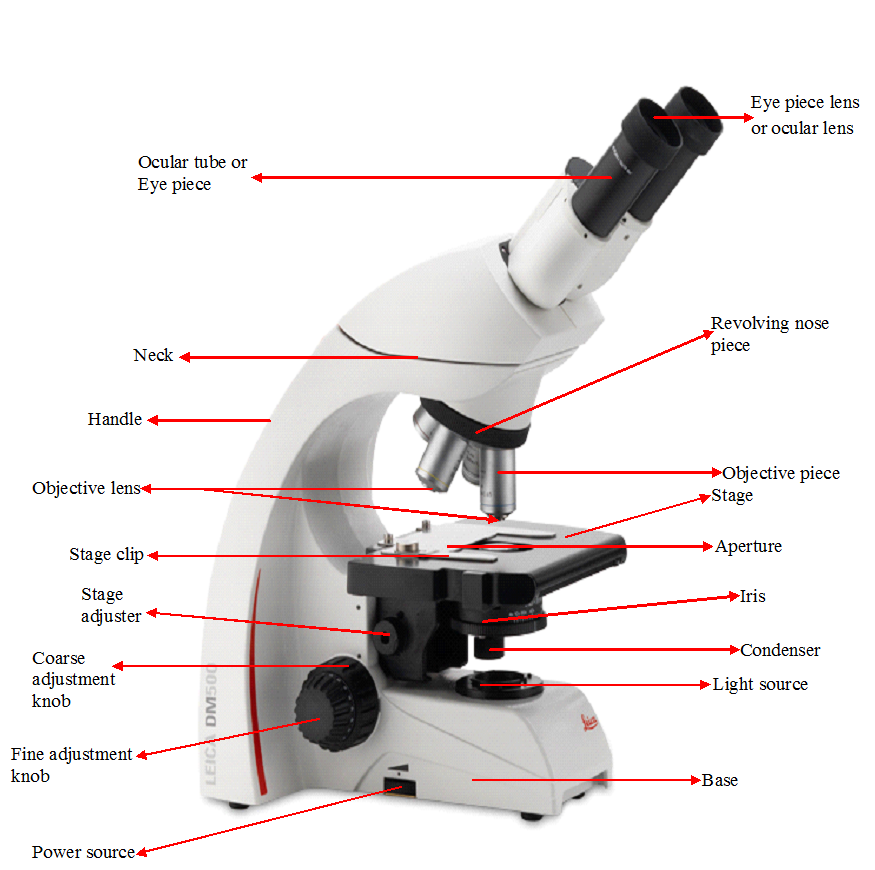
The microscope is a precision instrument composed of interconnected optical and mechanical components, each performing specific roles in image formation, illumination, and specimen manipulation (Figure 1). Its ocular and objective lenses work together to achieve magnification and resolution, while the nosepiece allows seamless switching between magnifications. Structural elements such as the arm, base, and body tube provide stability and alignment. The stage and its controls enable precise specimen positioning, while stage clips secure slides during observation. Focusing is achieved through coarse and fine adjustment knobs. Illumination is regulated by the illuminator, condenser, and diaphragm to optimize contrast and clarity. Together, these components ensure accurate microscopic observation.
Ocular lens (eyepiece): The ocular lens, commonly referred to as the eyepiece, is the uppermost optical component of the microscope through which the observer views the magnified image. Its primary function is to further enlarge the real, inverted image produced by the objective lens system so that it can be comfortably perceived by the human eye. Although it contributes significantly to total magnification (often 10× in standard microscopes), it does not independently generate an image; instead, it acts as a secondary magnifier within the optical pathway. The ocular lens is engineered to optimize visual clarity, field of view, and eye relief, ensuring that prolonged observation does not cause excessive strain. Many modern eyepieces also incorporate diopter adjustment mechanisms, allowing users to compensate for differences in vision between the two eyes. The cleanliness of the ocular lens is critical, as dust, oil, or smudges can distort the final image, reduce contrast, and impair resolution. In advanced microscopy systems, reticle eyepieces may also be included for measurement purposes, enabling users to estimate specimen dimensions directly within the field of view.
Objective lenses: The objective lenses are the primary magnifying components of the microscope and are responsible for forming the first, real image of the specimen. Positioned closest to the specimen, these lenses gather light transmitted or reflected from the sample and convert it into a magnified, inverted image. They are typically available in multiple magnification strengths, such as 4×, 10×, 40×, and 100× (oil immersion), each designed for specific levels of detail and resolution. The resolving power of an objective lens is determined by its numerical aperture, which governs its ability to distinguish between two closely spaced structures. Higher numerical apertures allow for greater detail and clarity but require precise focusing and proper illumination. The 100× oil immersion lens, in particular, requires the use of immersion oil to reduce light refraction and improve resolution at very high magnifications. Because objective lenses are positioned very close to the specimen, they are highly sensitive to damage from improper focusing or slide contact, making careful operation essential.
Nosepiece: The nosepiece, also known as the revolving turret, is the mechanical component that holds and supports the objective lenses. It is designed to rotate smoothly, allowing users to switch between different objective lenses without disturbing the alignment of the specimen. Each objective lens clicks into place to ensure accurate positioning along the optical axis. This precision is essential because even slight misalignment can result in blurred or partially illuminated images. The nosepiece must maintain mechanical stability while allowing fluid rotation, ensuring that changes in magnification are seamless and do not require complete refocusing. Its structural integrity directly influences the accuracy and efficiency of microscopic observation.
Body tube (head): The body tube is the cylindrical structure that connects the ocular lens to the objective lenses. Its primary function is to maintain the correct optical distance between these two systems, ensuring proper image formation and magnification. The body tube also ensures that light travels along a fixed path, preventing distortion and maintaining alignment between optical components. In modern microscopes, the body tube may be binocular or trinocular, allowing for two eyepieces or the attachment of cameras for imaging and documentation. The length and alignment of the body tube are critical for achieving accurate magnification calibration and image sharpness.
Arm (limb): The arm, also referred to as the limb, is the structural backbone of the microscope that connects the upper optical assembly to the base. It provides mechanical support and ensures stability for components such as the body tube, nosepiece, and stage. The arm also serves as a carrying handle, and proper handling requires one hand to grip the arm while the other supports the base to prevent tipping or damage. Structurally, the arm maintains alignment between optical and mechanical systems, which is essential for consistent image quality. Its rigidity reduces vibrations and contributes to overall instrument stability during use.
Base: The base is the foundational support structure of the microscope that bears its entire weight and ensures stability during operation. It prevents tipping and minimizes vibrations that could interfere with image clarity, especially at high magnifications. In modern microscopes, the base often houses the built-in illuminator and electrical components, making it both a structural and functional component. A heavy and well-balanced base is essential for maintaining steady focus and preventing accidental movement during observation. Its design contributes significantly to the safety, durability, and operational efficiency of the instrument.
Stage: The stage is the flat platform on which microscope slides are placed for observation. It contains a central aperture that allows light from the illuminator to pass through the specimen. In mechanical microscopes, the stage is designed for precise movement in horizontal and vertical directions, enabling controlled scanning of the specimen. This adjustability is essential for locating and examining specific regions of interest within a sample. The stage ensures that the specimen remains properly oriented and stable during observation, particularly at high magnifications where even slight movements can shift the image out of view.
Mechanical stage controls (stage adjuster): The mechanical stage controls, also known as stage adjusters, are knobs that enable precise movement of the stage in the X and Y directions. These controls allow the specimen to be moved smoothly left, right, forward, or backward without manually touching the slide. This precision is crucial when scanning across large or complex specimens, as it maintains focus and orientation while allowing systematic examination. Graduated scales may be included to record specimen coordinates, which is particularly useful in research and diagnostic applications where exact positioning must be documented.
Stage clips / slide holder: Stage clips or slide holders are small mechanical devices that secure the microscope slide firmly onto the stage. Their function is to prevent unwanted movement during observation, which is especially important at higher magnifications where even slight displacement can disrupt focus. By holding the slide in place, they ensure that the specimen remains stable while still allowing controlled movement via the mechanical stage. In more advanced microscopes, spring-loaded slide holders replace traditional clips to provide improved stability and ease of use.
Coarse adjustment knob: The coarse adjustment knob is a large focusing control used to bring the specimen into general focus. It operates by moving the stage or body tube rapidly over relatively large distances. This allows the user to locate the specimen and obtain an approximate image, usually under low-power magnification. Because it causes significant vertical movement, it must be used carefully to avoid contact between the objective lens and the slide. The coarse adjustment is always the first step in focusing before fine adjustments are made.
Fine adjustment knob: The fine adjustment knob is used for precise focusing after the specimen has been brought into approximate focus using the coarse adjustment knob. It moves the stage or optical system in very small increments, allowing subtle corrections that significantly enhance image sharpness and resolution. This knob is particularly important when using high-power or oil immersion objectives, where depth of field is extremely shallow. Proper use of the fine adjustment knob ensures clarity, detail, and accuracy in microscopic observation.
Condenser: The condenser is an optical component located beneath the stage that collects and focuses light from the illuminator onto the specimen. It concentrates the light into a sharp beam, improving resolution and enhancing image contrast. The condenser can often be adjusted vertically to optimize the focus of light depending on the magnification being used. Proper alignment of the condenser is essential for achieving uniform illumination and high-quality imaging, particularly in bright-field microscopy.
Diaphragm (iris diaphragm): The diaphragm is an adjustable aperture located beneath the condenser that controls the amount of light reaching the specimen. By opening or closing its aperture, it regulates brightness, contrast, and depth of field. A wide aperture allows more light and is useful for low magnification, while a narrow aperture increases contrast and improves visibility of fine details at high magnification. Proper adjustment of the diaphragm is essential for balancing illumination and achieving optimal image quality.
Illuminator (light source): The illuminator is the built-in light source of the microscope that provides illumination for specimen observation. It directs light upward through the condenser and stage, enabling visualization of transparent or semi-transparent specimens. Modern microscopes typically use LED or halogen bulbs, which offer consistent brightness and adjustable intensity. Without proper illumination, specimens would appear dark and indistinct, making observation impossible. The quality and stability of the illuminator directly affect image clarity and contrast.
Light control knob: The light control knob regulates the intensity of the illuminator. It allows users to increase or decrease brightness depending on the specimen type and magnification level. Proper light adjustment is essential to avoid overexposure, which can wash out specimen details, or underexposure, which can obscure fine structures. When used in combination with the diaphragm, it helps achieve optimal contrast and illumination balance.
Inclination joint: The inclination joint allows the microscope body to be tilted for more comfortable viewing. This feature improves ergonomics by enabling the user to adjust the viewing angle during prolonged observation sessions. It is especially useful in teaching and laboratory environments where extended periods of microscope use are common. However, excessive tilting must be avoided to prevent instability or spillage of liquid samples.
Revolving light intensity or filter system: Some microscopes include a filter system beneath the stage that allows the insertion of color filters or neutral density filters. These filters modify light quality to enhance contrast or reduce glare depending on the specimen type. This system improves image contrast and can be particularly useful in staining-based microscopy or when observing low-contrast specimens.
Each component of the microscope performs a specialized and essential function within a highly integrated system. From optical elements such as the ocular and objective lenses to mechanical structures like the stage, nosepiece, and adjustment knobs, every part contributes to image formation, clarity, stability, and precision. A thorough understanding of these components ensures correct usage, prevents mechanical damage, and enhances the quality of microscopic observation.
REFERENCES
Beck R.W (2000). A chronology of microbiology in historical context. Washington, D.C.: ASM Press.
Cheesbrough, M (2006). District Laboratory Practice in Tropical countries Part I Cambridge
Chung K.T, Stevens Jr., S.E and Ferris D.H (1995). A chronology of events and pioneers of microbiology. SIM News, 45(1):3–13.
Dictionary of Microbiology and Molecular Biology, 3rd Edition. Paul Singleton and Diana Sainsbury. 2006, John Wiley & Sons Ltd. Canada.
Glick B.R and Pasternak J.J (2003). Molecular Biotechnology: Principles and Applications of Recombinant DNA. ASM Press, Washington DC, USA.
Goldman E and Green L.H (2008). Practical Handbook of Microbiology, Second Edition. CRC Press, Taylor and Francis Group, USA.
Madigan M.T., Martinko J.M., Dunlap P.V and Clark D.P (2009). Brock Biology of microorganisms. 12th edition. Pearson Benjamin Cummings Publishers. USA.
Nester E.W, Anderson D.G, Roberts C.E and Nester M.T (2009). Microbiology: A Human Perspective. Sixth edition. McGraw-Hill Companies, Inc, New York, USA.
Prescott L.M., Harley J.P and Klein D.A (2005). Microbiology. 6th ed. McGraw Hill Publishers, USA.
Willey J.M, Sherwood L.M and Woolverton C.J (2008). Harley and Klein’s Microbiology. 7th ed. McGraw-Hill Higher Education, USA.
Discover more from Microbiology Class
Subscribe to get the latest posts sent to your email.