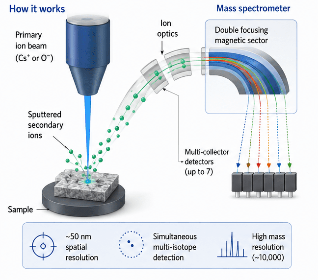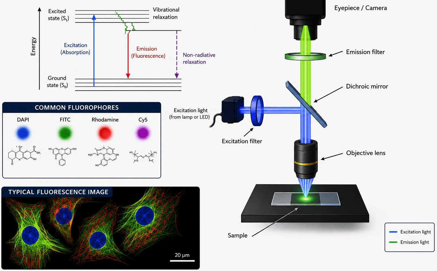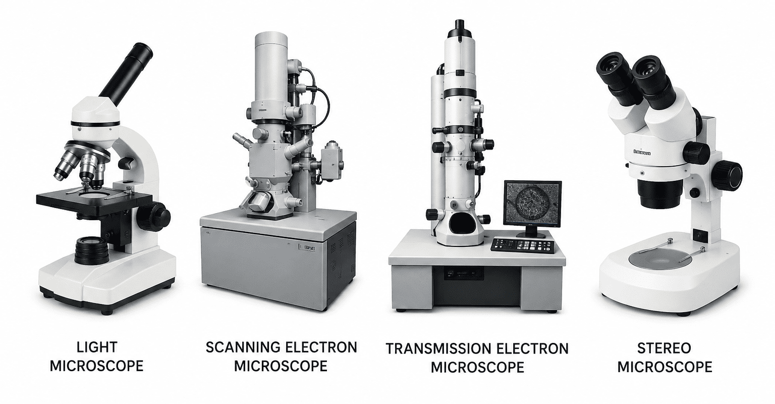

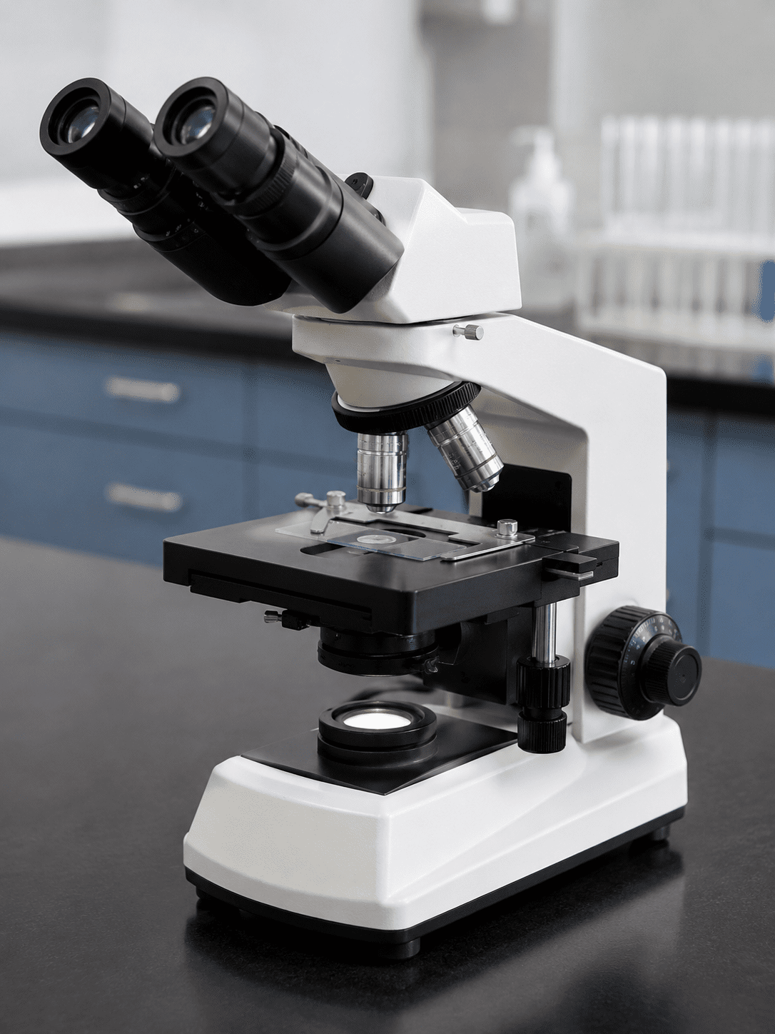
The microscope is one of the most indispensable instruments in microbiology and biomedical science. Its invention revolutionized scientific discovery by enabling researchers to observe microorganisms, cells, and other microscopic structures that are invisible to the naked eye. In microbiological investigations, microscopes play a central role in the identification, examination, and study of bacteria, fungi, protozoa, viruses, and other biological specimens. Because of this critical importance, microscopes must be handled carefully and maintained properly to ensure accuracy, durability, and continued effectiveness in laboratory work.
The microscope remains a vital instrument in microbiology, medicine, and biological sciences because it provides the means to visualize and study microscopic organisms and structures. Its usefulness and accuracy depend greatly on proper handling, cleaning, operation, storage, and maintenance. By following certain standard care guidelines, users can preserve the efficiency, reliability, and longevity of microscopes, thereby ensuring their continued value in laboratory investigations, scientific research, and educational activities.
Microscopes, whether compound light microscopes or electron microscopes, are delicate and expensive scientific instruments designed with precision optical and mechanical components. These instruments allow scientists, students, laboratory technologists, and researchers to magnify tiny objects and study their structural characteristics in detail. Without microscopes, the understanding of the microbial world and many biological processes would have remained impossible. The ability to diagnose infectious diseases, conduct biomedical research, analyze tissue samples, and study cellular functions depends heavily on the effective use of microscopes. Consequently, proper care and maintenance are essential responsibilities for every microscope user.
Proper care of the microscope begins with correct handling. A microscope should always be carried carefully using both hands: one hand holding the arm of the microscope and the other supporting its base. Carrying it carelessly or with one hand may result in accidental dropping, which can damage sensitive lenses, mirrors, mechanical parts, or the illumination system. Microscopes should also be placed on stable, clean, and dry laboratory benches away from the edge of the table to prevent falls and vibrations.
Cleanliness is another important aspect of microscope maintenance. Dust, oil, fingerprints, and chemical stains can reduce image quality and damage optical components. The lenses, especially the objective lenses and eyepieces, should only be cleaned with lens paper or a soft lint-free cloth designed specifically for optical instruments. Ordinary tissue paper, handkerchiefs, or rough materials should never be used because they can scratch the delicate glass surfaces. If immersion oil is used during microscopy, especially with the oil immersion objective lens, it should be cleaned immediately after use with an approved lens-cleaning solution to prevent the oil from hardening and damaging the lens.
Users should avoid touching the lenses directly with their fingers because fingerprints contain oils and dirt that can interfere with proper light transmission and image clarity. The microscope stage should also be kept clean and free from spilled stains, immersion oil, or specimen residues. Regular cleaning of all external surfaces helps to maintain the instrument in good working condition and prolongs its lifespan.
Correct operation of the microscope is equally important in ensuring its longevity. The coarse adjustment knob should be used carefully, particularly when focusing under high magnification, to prevent the objective lens from striking the slide. Sudden or forceful movements may crack the slide, damage the specimen, or scratch the objective lens. Fine adjustment knobs should be used for precise focusing, especially under higher magnifications. Mechanical parts such as stage clips, revolving nosepieces, and focusing knobs should never be forced if they appear stiff or resistant, as this may damage the instrument’s internal mechanisms.
Electrical safety must also be observed when using microscopes equipped with built-in illumination systems. Users should ensure that electrical cords are properly arranged and not left hanging where they can be pulled accidentally. After use, the microscope light should be switched off and allowed to cool before storage. Unnecessary prolonged illumination should be avoided because it can shorten the lifespan of the bulb and generate excess heat.
Proper storage is another major factor in microscope care. After use, the microscope should be returned to its storage cabinet or designated area and covered with a dust cover to protect it from dust, moisture, and environmental contaminants. Before storage, the low-power objective lens should be rotated into position, the stage lowered, and the power cord properly wrapped. Microscopes should be stored in dry environments because excessive humidity may encourage fungal growth on optical lenses, which can permanently damage them and reduce visibility.
Routine inspection and maintenance are necessary to ensure that microscopes remain functional and accurate.Laboratory personnel should regularly check for loose screws, damaged cords, dim illumination, or lens defects. Professional servicing and calibration may occasionally be required, especially for research-grade microscopes used in advanced scientific investigations. Preventive maintenance not only reduces repair costs but also ensures reliable and accurate laboratory results.
In teaching and research laboratories, all microscope users should be adequately trained on proper handling and maintenance procedures. Careless use of microscopes can lead to costly repairs, inaccurate observations, and interruption of laboratory activities. Since microscopes are shared instruments in many institutions, every user has a responsibility to preserve them for the benefit of others and for future scientific work.
As aforementioned, the microscope remains one of the most essential instruments in microbiology and biomedical laboratories because it enables the visualization and study of microscopic organisms and structures. Due to its delicate and expensive nature, proper care and maintenance are necessary to ensure accurate observations, long service life, and effective laboratory performance. Proper cleaning of lenses, careful handling during transportation, correct use of immersion oil, safe focusing procedures, appropriate storage, and regular maintenance are all important practices that every microscope user must observe. By following these guidelines consistently, laboratory personnel and students can preserve the efficiency, reliability, and durability of microscopes while ensuring high-quality scientific and diagnostic work in the laboratory.
Guidelines for proper care and maintenance of the microscope
Microscopes are precision instruments composed of sensitive optical, mechanical, and electrical components. Improper handling, poor cleaning habits, or careless usage can easily damage these parts and reduce the quality of observations. Therefore, every microscope user must follow standard operational and maintenance procedures to preserve the instrument and guarantee reliable laboratory results. Because of its importance and delicate construction, proper care and maintenance of the microscope are essential for ensuring its efficiency, durability, and accuracy during laboratory work.
The following guidelines explain in detail the proper care and maintenance procedures that should be observed when using microscopes in the microbiology laboratory.
Proper cleaning of the lens system
The lens system of the microscope is the most delicate and important component because it is responsible for magnifying and transmitting images. The ocular lenses (eyepieces) and objective lenses must always be properly cleaned before and after use to ensure clear visibility and accurate observation of specimens. Dust particles, fingerprints, immersion oil, stains, and moisture can accumulate on the lenses and interfere with image quality by causing blurred or distorted images.
Cleaning should only be carried out using approved lens tissue or soft lint-free cloths specifically designed for optical instruments. Ordinary tissue paper, cotton wool, handkerchiefs, or rough cloth materials should never be used because they can scratch the glass surfaces permanently. Approved lens cleaning solutions or solvents should also be used where necessary to remove stubborn stains or oil residues. In particular, after using the oil immersion objective lens, immersion oil should be carefully cleaned off immediately because dried oil can harden and damage the lens coating over time.
Regular cleaning of the lens system not only improves the quality of microscopic observations but also prolongs the lifespan of the microscope. Users should also avoid touching the lenses directly with their fingers because skin oils and dirt can reduce light transmission and affect image clarity.
Proper carrying and transportation of the microscope
Microscopes are fragile instruments that must be carried with extreme care. Improper transportation is one of the major causes of microscope damage in laboratories. Whenever a microscope is to be moved from one place to another, it should always be carried with both hands. One hand should firmly hold the arm of the microscope while the other hand supports the base. Carrying the microscope in this manner provides balance and reduces the risk of dropping it.
The microscope should never be swung, dragged, or carried carelessly with one hand because accidental falls may damage the lenses, stage, illumination system, or internal mechanical components. Sudden movements and vibrations should also be avoided during transportation. In addition, microscopes should only be placed on stable, flat, and vibration-free laboratory benches to prevent tipping or shaking during use.
Use of cover slips during wet preparation observation
Cover slips are very important when examining wet preparations or unstained specimens under the microscope. A wet preparation usually contains living microorganisms suspended in liquid, and without a cover slip, the specimen may spread unevenly or come into direct contact with the objective lens. The cover slip serves several important functions. First, it helps flatten the specimen into a thin, uniform layer that allows light to pass through effectively for better visualization.
Second, it protects the objective lens from contamination by water, stains, or biological material. Third, it prevents the formation of air bubbles that may interfere with microscopic observation. Failure to use a cover slip can result in damage to the objective lens, poor image quality, and inaccurate examination results. Therefore, cover slips should always be properly placed over wet preparations before viewing them under the microscope.
Proper use of immersion oil with the oil immersion objective lens
The oil immersion objective lens, usually the 100x objective, is specially designed for observing very small microorganisms such as bacteria. In order to achieve maximum magnification and resolution, immersion oil must always be used with this objective lens. Immersion oil works by reducing the refraction or bending of light as it passes from the glass slide into the air and then into the objective lens. Since the refractive index of immersion oil is similar to that of glass, more light enters the objective lens, thereby producing a brighter, sharper, and more detailed image.
When using the oil immersion objective, a small drop of immersion oil should be placed directly on the stained specimen slide. The oil immersion objective lens should then be carefully rotated into position until it makes contact with the oil and not the slide itself. Contact between the objective lens and the slide may scratch or damage the lens and break the slide. After observation, the immersion oil should immediately be cleaned off the objective lens and slide using lens tissue and approved cleaning solution. Leaving oil on the lens for prolonged periods can damage the lens coating and reduce optical efficiency.
Avoid unauthorized dismantling of microscope parts
Microscopes contain complex optical and mechanical systems that require professional handling and maintenance. Therefore, no part of the microscope should be removed, dismounted, or tampered with without the permission or supervision of the instructor, laboratory scientist, or technician in charge. Unauthorized dismantling may result in misalignment of optical components, loss of delicate parts, or damage to internal mechanisms. Since many microscope components are precisely calibrated, improper handling can reduce the instrument’s accuracy and functionality. If any part of the microscope appears faulty or damaged, it should immediately be reported to the responsible laboratory personnel for proper inspection and repair.
Careful use of adjustment knobs and stage controls
The coarse adjustment knob, fine adjustment knob, and stage adjustment controls are essential for focusing and positioning specimens during microscopic examination. These components should always be operated gently and carefully to prevent damage to the microscope and specimen slides. Excessive force while adjusting focus can cause the objective lens to crash into the slide, leading to cracked slides, damaged specimens, or scratched objective lenses.
This is especially important when using high-power objectives and the oil immersion lens because they operate very close to the specimen. The coarse adjustment knob should primarily be used under low-power magnification for rough focusing, while the fine adjustment knob should be used for precise focusing at higher magnifications. Smooth and controlled movements of the stage controls help maintain specimen stability and improve observation accuracy.
Use of a stable and obstruction-free bench
The microscope should always be placed on a strong, balanced, and clean laboratory bench during use. An unstable or uneven surface may cause vibrations that interfere with image quality or increase the risk of the microscope falling. The working area should also remain free from unnecessary materials, chemicals, books, bags, or obstructions that may accidentally come into contact with the microscope. Adequate working space allows the user to operate the microscope comfortably and safely. Furthermore, liquids should be kept away from the microscope to prevent accidental spills that may damage electrical or optical components.
Use of both eyes during observation
When observing specimens under the microscope, users are encouraged to keep both eyes open instead of closing one eye. Although only one eye may directly look through the ocular lens in monocular microscopes, keeping both eyes open reduces eye strain and fatigue during prolonged observation. In binocular microscopes, both eyes should naturally be used for viewing because this improves comfort, depth perception, and concentration. Continuous squinting or closing of one eye may lead to headaches, visual discomfort, and reduced observation efficiency, especially during extended laboratory sessions.
Cleaning using authorized materials and manufacturer’s instructions
Cleaning and maintenance procedures should only be performed using approved materials and according to the manufacturer’s recommendations. Different microscopes may have specific maintenance requirements depending on their design and optical system. Using unauthorized chemicals, abrasive materials, or inappropriate cleaning techniques can damage sensitive coatings, electrical systems, or moving components. Therefore, laboratory personnel should always follow the operational manuals and maintenance instructions provided by the manufacturer. Proper maintenance schedules should also be established for routine inspection and servicing of laboratory microscopes.
Proper storage of ocular and objective lenses
Objective lenses and ocular lenses that are not currently in use should be stored carefully in clean, dry, and tightly sealed containers. This prevents dust accumulation, moisture exposure, fungal growth, and accidental scratches. In humid environments, fungal contamination of lenses is a common problem that can permanently damage optical surfaces and reduce image clarity. Proper storage conditions help preserve the quality and lifespan of microscope lenses. Lens caps and protective covers should also be used whenever available.
Proper illumination and stage adjustment
Good illumination and proper stage adjustment are essential for achieving clear and accurate microscopic images. Insufficient or excessive light can reduce image quality and make observation difficult. Therefore, users should properly adjust the microscope condenser, diaphragm, mirror, or illumination intensity according to the specimen being viewed. The specimen slide should also be properly centered and secured on the stage before observation begins. Correct stage positioning ensures efficient focusing and accurate examination of the specimen. Proper lighting techniques enhance contrast, resolution, and visibility of microscopic structures.
Use of dust covers after use
After completing microscope work, the instrument should always be covered with its dust cover before storage. Dust particles can settle on the optical surfaces, illumination system, and mechanical components, thereby reducing efficiency and image quality. Dust accumulation over time may also contribute to scratches and contamination of sensitive parts. Covering the microscope protects it from dust, moisture, and environmental contaminants, especially in laboratories that are not frequently cleaned or are exposed to air pollutants.
Disconnecting the microscope from electricity source after use
Microscopes equipped with built-in electrical illumination systems should always be disconnected from the power supply after use. This is important for electrical safety, energy conservation, and prevention of damage to electrical components. Leaving the microscope connected unnecessarily may increase the risk of electrical faults, overheating, bulb damage, or fire hazards. The illumination should first be switched off and allowed to cool before unplugging the instrument. Electrical cords should also be arranged neatly to prevent tangling, pulling, or accidental damage.
REFERENCES
Beck R.W (2000). A chronology of microbiology in historical context. Washington, D.C.: ASM Press.
Cheesbrough, M (2006). District Laboratory Practice in Tropical countries Part I Cambridge
Chung K.T, Stevens Jr., S.E and Ferris D.H (1995). A chronology of events and pioneers of microbiology. SIM News, 45(1):3–13.
Dictionary of Microbiology and Molecular Biology, 3rd Edition. Paul Singleton and Diana Sainsbury. 2006, John Wiley & Sons Ltd. Canada.
Glick B.R and Pasternak J.J (2003). Molecular Biotechnology: Principles and Applications of Recombinant DNA. ASM Press, Washington DC, USA.
Goldman E and Green L.H (2008). Practical Handbook of Microbiology, Second Edition. CRC Press, Taylor and Francis Group, USA.
Madigan M.T., Martinko J.M., Dunlap P.V and Clark D.P (2009). Brock Biology of microorganisms. 12th edition. Pearson Benjamin Cummings Publishers. USA.
Nester E.W, Anderson D.G, Roberts C.E and Nester M.T (2009). Microbiology: A Human Perspective. Sixth edition. McGraw-Hill Companies, Inc, New York, USA.
Prescott L.M., Harley J.P and Klein D.A (2005). Microbiology. 6th ed. McGraw Hill Publishers, USA.
Willey J.M, Sherwood L.M and Woolverton C.J (2008). Harley and Klein’s Microbiology. 7th ed. McGraw-Hill Higher Education, USA.
Discover more from Microbiology Class
Subscribe to get the latest posts sent to your email.