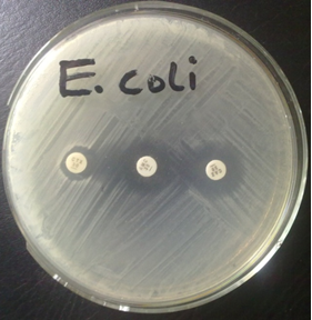


Antimicrobial resistance has become an important challenge in clinical microbiology. This is because bacteria can acquire mechanisms that reduce or eliminate the effectiveness of commonly used antibiotics. Among these mechanisms, the production of beta-lactamase enzymes is especially significant. Beta-lactamases enable bacteria to resist beta-lactam antibiotics such as penicillins by breaking the characteristic beta-lactam ring that is essential for the antibacterial activity of drugs such as penicillins and cephalosporins. Detecting this enzymatic activity is therefore valuable for understanding bacterial resistance and for supporting appropriate antimicrobial susceptibility assessment.
The nitrocefin test is a rapid biochemical method used to detect beta-lactamase production in bacterial isolates. Nitrocefin is a chromogenic cephalosporin designed to undergo a visible color change when its beta-lactam ring is hydrolyzed by a beta-lactamase enzyme. Before enzymatic hydrolysis, the reagent has a yellowish appearance. When a bacterium produces a beta-lactamase capable of acting on nitrocefin, cleavage of the beta-lactam ring produces a distinct red or pink coloration. This reaction provides a straightforward visual indication of enzymatic activity and can often be observed within a short period.
The usefulness of the nitrocefin assay lies in its direct detection of an important resistance mechanism rather than simply measuring bacterial growth in the presence of an antibiotic. This distinction is relevant because resistance can arise through several different biological processes, including alteration of antibiotic targets, reduced permeability, active efflux, or enzymatic inactivation. By specifically demonstrating beta-lactam hydrolysis, the test provides mechanistic information about the organism being examined.
The assay can be applied to bacterial cultures in laboratory investigations where rapid identification of beta-lactamase activity is required. Its relatively simple visual endpoint also makes it accessible for routine microbiological testing and educational laboratory work. However, interpretation requires appropriate controls and awareness of the limitations of the assay. A negative nitrocefin reaction does not necessarily establish complete susceptibility to every beta-lactam antibiotic, because bacteria may possess other resistance mechanisms or enzymes with limited activity against the substrate.
The nitrocefin assay illustrates the biochemical relationship between an antibiotic’s molecular structure, enzymatic degradation, and the resulting resistance phenotype. It provides a useful experimental approach for demonstrating how bacterial enzymes can neutralize antimicrobial compounds and highlights the importance of rapid resistance detection in microbiological research and clinical practice.
Principle of the nitrocefin test
The nitrocefin test is based on a simple biochemical principle: beta-lactamase enzymes can hydrolyze the beta-lactam ring of nitrocefin, producing a visible change in color. Unlike conventional antimicrobial susceptibility tests, which primarily indicate whether bacterial growth is inhibited, the nitrocefin assay provides a direct indication of enzymatic activity. The reaction therefore connects a bacterial resistance mechanism with a readily observable chemical response.
Structure and properties of nitrocefin
Nitrocefin is a chromogenic cephalosporin belonging to the beta-lactam family. Its molecular structure contains a four-membered beta-lactam ring, which is the structural feature recognized and attacked by beta-lactamases. Nitrocefin is particularly useful as a diagnostic substrate because its chemical structure has been modified to make enzymatic hydrolysis visually detectable. In its intact form, the compound has a relatively pale yellow coloration. This characteristic makes it possible to distinguish the unhydrolyzed substrate from the strongly colored product generated during the reaction.
The chromogenic nature of nitrocefin is central to its laboratory application. The substrate does not merely serve as a target for beta-lactamase; it also acts as an indicator of the reaction. Consequently, enzyme activity can be assessed without requiring complex analytical equipment. A bacterial isolate containing an active beta-lactamase can produce a visible response after exposure to the substrate.
Mechanism of beta-lactam ring hydrolysis
Beta-lactamases are enzymes that recognize beta-lactam compounds as substrates. During the reaction, the enzyme interacts with the beta-lactam ring and facilitates cleavage of the ring’s amide bond. This process is referred to as hydrolysis, because water participates in breaking the chemical bond. Once the beta-lactam ring is opened, the original molecular configuration of nitrocefin is disrupted and its chemical properties change.
This reaction represents an important bacterial defense mechanism. By destroying the beta-lactam structure, beta-lactamase can render susceptible beta-lactam antibiotics biologically ineffective. In the nitrocefin assay, however, the same enzymatic activity is deliberately exploited as a detection mechanism.
Basis of the color change
The visible color reaction occurs because hydrolysis alters the electronic structure of nitrocefin. The intact molecule appears yellow, whereas cleavage of its beta-lactam ring produces a hydrolyzed compound with a markedly different light-absorbing pattern. As a result, the reagent develops a red to pink coloration, indicating beta-lactamase activity.
Thus, the color transition is not simply an indicator added to the test; it is a direct consequence of the chemical transformation caused by the enzyme. The intensity and speed of the reaction can provide a practical indication of enzymatic activity, although interpretation should be performed with appropriate controls. The nitrocefin test converts an otherwise microscopic biochemical event enzymatic destruction of a beta-lactam ring into an easily recognizable laboratory signal.
Beta-lactamase production in bacteria
Beta-lactamase production is an important bacterial strategy for surviving exposure to beta-lactam antimicrobial agents. Beta-lactamases are enzymes produced by certain bacteria that inactivate beta-lactam antibiotics through hydrolysis of the beta-lactam ring, a structural feature required for the activity of these drugs. Once the ring is disrupted, the antibiotic loses its ability to interact effectively with bacterial cell-wall synthesis machinery. The production of these enzymes can therefore transform an otherwise susceptible bacterial population into one capable of persisting during antimicrobial treatment.
Beta-lactam resistance can develop through several complementary mechanisms. Enzymatic degradation is one of the most prominent, but bacteria may also modify the proteins targeted by beta-lactam antibiotics, reduce antibiotic entry through alterations in membrane permeability, or increase the removal of antimicrobial compounds through efflux systems. In Gram-negative organisms, changes in porin channels can restrict the movement of beta-lactams into the periplasmic space, while modifications of penicillin-binding proteins (PBPs) can decrease antibiotic affinity. When these mechanisms occur alongside beta-lactamase production, resistance may become substantially more pronounced.
Beta-lactamases are diverse and differ in their substrate preferences, molecular structures, and genetic origins. Penicillinases primarily hydrolyze penicillins, whereas broader-spectrum enzymes can act against multiple generations of beta-lactam antibiotics. Extended-spectrum beta-lactamases (ESBLs) are capable of hydrolyzing many penicillins and cephalosporins, making infections caused by their producers more difficult to treat. AmpC beta-lactamases possess activity against several cephalosporins and are notable because their expression may be inducible or associated with chromosomal or acquired resistance determinants. Another clinically important group is the carbapenemases, which can hydrolyze carbapenems, antibiotics often reserved for infections involving multidrug-resistant organisms. Some carbapenemases are metallo-beta-lactamases, requiring metal ions such as zinc for enzymatic activity.
The significance of beta-lactamase production extends beyond individual bacterial cells. These enzymes contribute to the emergence and dissemination of antimicrobial resistance within healthcare and community environments. Resistance genes may occur on plasmids, transposons, integrons, or other mobile genetic elements, allowing resistance determinants to move between bacteria. Consequently, organisms that acquire beta-lactamase genes can become reservoirs from which resistance spreads to other bacterial populations.
From a microbiological perspective, identifying beta-lactamase production assists in characterizing resistant isolates and interpreting antimicrobial susceptibility patterns. Clinically, detection can influence therapeutic decisions, infection-control measures, and surveillance of emerging resistance. Understanding beta-lactamase production is therefore essential for linking bacterial enzymology with the broader problem of antimicrobial resistance and for recognizing why rapid detection of resistance mechanisms remains a central objective of modern diagnostic microbiology.
Procedure for the nitrocefin test
The nitrocefin test is a rapid laboratory procedure used to determine whether a bacterial isolate produces beta-lactamase. The procedure is based on the ability of beta-lactamase enzymes to hydrolyze nitrocefin, producing a visible color reaction. Careful preparation and appropriate controls are necessary to obtain an interpretable result.
Materials and reagents required
The test requires a fresh bacterial culture, nitrocefin reagent or a commercially prepared nitrocefin test disc, sterile inoculating equipment, a clean glass slide or appropriate reaction surface, and sterile water or saline when suspension of the organism is necessary. Personal protective equipment, including laboratory coat and gloves, should be used according to institutional biosafety requirements. A known beta-lactamase-producing organism can serve as the positive control, while a bacterium known not to produce the enzyme can serve as the negative control.
Preparation of bacterial isolates
A well-isolated colony from a fresh, pure bacterial culture is selected for testing. The organism should be identified and maintained using appropriate microbiological procedures before the assay is performed. Using an excessively old, mixed, or contaminated culture may interfere with interpretation. A sufficient amount of bacterial growth is collected with sterile equipment and prepared according to the instructions supplied with the particular nitrocefin reagent or test format. The use of a fresh isolate helps ensure that the enzyme activity detected reflects the characteristics of the organism being investigated.
Application of nitrocefin
The bacterial material is brought into contact with the nitrocefin reagent. For a reagent-based assay, the recommended quantity of bacterial suspension or colony material is applied to the test reagent. When nitrocefin discs are used, the bacterial inoculum is placed directly onto the designated area of the disc. The manufacturer’s specified reaction conditions should be followed because reagent concentration, sample quantity, and reaction time can vary between commercial preparations. The test should be protected from unnecessary contamination and excessive environmental exposure during the reaction.
Observation and recording of results
Following application, the reaction is examined for a characteristic color change. Beta-lactamase-mediated hydrolysis of nitrocefin produces a rapid change from the reagent’s original yellowish appearance toward pink, red, or reddish-purple, depending on the test system. The reaction should be observed within the manufacturer’s recommended reading period. Results should be documented immediately, including the bacterial isolate, test date, reagent or kit used, observed color, and final interpretation. Delayed readings should not be substituted for the specified observation period because prolonged exposure may complicate interpretation.
Positive and negative controls
Controls are essential for validating the assay. The positive control should demonstrate the expected color transformation, confirming that the reagent is functional and capable of detecting beta-lactamase activity. The negative control should retain the original reagent appearance, demonstrating that spontaneous color development or nonspecific reactions are not responsible for the result. If either control produces an unexpected reaction, the test should be considered unreliable and repeated with fresh materials and appropriate controls. The nitrocefin procedure provides a rapid visual approach for demonstrating beta-lactamase activity while emphasizing the importance of standardized handling, controlled observation, and accurate documentation.
Interpretation of nitrocefin test results
Interpretation of the nitrocefin test depends primarily on the visible response produced after a bacterial isolate comes into contact with the chromogenic substrate. The test is designed to reveal beta-lactamase activity through hydrolysis of the beta-lactam ring in nitrocefin. Because the reaction produces a readily observable color change, results can generally be distinguished without complex instrumentation. Nevertheless, careful observation and appropriate controls are necessary to obtain a reliable interpretation.
A positive reaction is indicated by a rapid change in the nitrocefin reagent from its original yellow or pale yellow appearance to a distinct red or pink coloration. This transformation occurs when beta-lactamase produced by the bacterial cell hydrolyzes the beta-lactam ring of nitrocefin. The resulting structural alteration changes the optical properties of the compound, producing the characteristic red endpoint. A clearly developed color therefore indicates that the tested organism possesses enzymatic activity capable of hydrolyzing nitrocefin. The intensity and speed of the color development may vary according to the amount of enzyme produced and the conditions under which the test is performed.
A negative reaction occurs when no detectable color conversion takes place during the specified observation period. The reagent generally remains yellow or retains its initial coloration. Such a result suggests that the organism did not demonstrate detectable nitrocefin-hydrolyzing beta-lactamase activity under the test conditions. However, a negative result should not automatically be interpreted as proof that the bacterium is susceptible to all beta-lactam antibiotics. Resistance can result from mechanisms unrelated to beta-lactamase production, while some enzymes may have weak or variable activity toward the substrate.
Several factors can influence nitrocefin test results. The age and physiological condition of the bacterial culture may affect enzyme expression. Insufficient bacterial material can produce a weak reaction, whereas an abundant inoculum may produce a more pronounced response. Reagent quality is also important because nitrocefin can deteriorate when exposed to unsuitable storage conditions, excessive light, or prolonged environmental exposure. Temperature, reaction time, inoculum preparation, and the method used to apply the reagent can likewise influence the observed endpoint. Properly maintained positive and negative controls are therefore essential for distinguishing genuine enzymatic activity from technical variation.
False-positive reactions may occur when the reagent undergoes nonspecific degradation or when contamination introduces a beta-lactamase-producing organism into the test. Conversely, false-negative reactions may result from insufficient inoculum, low-level enzyme production, inactive or degraded reagent, inappropriate incubation conditions, or beta-lactamases with poor activity against nitrocefin. Consequently, unexpected results should be reassessed using suitable controls and, where necessary, an alternative confirmatory method. Accurate interpretation ultimately depends on combining the observed color reaction with sound laboratory technique and appropriate microbiological context.
Applications, advantages, and limitations of the nitrocefin test
The nitrocefin test has practical value in microbiology because it provides a direct approach for identifying beta-lactamase-producing bacteria. Its principal application is the detection of enzymatic activity responsible for the hydrolysis of beta-lactam compounds. When nitrocefin encounters a bacterium producing a compatible beta-lactamase, the substrate is hydrolyzed and produces a characteristic color change. This visible reaction allows laboratories to identify enzyme production without depending solely on bacterial growth patterns around antibiotic discs. The test can provide rapid evidence that a bacterial isolate possesses an important mechanism of beta-lactam resistance.
In clinical microbiology, nitrocefin testing can contribute to the characterization of bacterial isolates obtained from clinical specimens. Detecting beta-lactamase activity may assist laboratory personnel in recognizing organisms with reduced responsiveness to particular beta-lactam agents. The test can therefore complement antimicrobial susceptibility testing and other resistance-detection procedures. In research microbiology, the assay is useful for investigating enzyme activity, comparing bacterial isolates, studying resistance phenotypes, and examining the biological effects of beta-lactamase production. Its straightforward visual endpoint also makes it suitable for laboratory demonstrations of enzymatic antibiotic resistance.
One important advantage of the nitrocefin method is its rapidity. A detectable color reaction can develop within a short period when sufficient enzyme activity is present, allowing results to be obtained considerably faster than approaches that depend on extended bacterial growth. The assay is also relatively simple to perform and does not require highly complex instrumentation for basic visual interpretation. Furthermore, because the test detects enzymatic hydrolysis directly, it provides mechanistic information rather than merely indicating that bacterial growth is associated with antibiotic resistance. Compared with some more elaborate biochemical or molecular techniques, this simplicity can make nitrocefin testing convenient for preliminary screening.
Despite these strengths, the test has important limitations. Nitrocefin detects beta-lactamase activity toward its specific substrate; therefore, a negative result does not necessarily mean that the organism lacks all beta-lactam resistance mechanisms. Some enzymes may demonstrate weak activity against nitrocefin, while enzyme expression can vary between isolates. Test conditions, reagent quality, inoculum characteristics, and interpretation of subtle color changes can also influence the result. In addition, the assay does not by itself identify the precise beta-lactamase gene or fully characterize the enzyme involved.
For these reasons, confirmatory testing may be necessary when results are clinically significant, unexpected, or ambiguous. Antimicrobial susceptibility testing, inhibitor-based assays, phenotypic confirmatory methods, or molecular techniques may provide additional information. Thus, nitrocefin is best regarded as a rapid and useful tool for detecting beta-lactamase activity, while definitive resistance characterization may require complementary laboratory methods.
Further reading
Nordmann, P., Helsens, N., Kieffer, N., Tinguely, C., Greub, G., & Poirel, L. (2025). Rapid detection of β-lactamase activity using the rapid Amp NP test. Microbiology Spectrum, 13(4), e00782-24.
Khan, S., Sallum, U. W., Zheng, X., Nau, G. J., & Hasan, T. (2014). Rapid optical determination of β-lactamase and antibiotic activity. BMC Microbiology, 14, Article 84.
Levy, S. B. (1982). Microbial resistance to antibiotics: An evolving and persistent problem. The Lancet, 320(8289), 83–88.
Livermore, D. M., & Brown, D. F. J. (2001). Detection of β-lactamase-mediated resistance. Journal of Antimicrobial Chemotherapy, 48(Suppl. 1), 59–64.
Skov, R., Lonsway, D. R., Larsen, J., Larsen, A. R., Samulionienė, J., & Limbago, B. M. (2021). Evaluation of methods for detection of β-lactamase production in MSSA. Journal of Antimicrobial Chemotherapy, 76(6), 1487–1494.
Discover more from Microbiology Class
Subscribe to get the latest posts sent to your email.