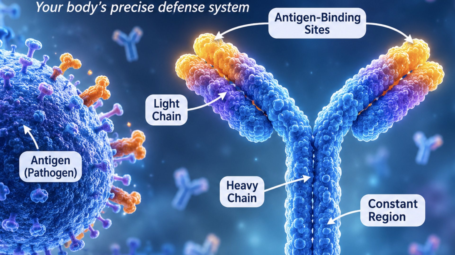

Immunoglobulin E (IgE) is a specialized class of antibody that forms an important component of the human immune system. IgE plays a dual role in immune defense, contributing both to allergic immune responses and to host protection against parasitic infections. It is one of the five major immunoglobulin classes, alongside IgG, IgA, IgM and IgD, and is distinguished by its unique involvement in allergic inflammation and immune responses against parasitic organisms. Although IgE represents the least abundant immunoglobulin in normal human circulation, its biological activity is remarkably significant. Even at extremely low serum concentrations, it can initiate powerful immune reactions through its interaction with specific receptors on immune cells.
The production of IgE is closely associated with the adaptive immune response, particularly the activity of B lymphocytes and T-helper 2 (Th2) cells. Following exposure to certain foreign substances, such as environmental allergens or parasite-derived antigens, activated B cells may undergo immunoglobulin class switching and differentiate into plasma cells that secrete IgE. This process is regulated by cytokines, especially interleukin-4 (IL-4) and interleukin-13 (IL-13), which facilitate the development of IgE-mediated immune responses.
One of the most distinctive characteristics of IgE is its strong affinity for Fc epsilon RI (FcεRI) receptors expressed on mast cells and basophils. Through this receptor interaction, IgE remains attached to the surface of these cells, effectively sensitizing them to subsequent antigen exposure. When an allergen binds and cross-links receptor-associated IgE molecules, it triggers cellular activation and the release of inflammatory mediators, including histamine, leukotrienes and prostaglandins. These substances contribute to the development of immediate hypersensitivity reactions, which may manifest as allergic rhinitis, bronchial asthma, urticaria and, in severe cases, anaphylaxis.
Beyond its association with allergic disorders, IgE also contributes to host protection against helminths, including parasitic worms that inhabit different tissues and organs of the human body. Its interaction with immune cells, particularly eosinophils, supports inflammatory and cellular mechanisms involved in parasite recognition and elimination. This function is especially relevant in tropical and subtropical environments, where exposure to helminth infections may be relatively frequent.
IgE occupies a distinctive position at the intersection of protective immunity and pathological inflammation. Its biological significance extends beyond allergy, reflecting the evolutionary development of immune mechanisms that recognize and respond to potentially harmful environmental and parasitic challenges. IgE is an antibody that is known to bind to host tissue cells (e.g. mast cells), and it is largely responsible for most hypersensitivity reactions (i.e. allergy) in the body.
Some of the symptoms of the allergic reactions mediated by IgE response to the invasion of antigens include anaphylactic shock, hay fever and asthma. IgE has a structure similar to the generalized immunoglobulin structure; and it is made up of two pairs of heavy and light chains that are joined by disulfide bonds. The type of allergic reaction mediated by IgE is known as Type I hypersensitivity reaction.
Type I hypersensitivity reaction which can also be called anaphylactic or atopic hypersensitivity is an IgE-mediated type of immediate allergic reaction that provides a systemic or local protective functions at various body sites especially on the skin. Immunoglobulin E is specifically bound to the Fc receptors found on mast cells; and it controls the activities of the mucosal associated lymphoid tissue (MALT) which provides protective functions around various mucous membrane surfaces in the body (inclusive of the nasal area, oral cavity and intestinal mucosal surfaces).
Production and regulation of immunoglobulin E
The production of immunoglobulin E is a highly regulated immunological process involving the coordinated activities of B lymphocytes, T-helper cells, cytokines, and antibody-producing plasma cells. Unlike other immunoglobulin classes, IgE synthesis is particularly associated with type 2 immune responses, which are essential for allergic inflammation and host defence against parasitic infections. Its production is controlled through a process known as immunoglobulin class-switch recombination, which enables activated B cells to change the antibody class they produce without altering antigen specificity. The process begins when a naïve B lymphocyte encounters and recognises a specific antigen through its surface-bound immunoglobulin receptors.
Following antigen recognition, the B cell internalises the antigen, processes it, and presents antigen-derived peptides to CD4+ T-helper cells through major histocompatibility complex class II (MHC II) molecules. Under appropriate immunological conditions, particularly those favouring type 2 immunity, T-helper 2 (Th2) cells provide essential signals that direct B-cell differentiation towards IgE production. One of the most important interactions involves the binding of CD40 ligand (CD40L), expressed on activated T-helper cells, to CD40 receptors on B lymphocytes. This interaction initiates intracellular signalling pathways necessary for antibody class switching.
Cytokines also exert a decisive influence on IgE synthesis. Interleukin-4 (IL-4) and interleukin-13 (IL-13) are the principal cytokines associated with IgE class switching. They activate signalling pathways, particularly the Janus kinase-signal transducer and activator of transcription 6 (JAK-STAT6) pathway, which promotes the expression of molecular components required for IgE production. These signals stimulate activation-induced cytidine deaminase (AID), an enzyme responsible for initiating class-switch recombination at the immunoglobulin heavy-chain gene locus. Consequently, activated B cells replace their original heavy-chain constant region with the epsilon (ε) region, enabling them to produce IgE antibodies.
Following successful class switching, B lymphocytes differentiate into IgE-secreting plasma cells or memory B cells. Plasma cells release IgE into the surrounding tissues and circulation, where the antibodies can bind to high-affinity FcεRI receptors on mast cells and basophils. This interaction enables the immune system to maintain allergen-specific responsiveness, allowing subsequent antigen exposure to trigger rapid immune reactions. IgE production is additionally influenced by genetic predisposition and environmental conditions. Genetic variations affecting immune regulation, epithelial barrier function, and cytokine signaling may contribute to differences in individual IgE responses.
Environmental exposures, including allergens, air pollutants, microbial diversity, dietary factors, and parasitic infections, can further modify IgE synthesis. In particular, helminth infections may stimulate pronounced type 2 immune responses, resulting in increased IgE production. IgE regulation represents a complex interaction between cellular immunity, cytokine-mediated signaling, genetic susceptibility, and environmental exposure. The precise coordination of these mechanisms determines the magnitude and specificity of IgE responses, influencing both protective immunity against parasites and the development of allergic disorders.
Structure and characteristics of immunoglobulin E
Immunoglobulin E is a specialized class of antibody that possesses distinctive structural and biochemical properties, enabling it to participate in highly regulated immune responses. Although it represents the least abundant immunoglobulin in normal human serum, its biological activity is considerable, particularly in allergic inflammation and host defence against parasitic organisms. The molecular organisation of IgE distinguishes it from other antibody classes and provides the structural basis for its interaction with immune cells, antigens and inflammatory mediators.
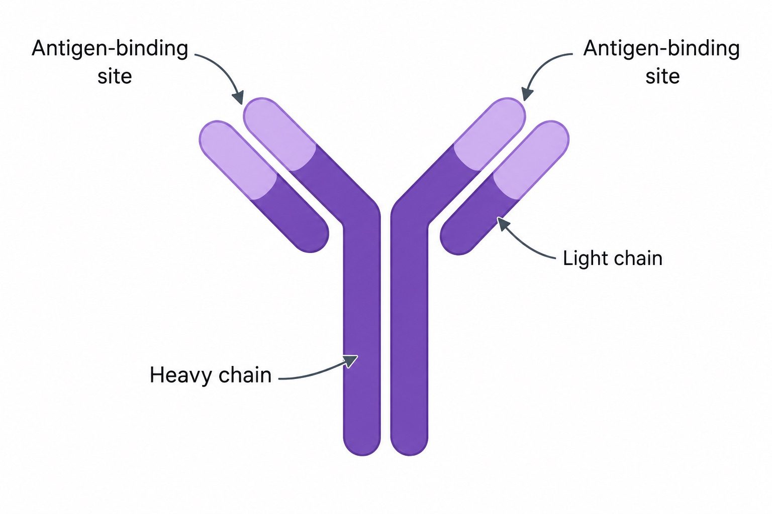
Structurally, IgE is a glycoprotein composed of four polypeptide chains: two identical heavy chains and two identical light chains, interconnected by disulphide bonds (Figure 1). Its heavy chains belong to the epsilon (ε) class, which determines its immunological identity and distinguishes it from immunoglobulins G, A, M and D. Unlike IgM, which may circulate as a pentamer, IgE exists predominantly as a monomer, giving it a relatively compact molecular configuration. Its molecular mass is approximately 190 kDa, reflecting its relatively extensive carbohydrate content and distinctive heavy-chain organization.
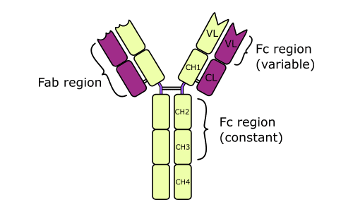
The IgE molecule possesses two principal functional regions: the antigen-binding fragment (Fab) and the crystallisable fragment (Fc). The Fab regions are situated at the terminal ends of the molecule and contain variable domains responsible for recognising and binding specific antigenic determinants, also known as epitopes. Each IgE molecule therefore has two antigen-binding sites, allowing it to interact with its corresponding antigen. The specificity of this interaction is determined by the variable regions of its heavy and light chains, which are generated through genetic rearrangement during B-cell development.
A particularly important structural feature of IgE is its epsilon heavy chain, which contains four constant domains (Cε1-Cε4). Unlike IgG, IgA and IgD, IgE lacks a conventional flexible hinge region. Instead, its additional constant domain contributes to the molecular architecture and influences the spatial arrangement of its functional regions. This configuration is important for receptor recognition and subsequent immune-cell activation.
The Fc portion of IgE has a strong affinity for the high-affinity Fc epsilon RI (FcεRI) receptors expressed predominantly on mast cells and basophils. Once IgE binds to these receptors, it remains associated with the cell surface, effectively sensitising these cells to subsequent exposure to specific allergens or antigens. When an antigen cross-links adjacent receptor-bound IgE molecules, intracellular signalling pathways are initiated, resulting in cellular activation and the release of histamine, leukotrienes and other inflammatory mediators.
In addition to FcεRI, IgE can interact with the low-affinity FcεRII (CD23) receptor, which is expressed on several immune-cell populations, including B lymphocytes. This interaction contributes to the regulation of IgE production and antigen presentation. The structural characteristics of IgE are closely linked to its biological functions, establishing it as a specialized antibody in allergic responses, immune regulation and protection against parasitic infections.
Biological functions of immunoglobulin E
IgE is a specialized antibody that performs important functions in immune regulation, particularly in allergic responses and protection against parasitic infections. Although it occurs in relatively small concentrations in the bloodstream, its biological activity is considerable because of its strong affinity for specific receptors on immune cells. IgE acts as an important link between antigen recognition and the activation of inflammatory mechanisms.
This linkage enables the immune system to respond rapidly to certain environmental substances and invading parasites. As aforesaid, IgE performs a dual biological role by participating in protective immunity against parasitic infections while also mediating allergic inflammation. Its activities demonstrate how a highly specialized antibody can contribute to host defense but, under certain conditions, also produce harmful immune responses.
Activation of mast cells and basophils: One of the principal biological functions of IgE is its ability to activate mast cells and basophils. These immune cells possess high-affinity receptors known as FcεRI, which bind to the constant region of IgE antibodies. When an allergen binds to and cross-links adjacent IgE molecules on the cell surface, it initiates a signalling cascade that triggers cellular activation and degranulation.
This process results in the release of biologically active substances that contribute to inflammation and immune defense. IgE has a high affinity for mast cells. Immunoglobulin E antibody is a cell membrane bound immunoglobulin that mediates hypersensitivity reactions in the body. IgE binds to the Fc region of basophils; and this result in the release of vasoactive substances (e.g. histamines) following the degranulation of mast cells. The bridging of two IgE molecules on mast cell membranes causes the release of granules and/or vasoactive substances (Figure 2).
IgE is mainly responsible for the mediation of immediate Type I Hypersensitivity reactions that cause hay fever, asthma, hives, and anaphylactic shock; and the immunoglobulin assist in defending the body against parasitic or helminthic infections. IgE bind with high affinity to Fc receptors on the surfaces of mast cells and/or basophils; and this binding activates the degranulation of the mast cells to release vasoactive substances inclusive of histamines, cytokines, prostaglandins and leukotrienes amongst others. These pharmacologically active substances mediates Type I hypersensitivity reactions in the body.
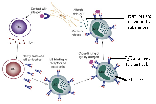
Regulation of allergic immune responses: IgE plays a central role in immediate allergic reactions, also known as Type I hypersensitivity. During initial exposure to an allergen, the immune system may produce allergen-specific IgE antibodies, which subsequently attach to mast cells and basophils. Upon repeated exposure, the same allergen interacts with these surface-bound antibodies, initiating an inflammatory response. This mechanism contributes to allergic manifestations such as sneezing, itching, swelling, skin rashes and respiratory difficulties. In severe cases, widespread IgE-mediated activation can contribute to anaphylaxis, a potentially life-threatening systemic reaction.
Release of histamine and inflammatory mediators: Another essential function of IgE is the stimulation of inflammatory mediator release. Following mast cell and basophil activation, preformed histamine is released alongside newly synthesised lipid mediators and cytokines. Histamine increases vascular permeability, promotes vasodilation and stimulates sensory nerve endings, producing characteristic allergic symptoms. Other mediators, including leukotrienes and prostaglandins, contribute to bronchoconstriction, mucus secretion and sustained inflammation. Through these mechanisms, IgE coordinates cellular and tissue responses during allergic reactions.
Defense against parasitic infections: IgE also contributes to host protection against parasitic organisms, particularly helminths such as roundworms, hookworms and tapeworms. During helminth infections, IgE production is commonly associated with T-helper 2 (Th2)-mediated immune responses. Parasite-specific IgE can facilitate interactions between parasites and immune effector cells, including eosinophils. These cells release toxic granule proteins that can damage parasite surfaces and contribute to parasite elimination. IgE therefore represents an important component of the immune mechanisms involved in anti-helminthic defense.
Protection of skin and mucosal surfaces: IgE-associated immune activity also contributes to defense at barrier tissues, including the skin and the mucosal surfaces of the respiratory and gastrointestinal tracts. These anatomical sites are common entry points for allergens and parasites. Through interactions with mast cells and other immune components, IgE participates in local inflammatory responses, helping the host recognise and respond to potentially harmful external agents. However, excessive activation of these mechanisms may also contribute to chronic allergic inflammation and tissue irritation.
IgE and allergic reactions
IgE is a key antibody involved in the development and regulation of allergic reactions, particularly Type I (immediate) hypersensitivity. This immune response occurs when the body mistakenly identifies otherwise harmless environmental substances, such as pollen, dust mites, certain foods, or animal-derived proteins, as potential threats. Following initial exposure to an allergen, the immune system may undergo sensitisation, during which B lymphocytes differentiate into plasma cells and produce allergen-specific IgE antibodies.
Once released into circulation, these antibodies attach to high-affinity Fc epsilon RI (FcεRI) receptors expressed on the surface of mast cells and basophils. These immune cells are particularly important in allergic inflammation because they act as reservoirs of biologically active mediators. During subsequent exposure to the same allergen, the allergen binds to and cross-links adjacent IgE molecules attached to these receptors. This interaction initiates intracellular signalling pathways that stimulate cellular activation and degranulation.
Degranulation results in the rapid discharge of preformed inflammatory substances, particularly histamine, alongside the synthesis and release of additional mediators, including leukotrienes, prostaglandins and cytokines. These substances produce characteristic allergic manifestations such as vasodilation, increased vascular permeability, mucus secretion, itching, tissue swelling and contraction of bronchial smooth muscles. The intensity and location of these responses determine the clinical presentation of allergic disease.
IgE-mediated hypersensitivity is associated with several common allergic disorders, including allergic rhinitis, bronchial asthma, atopic dermatitis (eczema) and food allergies. In allergic rhinitis, mediator release produces sneezing, nasal congestion and excessive mucus production. In asthma, inflammatory mediators contribute to airway narrowing, bronchial hyperresponsiveness and breathing difficulties. Food allergies may produce gastrointestinal disturbances, skin reactions and respiratory symptoms, depending on the allergen and the severity of the immune response.
In certain circumstances, excessive or widespread IgE-mediated activation can result in anaphylaxis, a potentially life-threatening systemic hypersensitivity reaction. Anaphylaxis may involve extensive vasodilation, a sudden decline in blood pressure, airway obstruction and cardiovascular compromise. It requires immediate medical intervention, usually involving intramuscular adrenaline (epinephrine).
Clinical importance and diagnostic applications of immunoglobulin E
Immunoglobulin E occupies an important position in clinical immunology because its concentration and allergen-binding activity provide useful insights into allergic sensitization, immune dysregulation and certain parasitic infections. Its clinical relevance extends beyond its involvement in hypersensitivity reactions, offering valuable information for disease investigation, risk assessment and therapeutic decision-making.
The measurement of total serum IgE provides an estimate of the overall circulating IgE concentration and may help identify immunological patterns associated with atopic disorders, parasitic diseases and selected immunodeficiency conditions. Nevertheless, an elevated total IgE concentration is not exclusively indicative of allergy, since several unrelated pathological and environmental factors can influence its production. Normal total IgE levels do not necessarily exclude an allergic condition.
Allergen-specific IgE testing provides a more targeted diagnostic approach by detecting antibodies directed against particular environmental, dietary or occupational allergens. These investigations help establish whether an individual has developed immunological sensitisation to a suspected allergen. Depending on the clinical circumstances, laboratory-based assays may be complemented by skin-prick testing and carefully documented exposure histories. Importantly, the detection of allergen-specific IgE demonstrates sensitisation rather than automatically confirming clinically significant allergic disease.
Correlation with the patient’s symptoms and exposure patterns is therefore essential. In regions where parasitic infections are prevalent, IgE measurements may also contribute to the investigation of helminth-associated immune responses. Certain worm infections stimulate substantial IgE production, frequently accompanied by peripheral eosinophilia. However, these findings are not sufficiently specific to establish parasitic infection independently and should be evaluated alongside epidemiological history, clinical manifestations and appropriate parasitological investigations.
Beyond its diagnostic applications, IgE has become an important therapeutic target in modern allergy management. Omalizumab, a monoclonal antibody directed against circulating IgE, reduces its availability for binding to high-affinity FcεRI receptors on mast cells and basophils. This intervention attenuates allergic inflammatory pathways and is used in selected conditions, including allergic asthma and chronic spontaneous urticaria, according to established clinical indications.
The clinical interpretation of IgE requires an integrated approach that considers laboratory measurements, immune mechanisms, environmental exposures and individual disease characteristics. Its greatest diagnostic value lies not in an isolated numerical result but in its ability to contribute to a broader understanding of allergic and parasite-associated immune responses.
References
Abbas A.K, Lichtman A.H and Pillai S (2010). Cellular and Molecular Immunology. Sixth edition. Saunders Elsevier Inc, USA.
Actor J (2014). Introductory Immunology. First edition. Academic Press, USA.
Bach F and Sachs D (1987). Transplantation immunology. N. Engl. J. Med. 317(8):402-409.
Jaypal V (2007). Fundamentals of Medical Immunology. First edition. Jaypee Brothers Medical Publishers (P) Ltd, New Delhi, India.
Levinson W (2010). Review of Medical Microbiology and Immunology. Twelfth edition. The McGraw-Hill Companies, USA.
Roitt I, Brostoff J and Male D (2001). Immunology. Sixth edition. Harcourt Publishers Limited, Spain.
Zon LI (1995). Developmental biology of hematopoiesis. Blood, 86(8): 2876–91.
Discover more from Microbiology Class
Subscribe to get the latest posts sent to your email.