
Immunoglobulin D (IgD) is one of the five major classes of antibodies found in the human immune system, alongside immunoglobulin G (IgG), immunoglobulin A (IgA), immunoglobulin M (IgM), and immunoglobulin E (IgE). It is a specialized glycoprotein that contributes to immune surveillance by helping the body recognize and respond to foreign substances. Although IgD occurs in relatively small quantities compared with other antibody classes, its biological importance is closely associated with the development, activation, and regulation of B lymphocytes. Its distinctive properties make it an important component of the adaptive immune system.
From a structural perspective, IgD is composed of two identical heavy chains and two identical light chains arranged in a characteristic Y-shaped configuration. The heavy chains belong to the delta (δ) class, which distinguishes IgD from other immunoglobulins. Each antibody molecule contains two antigen-binding regions responsible for recognizing specific molecular structures on foreign substances. These binding regions are supported by constant regions that contribute to the molecule’s biological characteristics. IgD has an approximate molecular mass of 180 kDa in its monomeric form, although variations may occur because of differences in glycosylation.
Its carbohydrate components influence molecular stability, distribution, and interactions with other biological molecules. The identification of IgD emerged from immunological investigations into unusual antibody proteins found in human blood. In 1965, researchers discovered a previously unrecognized immunoglobulin class in the serum of a patient with multiple myeloma. Subsequent investigations established its identity as a distinct antibody category. IgD was subsequently classified according to its unique delta heavy-chain structure. This discovery broadened scientific understanding of antibody diversity and demonstrated that immune protection involves several immunoglobulin classes with different structural and functional characteristics.
In the human body, IgD exists primarily in two forms: membrane-bound and secreted. Membrane-bound IgD is expressed on the surface of mature, naïve B lymphocytes, where it functions as part of the B-cell antigen receptor complex. It is commonly co-expressed with IgM and enables developing immune cells to detect and recognize specific antigens. Secreted IgD, on the other hand, is present in very low concentrations in the bloodstream and can also be found in certain mucosal secretions, particularly those associated with the respiratory tract.
The immunological significance of IgD extends beyond antigen recognition. IgD participates in the regulation of B-cell responsiveness, helping immune cells distinguish relevant antigenic signals and initiate appropriate defensive activities. Its interactions with other immune components may also contribute to mucosal immune surveillance and the regulation of inflammatory responses. Furthermore, IgD has been associated with immune interactions involving basophils and other innate immune cells.
Synthesis and production of immunoglobulin D
IgD is a specialized antibody produced primarily by mature B lymphocytes as part of the adaptive immune system. Its synthesis represents an important stage in B-cell differentiation and immune regulation. Unlike some other immunoglobulin classes, IgD is predominantly expressed on the surface of mature, naïve B cells, where it functions as an antigen-recognition receptor. Its production involves coordinated cellular development, genetic recombination, transcriptional regulation, and protein synthesis.
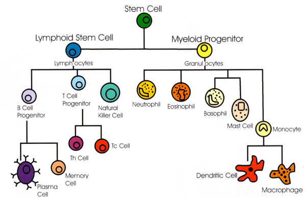
B-lymphocyte production of IgD: IgD originates from B lymphocytes, which develop from haematopoietic stem cells in the bone marrow. During B-cell maturation, these cells acquire the molecular machinery required to recognize specific antigens. Mature naïve B lymphocytes commonly co-express IgM and IgD on their plasma membranes. These antibodies possess identical antigen-binding specificity but differ in their constant regions. IgD production therefore contributes to the functional diversity of B-cell receptors and enables lymphocytes to participate effectively in immune surveillance.
B-cell development and maturation
The development of IgD is closely associated with the progression of B lymphocytes through distinct maturation stages. Immature B cells initially express membrane-bound IgM following successful immunoglobulin gene rearrangement. As these cells mature, they begin expressing IgD alongside IgM through alternative RNA processing mechanisms. This transition marks an important phase of immunological development, preparing mature naïve B cells to recognize foreign substances and initiate appropriate immune responses. The simultaneous expression of both immunoglobulins enhances the functional adaptability of B-cell populations.
Gene expression and class switching: IgD synthesis is controlled by the immunoglobulin heavy-chain gene located on chromosome 14 in humans. The gene contains a variable region and constant-region segments responsible for determining antibody specificity and class. IgD expression primarily occurs through alternative RNA splicing and polyadenylation, allowing the same rearranged variable-region gene to associate with either the IgM or IgD constant region. Unlike conventional antibody class-switch recombination, IgD expression does not generally require DNA-level class-switch rearrangement. This genetic arrangement enables B lymphocytes to produce IgD without changing their original antigen specificity.
Regulation of IgD synthesis: The production of IgD is influenced by several molecular and cellular regulatory mechanisms. Transcription factors, RNA-processing proteins, cytokine signals, and the developmental condition of B lymphocytes contribute to its expression. Alternative splicing determines the quantity of IgD produced relative to IgM, while cellular activation influences the functional behaviour of B cells. These regulatory processes maintain an appropriate balance of immunoglobulin expression and support effective immune responsiveness.
Secretion into the bloodstream and mucosal tissues: Although membrane-bound IgD is the predominant form associated with naïve B lymphocytes, a secreted form is also produced by differentiated antibody-secreting cells. Secreted IgD enters the circulation and can be detected in serum at relatively low concentrations. It is also associated with mucosal surfaces, particularly in the upper respiratory tract, where it participates in local immune defence. At these sites, IgD may interact with microbial antigens and contribute to the coordination of immune responses. Its distribution demonstrates that IgD production extends beyond conventional circulating antibodies and supports immune protection at important environmental interfaces.
Structure and characteristics of immunoglobulin D
IgD is a distinctive antibody isotype characterized by its specialized molecular architecture and important contribution to immune system regulation. Its structure is designed to facilitate antigen recognition, cellular communication, and the coordination of immune responses. Although IgD occurs in relatively low concentrations in the bloodstream compared with other immunoglobulin classes, its presence on the surface of mature naïve B lymphocytes makes it an important component of adaptive immunity.
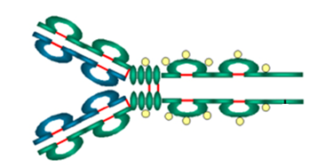
IgD possesses a characteristic Y-shaped molecular configuration consisting of four polypeptide chains: two identical heavy chains and two identical light chains (Figure 1). The heavy chains are relatively large and form the central framework of the antibody, while the light chains contribute to the antigen-recognition regions. Each light chain is associated with one heavy chain through covalent and non-covalent interactions, maintaining the structural stability of the molecule. The coordinated arrangement of these chains creates two functional arms responsible for binding specific antigens. This four-chain organization enables IgD to recognize a broad range of molecular structures while maintaining the characteristic architecture shared by conventional antibodies.

A defining structural feature of IgD is its delta (δ) heavy chain, which distinguishes it from other immunoglobulin isotypes. The δ heavy chain contains one variable domain and four constant domains, designated Cδ1, Cδ2, Cδ3, and Cδ4. The variable domain participates in antigen recognition, whereas the constant domains contribute to molecular organization, flexibility, and interactions with other components of the immune system. IgD also possesses an extended hinge region that provides considerable flexibility to its antigen-binding arms. This structural characteristic allows the antibody to accommodate antigens with different spatial arrangements and binding orientations.
IgD contains two antigen-binding sites located at the terminal regions of its Y-shaped structure. These sites are formed through the interaction of variable regions from the heavy and light chains. Their distinctive amino acid sequences determine the specificity and affinity of antigen recognition. Through these binding regions, IgD can identify particular molecular patterns present on foreign substances and other antigens. The recognition process contributes to the activation and regulation of B lymphocytes, helping initiate appropriate immune responses. The two binding sites provide the molecule with the capacity to interact with two identical or closely related antigenic determinants.
Disulfide bonds are essential structural elements that maintain the integrity and stability of IgD. These covalent linkages form between cysteine residues within and between the polypeptide chains. Intrachain disulfide bonds stabilize individual protein domains, while interchain disulfide bonds help connect the heavy and light chains into a functional antibody molecule. These linkages preserve the correct three-dimensional arrangement required for antigen recognition and molecular activity. Together with non-covalent interactions, disulfide bonds contribute to the durability and structural organization of IgD under physiological conditions.
IgD exists in two principal forms: membrane-bound IgD and secreted IgD. Membrane-bound IgD is expressed on the surface of mature naïve B lymphocytes, where it functions as part of the B-cell antigen receptor (BCR) complex. In this position, it works alongside IgM to facilitate antigen recognition and influence B-cell activation, maturation, and immune regulation. Its membrane attachment is supported by a hydrophobic transmembrane segment and a short intracellular region.
In contrast, secreted IgD is released into extracellular fluids and circulates in the bloodstream at relatively low concentrations. It can also be found in mucosal tissues, particularly in association with the upper respiratory tract. Secreted IgD exhibits structural adaptations that support its extracellular distribution and interactions with immune cells. The coexistence of these two forms enables IgD to participate in both localized cellular recognition and broader immune communication, demonstrating the functional significance of its specialized molecular structure.
Biological functions of immunoglobulin D
Immunoglobulin D is an essential component of the adaptive immune system, contributing to immune surveillance, B-lymphocyte regulation, antigen recognition, and the coordination of protective immune responses. Although it occurs in relatively small concentrations in the bloodstream compared with other immunoglobulin classes, IgD performs distinctive biological activities that support immunological balance and host defence. Its functions extend beyond conventional antibody activity, particularly through its involvement in B-cell signalling and interactions with innate immune cells.
Recognition of foreign antigens: IgD participates in the identification of foreign substances through its antigen-binding regions, which possess specificity for particular molecular structures. On the surface of mature naïve B lymphocytes, membrane-bound IgD acts as an antigen receptor, enabling these cells to recognize potentially harmful microorganisms, microbial proteins, and other foreign antigens. Antigen recognition initiates molecular signalling events that prepare B lymphocytes for subsequent immune activation. This recognition mechanism contributes to the discrimination of external threats and supports the development of antigen-specific immune responses.
Activation and regulation of B cells: One of the principal biological functions of IgD involves the modulation of B-lymphocyte activity. Together with immunoglobulin M (IgM), IgD is expressed on mature naïve B cells, where it functions as part of the B-cell receptor (BCR) complex. Following antigen engagement, the receptor initiates intracellular signalling pathways that influence B-cell activation, proliferation, and differentiation. IgD also contributes to maintaining B-cell responsiveness by helping regulate activation thresholds and cellular sensitivity to antigens. Through these mechanisms, it supports appropriate immune reactions while helping prevent unnecessary or excessive lymphocyte stimulation.
Participation in immune responses: IgD contributes to the coordination of adaptive immunity by supporting the early stages of antigen-dependent B-cell responses. Its receptor activity facilitates the recognition of antigens and promotes cellular signalling necessary for subsequent immunological events. Activated B lymphocytes may undergo clonal expansion, differentiate into antibody-producing plasma cells, or develop into memory B cells. Although IgD is not the principal antibody responsible for long-term circulating humoral protection, its involvement in B-cell activation provides an important foundation for effective antibody-mediated defence.
Interaction with basophils and other immune cells: Secreted IgD demonstrates immunological activities through interactions with selected innate immune cells, particularly basophils. Research indicates that IgD can bind to basophils and influence their functional responses. This interaction may stimulate the release of inflammatory mediators, including cytokines and antimicrobial substances, which contribute to immune communication and pathogen defence. IgD-associated signalling has also been investigated in relation to mast cells and other immune populations. These cellular interactions suggest that IgD participates in linking innate and adaptive immune mechanisms.
Contribution to mucosal immunity: IgD is also associated with immune protection at mucosal surfaces, particularly within the respiratory tract and upper airway tissues. IgD-producing B cells and locally secreted IgD may contribute to the recognition of inhaled microorganisms and environmental antigens. At these anatomical sites, IgD can participate in interactions with epithelial-associated immune cells and support local immune surveillance. Its potential involvement in regulating inflammatory responses and maintaining mucosal defense highlights its importance in protecting tissues exposed continuously to environmental microorganisms and foreign particles.
Clinical significance of immunoglobulin D
Immunoglobulin D has considerable clinical relevance because of its involvement in immune regulation, B-cell activation, and inflammatory responses. Although it represents a relatively small fraction of circulating immunoglobulins, alterations in its concentration may provide valuable information about underlying immunological abnormalities. Abnormally low IgD concentrations, commonly described as IgD deficiency, have been observed in certain individuals with impaired immune responses.
However, isolated IgD deficiency is generally considered uncommon and does not always indicate a clinically significant disorder. Its interpretation requires consideration of other immunoglobulin levels, clinical symptoms, and the patient’s overall immune status. Increased serum IgD concentrations may occur in association with chronic inflammatory conditions, recurrent infections, and certain immune-mediated disorders. One notable condition is hyperimmunoglobulin D syndrome (HIDS), a rare autoinflammatory disorder characterized by recurrent episodes of fever, lymph node enlargement, abdominal discomfort, skin manifestations, and elevated inflammatory markers.
This condition is commonly associated with mutations in the MVK gene, which encodes the enzyme mevalonate kinase. Although elevated IgD levels may support clinical suspicion, they are neither sufficiently sensitive nor specific to establish the diagnosis independently. IgD has attracted scientific interest because of its potential involvement in autoimmune and inflammatory diseases. Its interactions with immune cells, particularly basophils and other components of innate immunity, suggest that it may contribute to inflammatory signalling and immune modulation.
Altered IgD expression has also been investigated in conditions involving abnormal B-cell activity and persistent immune stimulation. From a diagnostic perspective, serum IgD measurement can complement other immunological investigations when evaluating unexplained inflammatory symptoms or suspected immune dysregulation. Nevertheless, IgD concentrations must be interpreted alongside clinical findings and additional laboratory parameters.
Laboratory detection and diagnostic applications of immunoglobulin D
Laboratory detection of Immunoglobulin D involves specialized analytical procedures designed to identify, quantify, and evaluate its concentration in biological specimens. Although IgD occurs in relatively small amounts in human serum, its measurement provides valuable information about B-lymphocyte activity, immune regulation, and certain pathological conditions. Serum samples are commonly employed for IgD assessment, with careful specimen handling and appropriate storage required to preserve analytical reliability.
Quantitative determination of IgD is generally performed using immunochemical techniques that depend on specific antigen-antibody interactions. These methods enable laboratories to establish circulating IgD concentrations and distinguish normal physiological variations from potentially abnormal immunological patterns. Enzyme-linked immunosorbent assay (ELISA) is particularly useful because of its specificity, sensitivity, and ability to detect low concentrations of IgD through enzyme-mediated colour development. Other immunoassay platforms may also be employed depending on laboratory facilities and diagnostic requirements.
Immunofixation electrophoresis provides an additional investigative approach, particularly when abnormal immunoglobulin production or monoclonal protein patterns are suspected. By separating serum proteins according to their electrophoretic properties and subsequently identifying specific immunoglobulin components, this technique assists in characterizing unusual IgD-associated protein profiles. Specialized testing may be necessary when conventional electrophoretic procedures fail to demonstrate distinct immunoglobulin abnormalities.
Clinical interpretation of IgD measurements requires consideration of laboratory reference intervals, patient characteristics, accompanying immunological findings, and presenting symptoms. Increased serum IgD concentrations may occur in certain inflammatory, infectious, and immunological disorders, including hyper-IgD syndrome. However, an elevated result alone cannot establish a definitive diagnosis. Thus, reduced IgD concentrations should be interpreted cautiously because their clinical significance is not always clearly established.
References
Abbas A.K, Lichtman A.H and Pillai S (2010). Cellular and Molecular Immunology. Sixth edition. Saunders Elsevier Inc, USA.
Actor J (2014). Introductory Immunology. First edition. Academic Press, USA.
Bach F and Sachs D (1987). Transplantation immunology. N. Engl. J. Med. 317(8):402-409.
Barrett J.T (1998). Microbiology and Immunology Concepts. Philadelphia, PA: Lippincott-Raven Publishers. USA.
Jaypal V (2007). Fundamentals of Medical Immunology. First edition. Jaypee Brothers Medical Publishers (P) Ltd, New Delhi, India.
Levinson W (2010). Review of Medical Microbiology and Immunology. Twelfth edition. The McGraw-Hill Companies, USA.
Roitt I, Brostoff J and Male D (2001). Immunology. Sixth edition. Harcourt Publishers Limited, Spain.
Zon LI (1995). Developmental biology of hematopoiesis. Blood, 86(8): 2876–91.
Discover more from Microbiology Class
Subscribe to get the latest posts sent to your email.