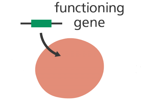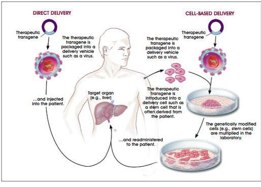


Ex vivo gene therapy, often referred to as a cell-based delivery approach, represents one of the most controlled and precise strategies within the broader field of gene therapy. At its core, ex vivo gene therapy involves the removal of cells from a patient (or, in some cases, a donor), the genetic modification of those cells under controlled laboratory conditions, and their subsequent reintroduction into the patient to achieve a therapeutic effect. This approach stands in contrast to in vivo gene therapy, where genetic material is delivered directly into the patient’s body without prior cellular manipulation outside the organism.
To fully appreciate the conceptual and practical significance of ex vivo gene therapy, it is essential to define the term “ex vivo” in a rigorous biological context. The phrase ex vivo (Latin for “out of the living”) refers to biological processes or experiments conducted on cells, tissues, or organs that have been removed from a living organism but are maintained in an environment that closely mimics physiological conditions. Unlike in vitro systems, which may involve simplified or artificial conditions (e.g., cell lines in culture), ex vivo systems aim to preserve the native characteristics, functionality, and microenvironmental context of the cells as much as possible. Thus, ex vivo gene therapy operates at the intersection of in vitro manipulation and in vivo relevance, leveraging the advantages of both.
In ex vivo gene therapy, cells are first isolated from the patient. These cells must possess several critical biological properties to be suitable for this approach. Most importantly, they must be capable of survival outside the body under laboratory conditions, maintaining viability and functionality during manipulation. They must retain the capacity to proliferate (if expansion is required), undergo efficient genetic modification, and successfully engraft or reintegrate into the host after reintroduction. Commonly used cell types in ex vivo systems include hematopoietic stem cells (HSCs), T lymphocytes, and, in some applications, mesenchymal stem cells or induced pluripotent stem cells (iPSCs). The choice of cell type is dictated by the disease target, therapeutic objective, and required duration of gene expression.
Once isolated, these cells are subjected to genetic engineering processes designed to introduce new genetic material commonly referred to as transgenes. A transgene is a gene that has been artificially introduced into a cell’s genome or maintained episomally to confer a new function or restore a defective one. These genes are typically selected based on their ability to correct genetic defects, produce a therapeutic protein, enhance immune function, or otherwise modify cellular behavior in a clinically beneficial manner.
The introduction of transgenes into target cells is achieved through specialized delivery systems, often involving viral vectors such as lentiviruses or retroviruses, or non-viral methods like electroporation or lipid-based transfection. The ex vivo setting provides a significant advantage in this regard: it allows for precise control over the conditions of gene transfer, including vector dosage, transduction efficiency, and selection of successfully modified cells. This level of control is difficult to achieve in in vivo systems, where biological barriers, immune responses, and off-target effects can limit efficiency and safety.
Another defining feature of ex vivo gene therapy is the opportunity for rigorous quality control prior to reintroduction into the patient. Modified cells can be extensively characterized to confirm successful gene integration or expression, assess functional outcomes, and ensure the absence of undesirable effects such as insertional mutagenesis or aberrant gene expression. This step is critical for minimizing risks and improving therapeutic reliability. In contrast, in vivo approaches lack this intermediate validation phase, as genetic modification occurs directly within the patient’s body.
A key conceptual distinction between ex vivo and in vivo gene therapy lies in the level of biological containment and predictability. Ex vivo gene therapy is inherently more contained because genetic manipulation occurs outside the body, reducing systemic exposure to vectors and limiting unintended interactions with non-target tissues. Furthermore, because only selected and validated cells are reintroduced, there is a greater degree of control over therapeutic outcomes. This makes ex vivo approaches particularly attractive for applications where safety, precision, and reproducibility are paramount.
Ex vivo gene therapy has been especially transformative in the field of hematological and immunological disorders. For example, in inherited blood disorders such as sickle cell disease or beta-thalassemia, hematopoietic stem cells can be harvested, genetically corrected, and reinfused to repopulate the patient’s bone marrow with functional cells. Similarly, in cancer immunotherapy, T cells can be engineered to express chimeric antigen receptors (CARs), enabling them to recognize and eliminate tumor cells with high specificity. These applications highlight the versatility of ex vivostrategies in both gene correction and functional enhancement contexts.
The success of ex vivo gene therapy is closely tied to the biological characteristics of the target cells. Cells that naturally circulate in the bloodstream or reside in regenerative compartments (such as the bone marrow) are particularly suitable because they can be readily harvested and reintegrated. Moreover, long-lived or self-renewing cells are preferred when sustained therapeutic effects are required. For instance, modifying stem cells allows for long-term expression of the therapeutic gene, as these cells can continuously generate progeny that inherit the genetic modification.
Another important consideration is the interaction between modified cells and the host environment upon reintroduction. The reinfused cells must be able to home to the appropriate tissue, survive immune surveillance, and integrate functionally within the existing cellular network. In some cases, patient preconditioning (e.g., through chemotherapy) is necessary to create space for the modified cells to engraft effectively. Although this aspect introduces additional complexity, it also underscores the systemic nature of ex vivo gene therapy as a coordinated interplay between engineered cells and the host organism.
Compared to in vivo gene therapy, ex vivo approaches generally exhibit a more favorable safety profile. The ability to screen and select modified cells reduces the likelihood of adverse effects, and the limited exposure of the patient to gene delivery vectors minimizes immune reactions. However, ex vivo gene therapy is not without challenges. The process can be technically demanding, time-intensive, and costly, requiring specialized infrastructure for cell isolation, cell culture, and genetic manipulation. Additionally, not all cell types are amenable to ex vivo modification, which limits the applicability of this approach for certain diseases.
Despite these challenges, advances in genome editing technologies, such as CRISPR-Cas systems, are rapidly expanding the potential of ex vivo gene therapy. These tools enable precise, targeted modifications of the genome, allowing not only the addition of transgenes but also the correction of specific mutations at their endogenous loci. This represents a significant evolution from earlier gene therapy strategies, which often relied on random integration of genetic material.
Ex vivo gene therapy is a sophisticated and highly controlled approach to genetic medicine that leverages the manipulation of patient-derived cells outside the body to achieve therapeutic outcomes. By combining the precision of laboratory-based genetic engineering with the biological relevance of patient-specific cells, this strategy offers a powerful platform for treating a wide range of diseases. Its defining features cell extraction, controlled genetic modification, rigorous validation, and reinfusion distinguish it from in vivo approaches and underpin its growing importance in modern biomedical science. As research continues to refine the efficiency, safety, and scalability of ex vivo techniques, this approach is poised to play an increasingly central role in the future of personalized medicine and targeted therapeutics.
Fundamental concept of ex vivo gene therapy
Ex vivo gene therapy is a cell-based genetic medicine strategy in which cells are isolated from a patient, genetically modified or otherwise genetically manipulated under controlled laboratory conditions, and subsequently returned to the patient to achieve a therapeutic effect. In contrast to in vivo gene therapy, where therapeutic genetic material is delivered directly into the patient’s tissues or cells, ex vivo approaches create an intermediate laboratory stage in which the target cells can be selected, modified, expanded, characterized, and subjected to quality-control procedures before administration.
The fundamental principle of ex vivo gene therapy is therefore the separation of genetic modification from its final therapeutic application. Patient-derived cells are first harvested from an appropriate tissue or biological compartment and maintained under optimized culture conditions. A therapeutic gene, gene-editing system, or other genetic construct is then introduced using an appropriate delivery method. Following modification, the cells can be evaluated for genetic modification efficiency, viability, phenotype, functionality, and potential unintended effects. Only cells meeting predefined quality and safety criteria are subsequently prepared for reintroduction into the patient.
This approach provides several important advantages. The genetic manipulation process can be performed under controlled and standardized conditions, allowing optimization of gene delivery and selection of appropriately modified cells before treatment. It also enables detailed characterization of the cellular product, potentially reducing exposure to unsuitable or inadequately modified cells. Furthermore, ex vivo strategies can be tailored to specific cell populations, making them particularly valuable when the therapeutic effect depends on modifying a defined cellular compartment.
However, ex vivo gene therapy also presents technical and clinical challenges, including efficient and stable genetic modification, maintenance of cell viability and function during culture, prevention of unwanted genetic alterations, and reproducible manufacturing at clinically relevant scales. The choice of gene-delivery platform viral or non-viral must be carefully matched to the therapeutic objective and safety requirements. Ex vivo gene therapy integrates cellular engineering, genetic modification, and controlled manufacturing to generate a personalized cellular therapeutic with the potential for precise and durable treatment.
Stepwise process of ex vivo gene therapy
Ex vivo gene therapy is a multistage therapeutic approach in which cells are removed from a patient, genetically modified under controlled laboratory conditions, evaluated for quality and safety, and subsequently returned to the patient. Each stage requires precise coordination to maintain cellular viability, achieve effective gene transfer, and minimize potential risks. Unlike in vivo approaches, the ex vivo strategy provides an opportunity to assess and manipulate the target cells before their administration, allowing greater control over the therapeutic process. The overall workflow therefore involves several interconnected stages, beginning with cell collection and continuing through genetic modification, quality assessment, expansion, and reinfusion (Figure 1).
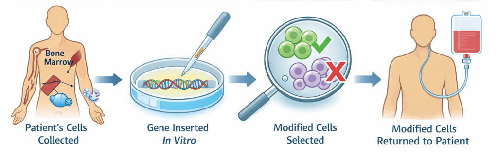
1. Patient cell isolation: Target cells are harvested from the patient using procedures such as bone marrow aspiration, blood collection, or tissue biopsy. The goal is to obtain a population of viable, functionally relevant cells.
2. Cell culture and expansion: The isolated cells are cultured under optimized laboratory conditions to maintain viability and, if necessary, to expand their numbers. This step ensures that sufficient cells are available for genetic modification.
3. Genetic modification (transfection/transduction): Cells are exposed to gene delivery systems, typically through transfection techniques, to introduce the therapeutic gene (transgene). This is a critical step where efficiency and precision must be carefully controlled.
4. Selection and screening of modified cells: Following genetic modification, cells are screened to identify those that have successfully incorporated and are expressing the transgene. This may involve molecular assays, protein expression analysis, or functional testing.
5. Quality control and safety assessment: Modified cells undergo rigorous quality control to ensure genetic stability, absence of contamination, and lack of undesirable mutations. This step is essential for minimizing clinical risks.
6. Preparation for reintroduction: Cells are prepared for reinfusion, which may include formulation, dosage standardization, and ensuring compatibility with the patient’s immune system.
7. Reintroduction into the patient: The genetically modified cells are reintroduced into the patient, typically via intravenous infusion or targeted delivery, depending on the therapy.
8. Engraftment and therapeutic action: Once inside the body, the cells must engraft, survive, and begin producing the therapeutic effect, such as protein production, immune activation, or tissue repair.
Target cell types in ex vivo gene therapy
One major limitation of ex vivo gene therapy is that, it is limited to cells that can be cultured ex vivo. A critical determinant of the success of ex vivo gene therapy is the selection of appropriate target cells. These cells must exhibit key biological characteristics, including viability outside the host, genetic modifiability, and the ability to engraft and function after reinfusion.
Several major cell types commonly targeted in ex vivo gene therapy include:
1. Bone marrow cells (Hematopoietic Stem Cells – HSCs): Bone marrow-derived cells, particularly hematopoietic stem cells (HSCs), are among the most widely used targets. These cells are self-renewing and capable of differentiating into multiple blood cell lineages, making them ideal for treating hematological disorders such as sickle cell disease, beta-thalassemia, and certain immunodeficiencies. Their ability to repopulate the bone marrow niche ensures long-term therapeutic benefit.
2. Muscle cells (myocytes): Muscle cells are often targeted in therapies aimed at treating muscular dystrophies, such as Duchenne muscular dystrophy. These cells are relatively stable and long-lived, allowing for sustained gene expression once successfully modified.
3. Liver cells (Hepatocytes): The liver plays a central role in metabolism and protein synthesis, making hepatocytes valuable targets for gene therapy addressing metabolic disorders. Their high biosynthetic capacity allows them to produce therapeutic proteins at systemic levels.
4. Fibroblasts: Fibroblasts are connective tissue cells that are easy to isolate and culture, making them convenient for experimental and clinical gene therapy applications. Although they may not integrate as broadly as stem cells, they are useful for localized or supportive therapeutic effects.
5. Immune cells (T lymphocytes): T cells are frequently used in cancer immunotherapy, where they are genetically engineered to express chimeric antigen receptors (CARs). These modified cells can recognize and destroy tumor cells, providing a highly targeted therapeutic approach.
The choice of cell type for ex vivo gene therapy depends on the disease pathology, desired duration of gene expression, and capacity for cellular integration and function within the host.
Gene delivery systems in ex vivo gene therapy
Gene delivery is a critical determinant of the efficiency, safety, and therapeutic potential of ex vivo gene therapy. In this approach, patient-derived cells are genetically modified outside the body before being expanded, characterized, and subsequently administered to the patient. Both viral and non-viral delivery systems can be employed, with the choice depending on the target cell type, therapeutic objective, and required duration of transgene expression. Although viral vectors offer high transduction efficiency and sustained gene expression, non-viral systems have attracted increasing interest because of their relatively favorable safety profile, manufacturing flexibility, lower immunogenic potential, and potential for scalable production.
Non-viral vectors used in ex vivo gene therapy and their advantages
Non-viral vectors are synthetic or physical delivery systems designed to introduce therapeutic nucleic acids into target cells without relying on viral components. In ex vivo gene therapy, these systems enable cells to be genetically modified outside the patient and subsequently returned to the body. Common approaches include lipid- and polymer-based nanoparticles, electroporation, microinjection, and other physical delivery methods. Compared with viral vectors, non-viral systems offer several potential advantages, including reduced immunogenicity, lower manufacturing complexity, greater flexibility in cargo size, and improved scalability. Their tunable properties also enable optimization for specific cell types and therapeutic applications, making them increasingly attractive for ex vivo cellular engineering.
These systems offer several advantages as follows:
- Reduced risk of immune activation
- Absence of viral pathogenicity
- Lower likelihood of insertional mutagenesis
- Greater flexibility in DNA size and composition
- Ease of large-scale production
Unlike viral vectors, which can trigger host immune responses and inflammatory reactions, non-viral systems are generally considered biocompatible and safer for clinical applications, particularly in controlled ex vivo environments. However, it is important to note that while non-viral vectors are safer, they often exhibit lower transfection efficiency and reduced gene expression duration compared to viral systems. This trade-off between efficiency and safety is a central consideration in gene therapy design.
Examples of non-viral vectors and techniques used in ex vivo gene therapy
These non-viral delivery systems provide versatile and safer alternatives to viral vectors in ex vivo gene therapy, although ongoing optimization is required to improve their efficiency, specificity, and long-term gene expression. The major non-viral vectors and delivery techniquesused in ex vivo gene therapy include:
1. Electroporation: A physical delivery method that uses short electrical pulses to create temporary pores in the cell membrane, allowing DNA or RNA molecules to enter the cell. It has high efficiency across a wide range of cell types, including primary cellsandstem cells.
2. Lipofection (lipid-based vectors/liposomes): Utilizes cationic lipidsto form complexes with DNA, facilitating entry into cells viamembrane fusionorendocytosis. There is low toxicity, ease of use, and suitability for repeated applications.
3. Polymer-Based Vectors (e.g., Polyethylenimine – PEI)
Synthetic cationic polymers bind to DNA and promote cellular uptake through endocytosis. Its advantage is that it has enhanced DNA protection and relatively stable gene delivery.
4. Calcium Phosphate Precipitation: DNA is precipitated with calcium phosphate, forming particles that are taken up by cells. The advantage here is that it is cost-effective and simple, suitable for laboratory-scale applications.
5. Naked DNA/plasmid DNA delivery: Direct introduction of free DNA molecules into cells without carriers. It is extremely safe with no additional vector-related toxicity.
6. Microinjection: This is the direct injection of DNA into the cytoplasm or nucleus using a fine needle. It is highly precise and ensures delivery into individual cells.
7. Nanoparticle-mediated delivery: It uses engineered nanoparticles (e.g., gold nanoparticles, lipid nanoparticles) to transport DNA into cells. It has improved cellular uptake, protection from degradation, and potential for targeted delivery.
8. Gene gun (biolistic delivery): In this method, DNA-coated particles are physically propelled into cells using high velocity. It is useful for hard-to-transfect cells and tissues.
9. Dendrimer-based vectors: It is a highly branched synthetic polymers that bind DNA and facilitate cellular uptake. The advantage is its controlled structure and efficient DNA condensation.
Mechanism of gene introduction in gene therapy: Transfection
The primary mechanism underlying ex vivo gene delivery using non-viral systems is transfection. Transfection refers to the deliberate introduction of exogenous nucleic acids, including DNA, messenger RNA (mRNA), small interfering RNA (siRNA), or other RNA molecules, into eukaryotic cells under controlled laboratory conditions. The objective is to introduce genetic material into target cells in a manner that enables transient or, in some applications, stable alteration of cellular function.
Following successful transfection, the introduced nucleic acid can interact with the cellular machinery to produce a desired biological effect. For example, delivered DNA can enter the nucleus and serve as a template for transcription, followed by translation of the encoded protein. mRNA can bypass the requirement for nuclear transcription and can be directly translated into a therapeutic protein in the cytoplasm. In contrast, nucleic acids such as siRNA can modulate gene expression through sequence-specific mechanisms that promote degradation or suppression of target mRNA. These different approaches allow transfection to be adapted to a wide range of therapeutic and experimental applications.
In ex vivo gene therapy, transfection is particularly useful because cells can be genetically modified outside the patient’s body under controlled conditions before being evaluated, expanded when appropriate, and subsequently administered back to the patient. Depending on the therapeutic objective, transfected cells may be engineered to produce therapeutic proteins, restore or compensate for defective gene function, modify cellular signaling pathways, or enhance specific immune responses. For example, immune cells can be genetically modified to express receptors or other proteins that alter their ability to recognize and respond to specific targets.
Transfection can be achieved using several non-viral approaches, broadly classified as chemical, physical, or nanoparticle-based methods. Chemical approaches commonly use lipid-based or polymer-based carriers to facilitate nucleic-acid uptake by cells. Physical approaches can employ techniques such as electroporation, in which a brief electrical pulse transiently increases cell-membrane permeability and facilitates nucleic-acid entry. The choice of method depends on factors such as cell type, nucleic-acid cargo, desired duration of expression, transfection efficiency, and cell viability.
Transfection differs fundamentally from transduction, in which genetic material is delivered using viral vectors. Although viral vectors can provide highly efficient gene transfer in many cell types, non-viral transfection offers important advantages for ex vivo applications, including the absence of viral replication, greater flexibility in the type of nucleic-acid cargo that can be delivered, and greater control over the duration and extent of genetic modification. Transfection represents an important platform for controlled, non-viral genetic engineering of cells for both research and therapeutic applications.
Non-viral transfection techniques in ex vivo gene therapy
Several non-viral transfection methods are utilized in ex vivo gene therapy, each with distinct mechanisms and applications. Each of these techniques operates through distinct biophysical or biochemical mechanisms, and the choice depends on factors such as cell type, desired efficiency, and clinical applicability. These non-viral transfection methods are:
1. Electroporation: Electroporation involves the application of short, high-voltage electrical pulses to cells, temporarily disrupting the cell membrane and allowing DNA molecules to enter. It is highly effective for a wide range of cell types, including stem cells and immune cells, but must be carefully optimized to avoid cell damage.
2. Lipofection (lipid-mediated transfection): Lipofection uses lipid-based carriers (liposomes) to encapsulate DNA and facilitate its entry into cells via membrane fusion. This method is widely used due to its simplicity and low toxicity, making it suitable for many ex vivo applications.
3. Microinjection: In microinjection, DNA is directly injected into individual cells using a fine glass needle. This method provides high precision but is labor-intensive and not suitable for large-scale applications.
4. Naked DNA / plasmid delivery: This technique involves the direct introduction of plasmid DNA into cells without carriers. While it is simple and safe, its efficiency is relatively low, limiting its use to specific contexts.
5. Calcium phosphate precipitation: This classical method uses calcium phosphate-DNA complexes to facilitate DNA uptake by cells. Although cost-effective, it is less commonly used today due to variable efficiency.
6. Liposomes and nanocarriers: Advanced liposomal systems and nanoparticles enhance DNA delivery by improving cellular uptake and protection of genetic material. These systems are increasingly used in modern gene therapy research.
Advantages of ex vivo gene therapy
Ex vivo gene therapy has the following advantages:
- There is high level of control over genetic modification.
- It allows the ability to perform extensive quality checks.
- It has a reduced immune response risk compared to in vivo gene therapy.
- There is usually a greater safety and predictability.
Disadvantages of ex vivo gene therapy
The following are the limitations of ex vivo gene therapy:
- Ex vivo gene therapy is technically complex and resource-intensive.
- It is limited to cells that can be cultured ex vivo.
- There is usually a potential for low transfection efficiency (non-viral methods).
- It requires specialized clinical infrastructure.
Limitations and optimization challenges of non-viral gene delivery systems
Although non-viral vectors are generally considered safer than viral vectors, they are often characterized by relatively low transfection efficiency and limited target specificity. Many non-viral delivery approaches lack the inherent biological mechanisms that viruses possess for efficient cell entry, nuclear targeting, and gene integration. As a result, the transformation of host cells with therapeutic DNA can be inconsistent, and in some cases, insufficient to achieve a meaningful therapeutic outcome. Additionally, certain non-viral techniques operate in a relatively non-specific manner, leading to heterogeneous gene uptake across cell populations, which may compromise the overall effectiveness of the therapy.
Despite these limitations, non-viral gene delivery systems are increasingly being developed and adopted as viable alternatives to viral-based methods, largely due to the safety concerns associated with viral vectors. Viral systems, while efficient, carry risks such as immunogenicity, inflammatory responses, and insertional mutagenesis, which can lead to unintended genetic alterations or adverse clinical effects. In contrast, non-viral systems offer improved biocompatibility, reduced risk of immune activation, and greater flexibility in terms of DNA cargo size and composition. These advantages make them particularly attractive for ex vivo gene therapy, where controlled conditions can partially compensate for their lower efficiency.
A major focus in advancing non-viral gene delivery lies in overcoming intracellular and extracellular barriers that limit gene transfer efficiency. Within the host cell, several obstacles impede successful delivery, including cell membrane permeability, endosomal entrapment, cytoplasmic degradation, and inefficient nuclear transport of the introduced DNA. Furthermore, cellular defense mechanisms may recognize and degrade foreign genetic material, further reducing gene expression levels. A detailed understanding of these biological barriers is essential for the rational design of more efficient delivery systems that can enhance cellular uptake, improve intracellular trafficking, and ensure effective gene expression within target cells.
Equally important is the requirement for sustained and functional expression of the delivered therapeutic gene. Following successful delivery, the introduced DNA must remain stable and transcriptionally active within the host cell to produce the desired gene products, such as enzymes or functional proteins. This sustained expression is critical for achieving long-term therapeutic benefits, particularly in the treatment of genetic disorders where continuous correction of a defective gene is required. Without adequate and persistent expression, the therapeutic effect may be transient or insufficient, undermining the overall success of the gene therapy intervention.
References
Sayed, N., Allawadhi, P., Khurana, A., Singh, V., Navik, U., Pasumarthi, S. K., Khurana, I., Banothu, A. K., Weiskirchen, R., & Bharani, K. K. (2022). Gene therapy: Comprehensive overview and therapeutic applications. Life Sciences, 294, 120375.
Rashid, R. A., & Ankathil, R. (2020). Gene therapy: An updated overview on the promising success stories. Malaysian Journal of Pathology, 42(2), 171–185.
Alberts B, Bray D, Johnson A, Lewis J, Raff M, Roberts Kand Walter P (1998). Essential Cell Biology: An Introduction to the Molecular Biology of the Cell. Third edition. Garland Publishing Inc., New York.
Dale J (2003). Molecular genetics of bacteria. Jeremy W. Dale and Simon Park (4th eds.). John Wiley & Sons Ltd, West Sussex, UK.
Edelstein, M. L., Abedi, M. R., Wixon, J., and Edelstein, R. M. (2004). Gene therapy clinical trials worldwide 1989-2004—an overview. J Gene Med, 6: 597-602.
Ferrua, F.; Brigida, I.; Aiuti, A. (2010). Update on gene therapy for adenosine deaminase-deficient severe combined immunodeficiency. Current Opinion in Allergy and Clinical Immunology. 10 (6): 551–556.
Gardlík R, Pálffy R, Hodosy J, Lukács J, Turna J, Celec P; Pálffy; Hodosy; Lukács; Turna; Celec (2005). Vectors and delivery systems in gene therapy. Med Sci Monit. 11 (4): RA110–21.
Horn PA, Morris JC, Neff T, Kiem HP; Morris; Neff; Kiem (2004). Stem cell gene transfer—efficacy and safety in large animal studies. Mol. Ther. 10 (3): 417–31.
S Li and L Huang (2000). Nonviral gene therapy: promises and challenges. Gene Therapy, 7:31-34. www.nature.com/gt
Salmons B, Günzburg WH; Günzburg (1993). Targeting of retroviral vectors for gene therapy. Hum Gene Ther. 4 (2): 129–41.
Sambrook, J., Russell, D.W. (2001). Molecular Cloning: a Laboratory Manual, 3rd edn. Cold Spring Harbor Laboratory Press, New York.
Sheridan C (2011). Gene therapy finds its niche. Nature Biotechnology. 29 (2): 121–128.
Vannucci, L; Lai, M; Chiuppesi, F; Ceccherini-Nelli, L; Pistello, M (2013). Viral vectors: A look back and ahead on gene transfer technology. The new microbiologica. 36 (1): 1–22.
Discover more from Microbiology Class
Subscribe to get the latest posts sent to your email.