Ethidium bromide (EtBr) is a fluorescent intercalating agent widely used in molecular biology to visualize nucleic acids DNA and RNA during gel electrophoresis. Its utility is based on its ability to insert itself between adjacent base pairs of double-stranded DNA, a process known as intercalation. This interaction significantly alters its fluorescence properties, making it highly useful for detecting DNA fragments after electrophoretic separation. In gel electrophoresis, DNA samples are loaded into an agarose gel matrix and subjected to an electric field. Because DNA is negatively charged due to its phosphate backbone, it migrates toward the positive electrode. Smaller DNA fragments move faster through the gel pores than larger ones, resulting in size-based separation. However, after separation, DNA is not inherently visible. This is where ethidium bromide plays a critical role.
Ethidium bromide fluoresces strongly under ultraviolet (UV) light when bound to DNA. It absorbs UV radiation (typically around 300 nm) and emits orange-red fluorescence (around 590 nm). This property allows researchers to visualize DNA bands as distinct glowing regions on the gel when placed on a UV transilluminator. The intensity of fluorescence is roughly proportional to the amount of DNA present, enabling both qualitative and semi-quantitative analysis.
There are two common methods of using ethidium bromide in gel electrophoresis: pre-staining and post-staining. In pre-staining, EtBr is added directly to the agarose gel before it solidifies or to the running buffer, allowing DNA to bind the dye during electrophoresis. In post-staining, the gel is soaked in an ethidium bromide solution after electrophoresis. Pre-staining is faster, while post-staining often provides sharper bands with lower background fluorescence.
Despite its widespread use, ethidium bromide is a potent mutagen because it can intercalate into DNA in living organisms, potentially causing frameshift mutations. For this reason, it must be handled with care, including the use of gloves (double gloves), protective eyewear, and proper waste disposal protocols. EtBr is a key molecular biology reagent that enables visualization of DNA in gel electrophoresis by binding to nucleic acids and fluorescing under UV light (Figure 1), although its mutagenic nature necessitates careful handling and disposal.

Intercalating agent
An intercalating agent is a type of molecule that can insert itself between adjacent base pairs of double-stranded nucleic acids, such as DNA or RNA, without forming covalent bonds. This process is known as intercalation, and it typically occurs because the intercalating molecule has a flat, planar structure that allows it to slide between stacked nucleotide bases. When an intercalating agent binds to DNA, it alters the physical properties of the molecule. It can cause local unwinding and elongation of the DNA helix, which may interfere with essential biological processes such as replication and transcription. Because of this, many intercalating agents are biologically active and can be toxic or mutagenic.
In laboratory settings, intercalating agents are commonly used as fluorescent stains for nucleic acid detection. A well-known example is ethidium bromide, which fluoresces strongly when bound to DNA under UV light, enabling visualization of DNA fragments in gel electrophoresis. Other examples include SYBR dyes and acridine orange, which serve similar purposes with varying binding preferences and fluorescence characteristics. Intercalating agents are DNA-binding compounds that insert between base pairs, often affecting DNA structure and enabling molecular detection techniques.
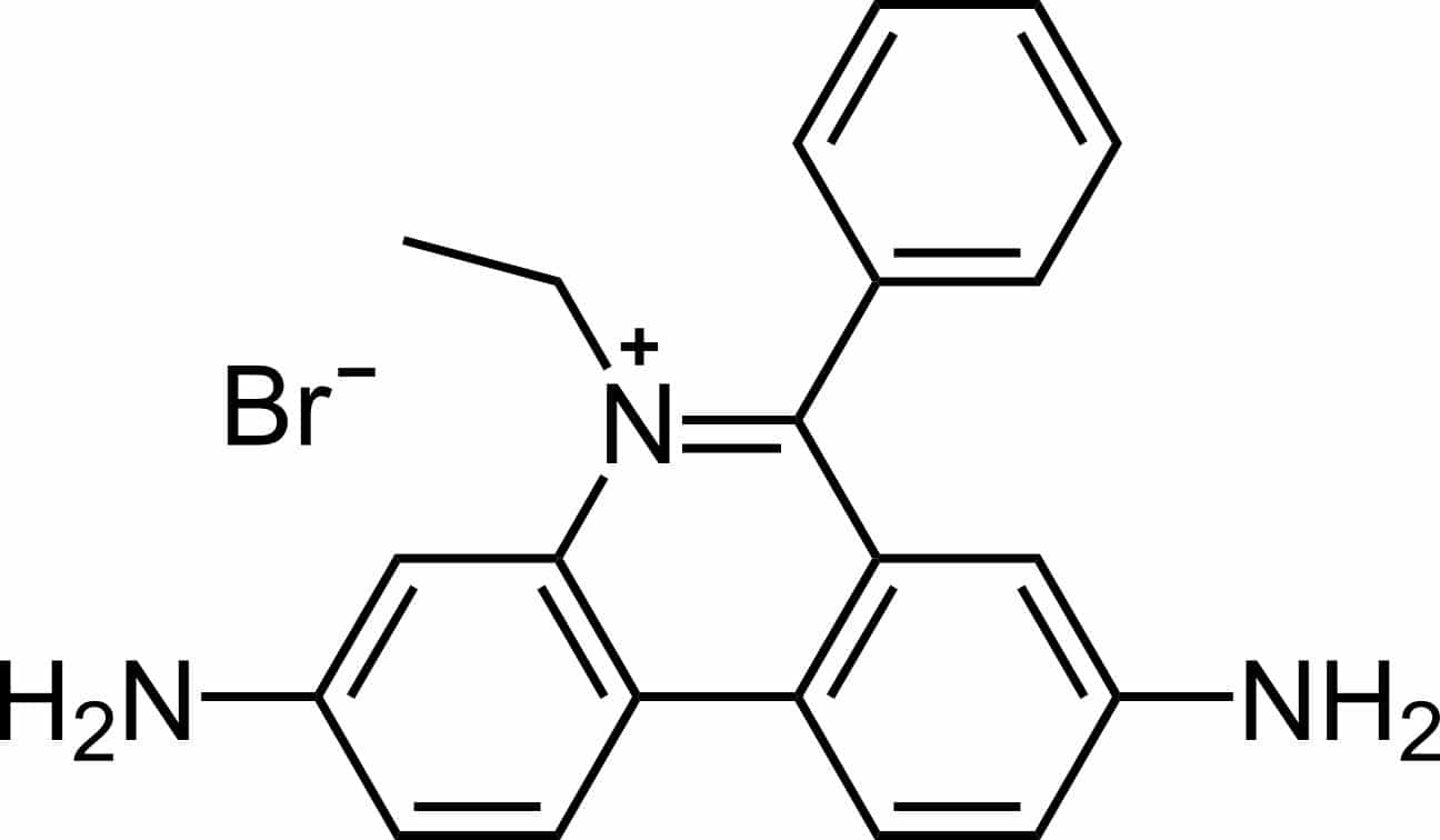
Chemical Structure and Molecular Features of Ethidium Bromide (EtBr)
Chemically, EtBr is classified as a phenanthridinium dye and has the molecular formula C21H20BrN3. Its structure consists of a tricyclic phenanthridine ring system fused into a rigid, planar aromatic framework (Figure 2), which is responsible for its ability to interact strongly with DNA. Attached to this aromatic core are amino substituents and an ethyl group, while the positively charged nitrogen atom forms the phenanthridinium cation; bromide serves as the counterion.
The planar geometry of ethidium bromide is central to its biological behavior. Because of its flat aromatic rings, the molecule can insert between adjacent base pairs of double-stranded DNA through a process known as intercalation. This interaction stabilizes the dye-DNA complex and significantly enhances fluorescence. In aqueous solution, free ethidium bromide exhibits relatively weak fluorescence, but upon binding to nucleic acids, fluorescence intensity increases markedly, producing bright orange-red emission under ultraviolet illumination.
The positive charge on the molecule also promotes electrostatic attraction to the negatively charged phosphate backbone of DNA. These combined structural features, planarity, aromaticity, and cationic character make ethidium bromide highly effective for visualizing DNA fragments during agarose gel electrophoresis.

Safer alternatives to ethidium bromide in DNA visualization
Due to its carcinogenic and mutagenic properties, many laboratories are progressively replacing EtBr with safer nucleic acid stains. EtBr is well documented as a potent mutagen, and this characteristic introduces significant occupational health and environmental risks during routine handling, including exposure during gel preparation, staining procedures, and waste disposal. This shift is driven by the need to improve laboratory safety without compromising the sensitivity and reliability of DNA visualization in gel electrophoresis.
Among the most widely adopted alternatives are SYBR Safe and GelRed. These dyes function similarly to ethidium bromide in that they bind to nucleic acids and fluoresce under ultraviolet or blue-light excitation, enabling visualization of DNA bands after electrophoretic separation. However, they are designed to have significantly reduced ability to penetrate biological membranes or intercalate into DNA within living cells, thereby lowering their mutagenic and cytotoxic potential.
SYBR Safe is commonly used with blue-light transilluminators, which further reduces DNA damage compared to UV exposure and improves downstream applications such as cloning or sequencing. GelRed, on the other hand, is a high-sensitivity dye that is structurally engineered as a dimeric molecule, preventing it from crossing cell membranes while maintaining strong fluorescence upon DNA binding. Despite their advantages, these safer dyes may differ slightly in cost, excitation requirements, or band intensity compared to ethidium bromide, which can influence laboratory choice depending on experimental needs. Nonetheless, their adoption reflects a broader trend in molecular biology toward safer, more environmentally responsible laboratory practices while preserving analytical performance.
Spectral properties, sensitivity, and analytical limitations of ethidium bromide
Ethidium bromide exhibits well-defined photophysical behavior that underpins its application in nucleic acid detection. It shows ultraviolet absorption maxima at approximately 300 nm and 360 nm, enabling efficient excitation under standard laboratory UV light sources used in gel documentation systems. In addition, the dye can participate in indirect excitation through energy transfer from nucleic acids absorbing at around 260 nm, thereby improving fluorescence output in DNA-containing samples.
Upon excitation, ethidium bromide emits fluorescence in the visible spectrum, producing a characteristic orange-yellow signal centered at approximately 590 nm. A key feature of this emission is its dependence on the chemical environment. In aqueous solution, the dye displays relatively weak fluorescence due to rapid non-radiative energy dissipation. However, when bound to nucleic acids, its rotational freedom is restricted, leading to a substantial increase in fluorescence quantum yield. This environmental sensitivity forms the basis for its use in detecting nucleic acids in electrophoretic systems.
From an analytical standpoint, ethidium bromide offers high sensitivity, with a detection threshold typically in the range of 1-5 ng of DNA per band under optimized imaging conditions. This level of sensitivity is sufficient for routine molecular biology workflows, including PCR product verification, plasmid analysis, and restriction fragment profiling. The dye also integrates flexibly into experimental workflows, as it can be incorporated into agarose gels prior to casting, added to running buffers for in-process staining, or applied post-electrophoresis through gel immersion techniques. Each method presents trade-offs between convenience, background fluorescence, and band resolution.
Despite its strong performance in agarose-based systems, ethidium bromide exhibits notable limitations depending on nucleic acid structure. Its fluorescence enhancement is significantly greater for double-stranded DNA compared to single-stranded DNA or RNA. In the case of ssDNA and RNA, staining efficiency decreases substantially, often requiring approximately an order of magnitude higher nucleic acid concentration to achieve comparable signal intensity. This structural dependence can restrict its utility in applications involving denatured nucleic acids or transcript analysis.
Gel matrix composition further influences detection sensitivity. In polyacrylamide gel electrophoresis (PAGE), ethidium bromide fluorescence is partially quenched by interactions with the polymer network, resulting in a marked reduction in signal intensity. Sensitivity losses of approximately 10-20 fold relative to agarose gels are commonly observed, which can limit its effectiveness for resolving low-abundance or small DNA fragments in high-resolution separations. These characteristics define both the strengths and constraints of ethidium bromide as a nucleic acid stain. While it remains highly sensitive, operationally versatile, and widely validated across molecular biology applications, its performance is context-dependent, particularly with respect to nucleic acid structure and gel matrix composition.
Detection of DNA/RNA using ethidium bromide in agarose gel electrophoresis
Safety considerations when using ethidium bromide
Caution: Ethidium bromide (EtBr) is a potent mutagen and must be handled using appropriate laboratory safety procedures. Always wear nitrile gloves, laboratory coats, and protective eyewear when preparing, handling, staining, or disposing of EtBr-containing solutions. Work in designated staining areas where possible and dispose of contaminated materials according to institutional hazardous waste regulations. Ethidium bromide solutions and gels should never be discarded directly into general laboratory waste or drains without approved decontamination procedures.
Method I: Incorporation of ethidium bromide into the gel and running buffer (pre-staining method)
This method allows DNA visualization immediately after electrophoresis because the nucleic acid becomes stained during migration through the gel matrix. It is rapid, convenient, and commonly used for routine molecular biology applications.
A. Preparation of ethidium bromide containing agarose gel
- Prepare the agarose gel according to the standard gel preparation protocol using the selected electrophoresis buffer (TAE or TBE).
- Heat until the agarose dissolves completely and allow the solution to cool to approximately 60-70°C before adding the stain. Cooling prevents thermal degradation and minimizes uneven dye distribution.
- Add ethidium bromide to obtain a final concentration of 0.5 µg/mL.
- Example preparation:
Using a 10 mg/mL stock solution, add 5 µL of stock per 100 mL of molten gel.
- Example preparation:
- Mix gently to ensure homogeneous distribution while avoiding bubble formation.
- Insert the comb and pour the stained agarose into the casting tray, and allow the gel to solidify completely.
B. Preparation of electrophoresis running buffer
- Prepare sufficient electrophoresis buffer (TAE or TBE) to fill both the gel chamber and reservoirs.
- Supplement the buffer with ethidium bromide to a final concentration of 0.5 µg/mL.
- Example: add 5 µL of 10 mg/mL EtBr stock per 100 mL buffer.
- Mix thoroughly before introducing into the electrophoresis apparatus.
C. Electrophoresis and visualization
- Load DNA or RNA samples into the gel wells following standard loading procedures.
- Run electrophoresis under the desired voltage conditions until adequate fragment separation is achieved.
- Upon completion, carefully remove the gel and place it on transparent plastic wrap or a UV-compatible viewing surface.
- Visualize under a UV transilluminator (approximately 300 nm wavelength).
- DNA bands will appear as bright orange fluorescent bands against a faint orange background.
Under optimized conditions, this method typically enables detection of approximately 5 ng of DNA per band. If excessive background fluorescence reduces contrast, the gel may be briefly destained using distilled water or 1 mM MgSO₄ solution to improve band visibility and overall sensitivity.
Alternative approach: ethidium bromide in the gel only
As an alternative to full incorporation, ethidium bromide may be added to the gel while omitted from the running buffer. Because ethidium bromide carries a positive charge, it migrates toward the cathode during electrophoresis, opposite to the movement of negatively charged nucleic acids. Despite this migration, sufficient dye generally remains associated with DNA molecules throughout the separation process to permit visualization. This approach reduces overall ethidium bromide consumption and decreases hazardous buffer waste generation. However, regions of elevated background fluorescence may remain where unbound dye has not migrated completely out of the gel. For routine applications, this method often provides adequate sensitivity while minimizing chemical usage.
Method II: Post-electrophoresis ethidium bromide staining (post-staining method)
Post-staining involves staining the gel after electrophoretic separation and may provide improved contrast and lower background fluorescence.
A. Preparation of staining solution
- Prepare enough staining solution containing 0.5 µg/mL ethidium bromide in either distilled water or electrophoresis buffer.
- Ensure sufficient volume to completely submerge the gel.
- Store the staining solution in a dark container at room temperature; when protected from light, it remains stable for approximately 1-2 months.
B. Staining procedure
- After electrophoresis is completed, carefully transfer the gel into the staining tray.
- Completely immerse the gel in the staining solution.
- Incubate for 15-30 minutes, depending on gel thickness and desired staining intensity.
- Gently agitate if necessary to promote uniform staining.
C. Visualization of stained gels
- Remove the gel from the staining solution and place it on plastic wrap or an imaging platform.
- Observe under 300 nm UV illumination.
- Properly stained nucleic acid bands will appear as bright orange fluorescent bands against a pale orange background, allowing assessment of fragment presence, integrity, and relative abundance.
References
Galindo-Murillo, R., & Cheatham, T. E. (2021). Ethidium bromide interactions with DNA: An exploration of a classic DNA–ligand complex with unbiased molecular dynamics simulations. Nucleic Acids Research, 49(7), 3735–3747.
Bourzac, K. M., LaVine, L. J., & Rice, M. S. (2003). Analysis of DAPI and SYBR Green I as alternatives to ethidium bromide for nucleic acid staining in agarose gel electrophoresis. Journal of Chemical Education, 80(11), 1292.
Borst, P. (2005). Ethidium DNA agarose gel electrophoresis: How it started. IUBMB Life, 57, 745–747.
Waring, M. J. (1965). Complex formation between ethidium bromide and nucleic acids. Journal of Molecular Biology, 13, 269–282.
Madigan M.T., Martinko J.M., Dunlap P.V and Clark D.P (2009). Brock Biology of Microorganisms, 12th edition. Pearson Benjamin Cummings Inc, USA.
McPherson M and Moller S (2002). PCR: The Basics. 2nd edition. Taylor and Francis Group. New York, USA.
Sambrook, J., Russell, D.W. (2001). Molecular Cloning: a Laboratory Manual, 3rd edn. Cold Spring Harbor Laboratory Press, New York.
Synder L, Peters J.E, Henkin T.M and Champness W (2013). Molecular Genetics of Bacteria. Fourth edition. American Society of Microbiology Press, USA.
Tamarin Robert H (2002). Principles of Genetics. Seventh edition. Tata McGraw-Hill Publishing Co Ltd, Delhi.
Discover more from Microbiology Class
Subscribe to get the latest posts sent to your email.