AIM: To provide information on the different white blood cells (neutrophils, basophils, eosinophils, lymphocytes, monocytes) present in the cerebrospinal fluid (CSF) specimen.
MATERIAL/APPARATUS: CSF specimen, methylene blue stain, microscope, glass slide, water, Bunsen burner, inoculating loop, immersion oil.
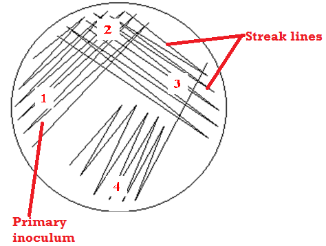
METHOD/PROCEDURE FOR DIFFERENTIAL COUNT OF CSF SAMPLE
- Place a drop of the CSF specimen on a clean glass slide.
- Spread the sample on the slide using a sterilized inoculating loop and allow to dry.
- Heat-fix the smear on the glass slide.
- Flood the slide with methylene blue stain.
- Allow for 5 minutes. Then wash off the methylene blue stain with water.
- Allow the slide to dry completely.
- Add a drop of immersion oil on the slide.
- View the slide under the microscope using the ×100/oil immersion objective lens.
- Systematically examine the slide and count the different white cells seen in each field.
REPORTING OF THE RESULT: Report the number of the different white blood cells seen under the microscope.
NOTE: Differential white cell count can be performed using 3 types of stains:
- Methylene blue stain.
- Giemsa stain.
- Leishma stain.
References
Basic laboratory procedures in clinical bacteriology. World Health Organization (WHO), 1991. Available from WHO publications, 1211 Geneva, 27-Switzerland.
Beers M.H., Porter R.S., Jones T.V., Kaplan J.L and Berkwits M (2006). The Merck Manual of Diagnosis and Therapy. Eighteenth edition. Merck & Co., Inc, USA.
Biosafety in Microbiological and Biomedical Laboratories. 5th edition. U.S Department of Health and Human Services. Public Health Service. Center for Disease Control and Prevention. National Institute of Health. HHS Publication No. (CDC) 21-1112.2009.
Cheesbrough M (2010). District Laboratory Practice in Tropical Countries. Part I. 2nd edition. Cambridge University Press, UK.
Cheesbrough M (2010). District Laboratory Practice in Tropical Countries. Part 2. 2nd edition. Cambridge University Press, UK.
Collins C.H, Lyne P.M, Grange J.M and Falkinham J.O (2004). Collins and Lyne’s Microbiological Methods. Eight edition. Arnold publishers, New York, USA.
Disinfection and Sterilization. (1993). Laboratory Biosafety Manual (2nd ed., pp. 60-70). Geneva: WHO.
Garcia L.S (2010). Clinical Microbiology Procedures Handbook. Third edition. American Society of Microbiology Press, USA.
Garcia L.S (2014). Clinical Laboratory Management. First edition. American Society of Microbiology Press, USA.
Fleming, D. O., Richardson, J. H., Tulis, J. I. and Vesley, D. (eds) (1995). Laboratory Safety: Principles and practice. Washington DC: ASM press.
Dubey, R. C. and Maheshwari, D. K. (2004). Practical Microbiology. S.Chand and Company LTD, New Delhi, India.
Gillespie S.H and Bamford K.B (2012). Medical Microbiology and Infection at a glance. 4th edition. Wiley-Blackwell Publishers, UK.
Discover more from Microbiology Class
Subscribe to get the latest posts sent to your email.