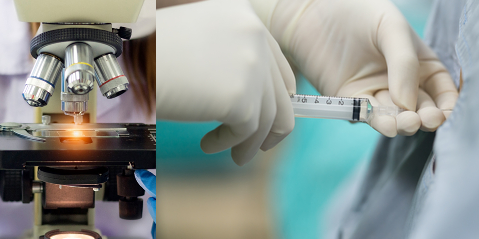
Cerebrospinal fluid (CSF) samples are obtained with extra precautions using a technique called lumbar puncture. Lumbar puncture is defined a medical procedure in which a sterile needle is inserted into the spinal canal (of a patient suspected of having a central nervous system infection) to obtain cerebrospinal fluid sample for microbiological and/or other medical/diagnostic testing.
Another name for lumber puncture is spinal tap. During a lumbar puncture, a sterile needle is inserted into the space between two lumbar bones (vertebrae) of the patient to remove a sample of cerebrospinal fluid required for bacteriological studies to investigate infections of the brain or spine or central nervous system (CNS). The microscopical examination of CSF specimen has different aspects to it. Some of the microscopical techniques involved in the analysis of CSF sample include:
- Gram smear of the CSF sample
- Cell count of the cells present in the CSF sample
- Differential count of the cells present in the sample
These microscopical techniques are important in the analysis of CSF specimen and each has its own role/significance in deciphering the presence of bacteria in a CSF sample. Their methods are expanded below.
However, before embarking on any of the above methods by which a CSF specimen can be analyzed microscopically, it is very important to also observe and report the appearance of the CSF specimen(s) first.
The appearance of a CSF specimen can be reported based on whether the CSF specimen is:
- Clear – normal CSF sample appears clear, bright, and colourless.
- Cloudy or purulent – This indicates the presence of pus cells that is suggestive of pyogenic bacterial meningitis.
- Bloody – The presence of blood in CSF specimen may be due to a bloody/traumatic lumbar puncture.
- Contains clots – The presence of blood clots in a CSF sample indicates a high protein concentration with increased fibrinogen.
References
Basic laboratory procedures in clinical bacteriology. World Health Organization (WHO), 1991. Available from WHO publications, 1211 Geneva, 27-Switzerland.
Beers M.H., Porter R.S., Jones T.V., Kaplan J.L and Berkwits M (2006). The Merck Manual of Diagnosis and Therapy. Eighteenth edition. Merck & Co., Inc, USA.
Biosafety in Microbiological and Biomedical Laboratories. 5th edition. U.S Department of Health and Human Services. Public Health Service. Center for Disease Control and Prevention. National Institute of Health. HHS Publication No. (CDC) 21-1112.2009.
Cheesbrough M (2010). District Laboratory Practice in Tropical Countries. Part I. 2nd edition. Cambridge University Press, UK.
Cheesbrough M (2010). District Laboratory Practice in Tropical Countries. Part 2. 2nd edition. Cambridge University Press, UK.
Collins C.H, Lyne P.M, Grange J.M and Falkinham J.O (2004). Collins and Lyne’s Microbiological Methods. Eight edition. Arnold publishers, New York, USA.
Disinfection and Sterilization. (1993). Laboratory Biosafety Manual (2nd ed., pp. 60-70). Geneva: WHO.
Garcia L.S (2010). Clinical Microbiology Procedures Handbook. Third edition. American Society of Microbiology Press, USA.
Garcia L.S (2014). Clinical Laboratory Management. First edition. American Society of Microbiology Press, USA.
Fleming, D. O., Richardson, J. H., Tulis, J. I. and Vesley, D. (eds) (1995). Laboratory Safety: Principles and practice. Washington DC: ASM press.
Dubey, R. C. and Maheshwari, D. K. (2004). Practical Microbiology. S.Chand and Company LTD, New Delhi, India.
Gillespie S.H and Bamford K.B (2012). Medical Microbiology and Infection at a glance. 4th edition. Wiley-Blackwell Publishers, UK.
Discover more from Microbiology Class
Subscribe to get the latest posts sent to your email.