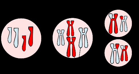
Meiosis unlike mitosis is the type of cell division that occurs during the formation of gamete cells (e.g. spermatozoa and ova). It only occurs in reproductive cells; and meiosis results in the formation of a diploid cell that gives rise to four haploid cells, with each having a single set of chromosomes. Diploid cells are cells that have two sets or copies of chromosomes. Each haploid cell (i.e. one copy of the chromosome) formed during meiosis can afterward fuse with the gamete of the opposite sex during sexual reproduction to form new diploid cell.
Though meiosis and mitosis are similar in their mode of division, meiosis occurs only in reproductive cells during sexual reproduction while mitosis occurs during asexual reproduction. Meiosis involves two stages: meiosis I (the first meiotic division) and meiosis II (the second meiotic division). Each of the divisions in meiosis exhibits the prophase, metaphase, anaphase and telophase stages that are characteristics of cell division by mitosis. At the end of meiosis I and meiosis II, the initial diploid cell will be changed into four haploid cells.
The first meiotic division comprises prophase I, metaphase I, anaphase I and telophase I while the second meiotic division (which is analogous to mitosis) consists of prophase II, metaphase II, anaphase II and telophase II. Meiosis I differs markedly from mitosis which exhibit similar characteristic with meiosis II. Meiosis is a reductive type of cell division because the chromosomes of the parent cell (which is diploid) is reduced by half or halved (i.e. as haploid cells); and each of the haploid cells receives a complete set of new chromosome. (Chromosome number is not halved during cell division by mitosis as is obtainable in meiosis).
The major role of meiosis in the life cycle of microbial cells and other eukaryotic organisms is to ensure that the diploid cells of the parent cell are halved during sexual reproduction. Only meiosis I, which is different from mitosis, shall be highlighted in this unit. (Meiosis II is a mitotic division of each of the haploid cells produced in Meiosis I). The interval between the two stages of meiosis (i.e. meiosis I and meiosis II) is known as interkinesis or interphase II. After the first meiotic cell division (meiosis I), interkinesis occurs prior to the start of meiosis II.
- Prophase I: This stage is characterized by the development of the spindle fiber, and the breakdown of the nuclear membrane of the chromosomes. The chromosomes appear at this stage of meiotic cell division as sister chromatids; and they are visible under the light microscope. There is a complete disintegration of the nucleolus and nuclear envelop or membrane at this stage of division. Prophase I is similar in to prophase in mitotic cell division.
- Metaphase I: Metaphase I is characterized by the alignment of the chromosome pairs on either side of the metaphase plate. And this is different from metaphase in mitotic cell division in which all the chromosomes are aligned to the metaphase plate in no particular arrangement. Crossing over occurs at this stage of meiotic cell division; and it is characterized by the formation of a layer at the equator of the microtubule spindle by the homologous pairs of chromosomes or non-sister chromatids. During crossing over, the arms of the chromatids overlap and momentarily fuse together to form a single larger circular molecule or chromosome. Crossing over is a genetic recombination process in which the exchange of hereditary information occurs between two linear duplexes of chromosomes. Crossing over occurs in meiosis between non-sister chromatids in homologous chromosomes; and it is lacking in mitotic cell division.
- Anaphase I: In anaphase I, the microtubule spindle fibers contract, pulling the homologous pairs of chromosome away from each other and toward each pole of the cell. Basically, the two chromosomes in each homologous pair move to opposite poles of the spindle fiber.
- Telophase I: Telophase I is the meiotic cell division in which each of the two sets of chromosomes becomes enclosed by a nuclear membrane. The chromosome does not disappear at this stage, and there is no reformation of nuclear membrane as well. Cytokinesis occurs in telophase I. Cytokinesis is the changes that normally occur in the cytoplasm of a cell during cell division, and it excludes nuclear division. Examples of such changes include the synthesis of new materials for the cell wall of each progeny and distribution of organelles in the cell. Each daughter cell has a single set of chromosomes that is half the total number of chromosome in the parent cell.
Table 1. DIFFERENCES BETWEEN MITOSIS AND MEIOSIS
| S/No | Mitosis | Meiosis |
| 1. | It occurs in the division of somatic or body cells. | It occurs in gamete cells. |
| 2. | Mitosis does not introduce variation in the genetic makeup of an organism. | Meiosis introduces variation in the genetic makeup of an organism. |
| 3. | It starts with the zygote after fertilization and continues throughout the life of the organism. | It occurs only after the age of maturity especially during the formation of gamete cells. |
| 4. | No crossing over occurs during mitosis. | Crossing over occurs during meiosis. |
| 5. | Chiastmata is not formed during mitosis. | Chiastmata is formed during meiosis. |
| 6. | During mitosis, there is no association of homologous chromosomes. | During meiosis, homologous chromosomes associate. |
| 7. | In mitosis, two daughter cells are usually formed in one cycle. | In meiosis, four daughter cells are formed in one cycle. |
| 8. | One nuclear division and one cytoplasmic division occur per cycle during mitosis. | Two cytoplasmic divisions and two nuclear divisions occur per cycle during meiosis. |
References
Alberts B, Bray D, Lewis J, Raff M, Roberts K and Watson J.D (2002). The molecular Biology of the Cell. Fourth edition. New York, Garland, USA.
Berg JM, Tymoczko JL, Stryer L (2002). Biochemistry (5th ed.). New York, NY: W. H. Freeman.
Brooks G.F., Butel J.S and Morse S.A (2004). Medical Microbiology, 23rd edition. McGraw Hill Publishers. USA.
Campbell, Neil A.; Brad Williamson; Robin J. Heyden (2006). Biology: Exploring Life. Boston, Massachusetts: Pearson Prentice Hall.
Cooper G.M and Hausman R.E (2004). The cell: A Molecular Approach. Third edition. ASM Press.
Dale J (2003). Molecular genetics of bacteria. Jeremy W. Dale and Simon Park (4th eds.). John Wiley & Sons Ltd, West Sussex, UK. Pp.
Karp, Gerald (2009). Cell and Molecular Biology: Concepts and Experiments. John Wiley & Sons.
Lodish H, Berk A, Matsudaira P, Kaiser C.A, Kreiger M, Scott M.P, Zipursky S.L and Darnell J (2004). Molecular Cell Biology. Fifth edition. Scientific American Books, Freeman, New York, USA.
Madigan M.T., Martinko J.M., Dunlap P.V and Clark D.P (2009). Brock Biology of microorganisms. 12th edition. Pearson Benjamin Cummings Publishers. USA. Pp.795-796.
Maton, Anthea (1997). Cells Building Blocks of Life. New Jersey: Prentice Hall.
Discover more from Microbiology Class
Subscribe to get the latest posts sent to your email.