Cell division is simply defined as the general way in which a microbial cell or a living organism (the parent cell in this case) reproduces itself in order to generate new offspring. It is the process in which parent or mother cells share their genetic materials (or DNA) with their progeny; and in some cases the offspring look like their parent cells while in other instances the progeny are not identical with their parents. Cell division is an important process in microbial cells because DNA replication only occurs in cells that are actively dividing.
MITOSIS
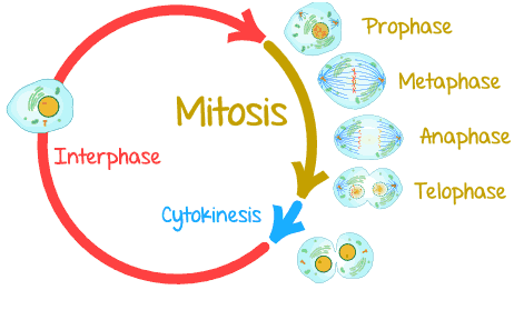
Mitosis is the type of nuclear or cell division in which the mother cell divides into two identical daughter cells. It occurs mainly in asexual type of reproduction. Mitosis is generally a sequence of events that occurs during the reproduction or cell division of eukaryotic cells that produces two genetically similar or identical nuclei that resemble their parent cells. Mitosis is characterized by several stages including interphase, anaphase, prophase, metaphase and telophase; all of which play critical role in the asexual division of eukaryotic cells. The four phases of mitosis are prophase, metaphase, anaphase and telophase.
Interphase
Interphase is the stage in which the eukaryotic cell is not experiencing anyform of cell division. A eukaryotic cell is always at a stage of interphase before it enters mitosis for cell division to occur. Interphase is a stage of a eukaryotic cell between divisions; and the partitioning of the mother cell does not occur at this phase. DNA replication occurs at this stage of interphase. Interphase perhaps is usually the first stage that precedes cell division in a living cell; and after this stage of interphase, the first stage of mitosis – which is known as prophase begins.
Prophase
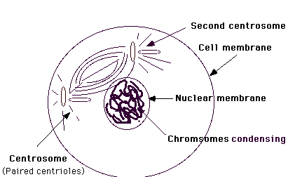
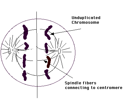
Prophase is the first stage of mitosis; and it is the stage in which the chromosomes of the parent cell condense in order to separate into two sister chromatids (Figure 1). Under the microscope, the two sister chromatids are seen as individual cells; and each moves towards the equator of the cell. A cell first replicates its entire DNA every time it divides, and the chromosomes also divide during this time to form sister chromatids which eventually separates and becomes individual cells of their own.
This stage of mitosis is generally known as prophase. Mitotic spindle is formed at this stage of prophase. The center of the chromosome is known as centromere; and it differentiates the two sister chromatids of the chromosome. Centromere is defined as the point of adhesion or attachment of two chromatids in a chromosome. It is formed during cell division in cells.
Metaphase
Metaphase is the stage of mitosis in which the chromosomes are arranged in the center of the mitotic spindle fiber (Figure 2). It is usually the second stage of mitosis. At this stage, the nuclear envelope of the nucleus breaks down and disappears. And a metaphase plate in which the chromosomes attaches to is formed at this stage. During metaphase, the chromosomes become visible and can pick up stains.
The chromosomes reach their maximum degree of coiling and condensation; and they can be counted at this stage. Metaphase is the third phase of mitosis. During metaphase, the chromosomes of the cell align themselves in the middle of the cell, and this leads to the formation of a type of cellular structure that resembles a tug of war. In metaphase, the chromosomes of the cell are at their most highly coiled and condensed state; and the chromosomes are aligned at the metaphase plate.

Anaphase
Anaphase is the third stage of mitosis. It is the stage of mitosis in which each of the sister chromatids move in opposite direction towards the ends of the spindle pole as the centromere of each chromosome splits into two (Figure 3). It is the stage in mitosis or meiosis following metaphase in which the daughter chromosomes move away from each other to opposite ends of the cell. During anaphase the spindle apparatus pulls the two sister chromatids of each chromosome apart and drags them toward opposite poles of the cell as shown in Figure 3.
Telophase
Telophase is the fourth stage of mitosis; and it is the stage of cell division after anaphase. At this stage, nuclear membrane or envelop assembles around each sister chromatids to form two new nuclei, with each having a new nucleolus (Figure 4). The chromatids become less visible at this stage since the nucleolus reappears and the sister chromatids become enveloped to from two new nuclei. And the separated chromatids are now known as chromosomes. The cell can now replicate its DNA again during interphase and the mitosis cycle continues (Figure 5).

During telophase, the sister chromatids arrive at the poles; and they start to uncoil and lose their morphology. Telophase is the process that separates the duplicated genetic material carried in the nucleus of a parent cell into two identical daughter cells. Cell division becomes complete once this stage of telophase is reached. Telophase eventually passes into the next interphase as the nuclear envelopes and nucleoli of the cell reforms and the chromatin becomes diffused.
Cytokinesis (cytoplasmic division) as shown in Figure 5 follows and occurs concurrently with telophase. Cytokinesis is simply defined as the division of the cytoplasm. And it includes those other activities that occur during the cell division (with the exception of nuclear division) of a parent eukaryotic cell into its progeny or daughter cells.
References
Alberts B, Bray D, Lewis J, Raff M, Roberts K and Watson J.D (2002). The molecular Biology of the Cell. Fourth edition. New York, Garland, USA.
Berg JM, Tymoczko JL, Stryer L (2002). Biochemistry (5th ed.). New York, NY: W. H. Freeman.
Brooks G.F., Butel J.S and Morse S.A (2004). Medical Microbiology, 23rd edition. McGraw Hill Publishers. USA.
Campbell, Neil A.; Brad Williamson; Robin J. Heyden (2006). Biology: Exploring Life. Boston, Massachusetts: Pearson Prentice Hall.
Cooper G.M and Hausman R.E (2004). The cell: A Molecular Approach. Third edition. ASM Press.
Dale J (2003). Molecular genetics of bacteria. Jeremy W. Dale and Simon Park (4th eds.). John Wiley & Sons Ltd, West Sussex, UK. Pp.
Karp, Gerald (2009). Cell and Molecular Biology: Concepts and Experiments. John Wiley & Sons.
Lodish H, Berk A, Matsudaira P, Kaiser C.A, Kreiger M, Scott M.P, Zipursky S.L and Darnell J (2004). Molecular Cell Biology. Fifth edition. Scientific American Books, Freeman, New York, USA.
Madigan M.T., Martinko J.M., Dunlap P.V and Clark D.P (2009). Brock Biology of microorganisms. 12th edition. Pearson Benjamin Cummings Publishers. USA. Pp.795-796.
Maton, Anthea (1997). Cells Building Blocks of Life. New Jersey: Prentice Hall.
Discover more from Microbiology Class
Subscribe to get the latest posts sent to your email.