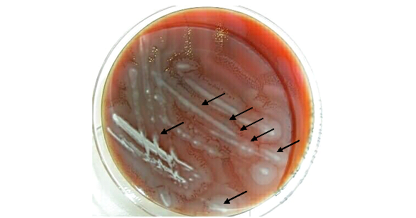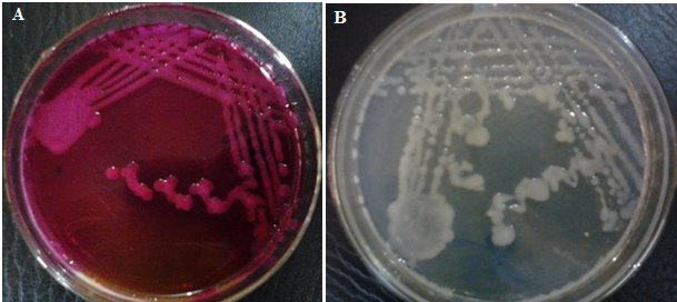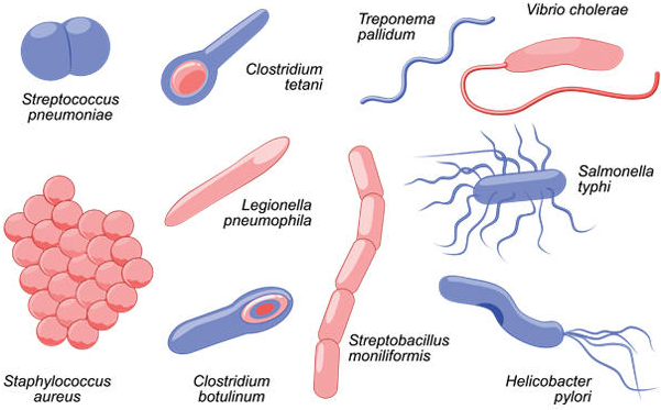

Inoculation technique is a fundamental procedure in microbiology that involves the deliberate introduction of microorganisms into a suitable culture medium under controlled laboratory conditions to support their growth, maintenance, observation, and examination. It provides a practical means of studying microbial life by creating an environment in which bacteria, fungi, and other microorganisms can develop characteristics that are useful for scientific investigation. The technique requires careful handling of microbial specimens, appropriate selection of culture media, and adherence to established laboratory procedures to obtain reliable and meaningful results.
In microbiological practice, inoculation serves as a bridge between the collection of a microbial specimen and its subsequent cultivation and analysis. Microorganisms are often invisible to the naked eye in their natural state; therefore, transferring them into a favourable growth environment makes it possible to observe their development and investigate their biological properties. Culture media may be prepared in solid, semi-solid, or liquid forms, depending on the organism under investigation and the objectives of the experiment. Each medium provides particular nutrients and environmental conditions that influence microbial growth and the expression of observable characteristics.
The significance of inoculation extends beyond the simple transfer of microorganisms from one location to another. It establishes the foundation for controlled microbial investigation, allowing laboratory personnel to examine growth patterns, colony morphology, pigmentation, and other characteristics that may contribute to microbial differentiation. When performed correctly, inoculation also helps maintain the integrity of a specimen by minimising the introduction of unwanted organisms. The application of aseptic principles is therefore essential, as contamination can alter culture characteristics, compromise experimental findings, and produce misleading interpretations.
Another important purpose of inoculation is to facilitate the isolation and propagation of microorganisms for further examination. Different inoculation methods are selected according to the nature of the specimen, the type of microorganism, and the intended outcome. For instance, streak inoculation can be used to separate individual colonies on solid media, whereas broth inoculation supports microbial multiplication in a liquid environment. These approaches enable investigators to obtain cultures suitable for morphological assessment, biochemical testing, and other identification procedures. The choice of technique must correspond to the requirements of the investigation to ensure that the resulting observations are scientifically useful.
In clinical microbiology, inoculation contributes to the investigation of infectious diseases by enabling microorganisms recovered from appropriate patient specimens to be cultivated and examined. The resulting cultures may support the identification of potential pathogens and guide further diagnostic testing. In food and environmental microbiology, inoculation is employed to investigate microbial contamination, assess hygiene conditions, and monitor the microbiological quality of different materials. Research laboratories also depend on this technique to study microbial physiology, evaluate antimicrobial substances, and explore interactions between microorganisms and their environments.
Inoculation promotes consistency and reproducibility in microbiological experiments when the procedure is carried out using suitable equipment, standardised methods, and appropriate incubation conditions. Accurate labelling, proper specimen handling, and careful documentation are equally important for maintaining traceability throughout the investigation. Collectively, these practices establish inoculation as an indispensable component of microbiological science, supporting the systematic cultivation, examination, and interpretation of microorganisms across diagnostic, educational, industrial, and research settings.
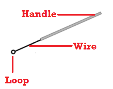
As aforementioned, inoculation is a microbiology technique which is used to introduce or place specimens and microbial cultures on or into a culture medium. The organism to be transferred into or onto the surface of the culture media (inclusive of solid and liquid or broth media) is known as inoculum. Inoculation is performed in the microbiology laboratory with an important piece of apparatus known as the inoculating loop (Figure 1). For example, inoculation technique allows microbiologists to obtain pure cultures of microorganisms through a process known as streaking.
Types of inoculation techniques in microbiology
Inoculation techniques are essential procedures in microbiology that involve introducing microorganisms into suitable culture media to facilitate their growth, isolation, identification, and examination. These techniques differ according to the nature of the microorganism, the purpose of the investigation, and the type of culture medium employed.
Proper inoculation enables microbiologists to obtain pure cultures, estimate microbial populations, investigate physiological characteristics, and assess responses to antimicrobial agents. The selection of an appropriate technique depends on the desired outcome and the characteristics of the sample. Common inoculation techniques include the streak plate, spread plate, pour plate, broth inoculation, stab inoculation, and lawn culture techniques.
1. Streak plate technique
The streak plate technique is a microbiological method used to separate individual microorganisms from a mixed or concentrated culture to obtain isolated colonies on the surface of solid agar medium. It is based on the principle of progressive dilution, in which microorganisms are distributed across successive sections of an agar plate, reducing the number of cells deposited in each area.
The procedure involves transferring a small quantity of microbial culture onto the agar surface using a sterile inoculating loop (Figure 2). The loop is moved across designated sections in a systematic pattern, spreading the organisms progressively. After inoculation, the plate is incubated under appropriate conditions to allow visible colonies to develop. Each well-isolated colony may originate from a single microbial cell or a cluster of cells.
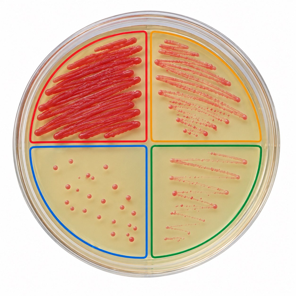
This technique is particularly valuable in diagnostic laboratories, where pure cultures are required for accurate microbial identification and antimicrobial susceptibility testing. It is also widely applied in environmental microbiology, food testing, and research involving mixed microbial populations. Its effectiveness depends on proper streaking patterns, suitable culture media, and strict aseptic practices. Excessive pressure or dense streaking may prevent adequate separation of colonies.
2. Spread plate technique
The spread plate technique is a quantitative microbiological method in which a measured volume of microbial suspension is distributed evenly over the surface of a pre-solidified agar medium. Its principal purpose is to obtain surface-grown colonies and determine the concentration of viable microorganisms in a sample.
In this method, an appropriate volume of diluted microbial suspension is placed onto the agar surface and spread using a sterile spreader (Figure 3). The sample is distributed gently to achieve relatively uniform coverage without damaging the agar. Following incubation, viable microorganisms develop into visible colonies that can be counted when the plate contains a suitable number of distinct colonies.
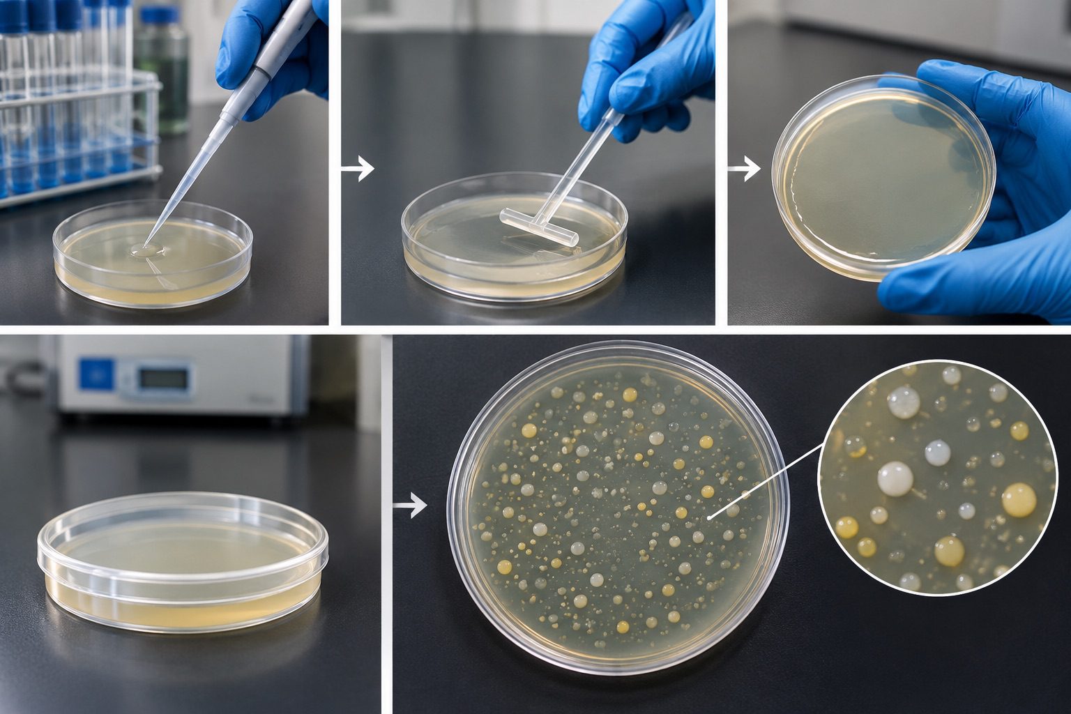
The spread plate technique is widely used in food microbiology, water quality assessment, pharmaceutical testing, and environmental investigations. It enables researchers to estimate viable microbial populations, commonly expressed as colony-forming units per millilitre (CFU/mL) or per gram (CFU/g), depending on the original sample. Since microorganisms grow on the agar surface, colony morphology can also be examined. Accurate results depend on appropriate serial dilution, reliable volume measurement, even spreading, and proper incubation. This method is especially suitable when surface colony observation and quantitative enumeration are required.
3. Pour plate technique
The pour plate technique is a microbiological procedure used to cultivate microorganisms by incorporating a measured sample into molten agar before the medium solidifies. Unlike the spread plate method, which confines growth mainly to the surface, the pour plate technique allows colonies to develop both within the agar and on its surface. The procedure begins with transferring a measured volume of a suitably diluted microbial suspension into a sterile Petri dish (Figure 4). Molten agar, cooled to a temperature appropriate for the target organisms, is added and mixed gently with the sample. Once the medium solidifies, the plate is incubated under suitable conditions. Microorganisms present in the sample form colonies at different depths within the agar or on its surface.
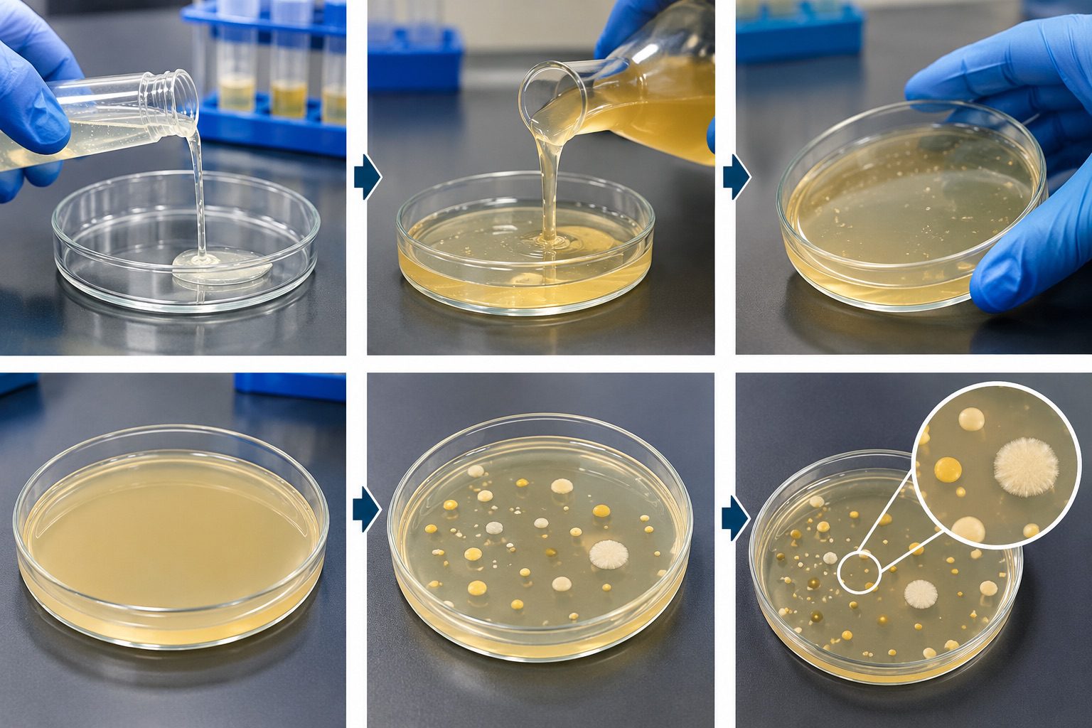
This method is useful for estimating viable microbial populations in food, water, and other samples. Colonies growing inside the agar may appear smaller than surface colonies because oxygen availability and nutrient diffusion differ with depth. The technique can accommodate organisms that tolerate the conditions associated with incorporation into agar, but heat-sensitive microorganisms may be injured by the warm medium. Careful temperature control, accurate dilution, and consistent mixing are therefore essential. The pour plate method provides a practical approach to microbial enumeration when both surface and subsurface colony development are acceptable.
4. Broth inoculation technique
Broth inoculation is a cultivation method in which microorganisms are introduced into a sterile liquid nutrient medium to support multiplication under controlled conditions. The medium contains essential nutrients, such as carbon sources, nitrogen compounds, minerals, and other growth factors required by the target organism. In this technique, a suitable inoculum is transferred aseptically into a sterile broth container using an appropriate sterile instrument (Figure 5).
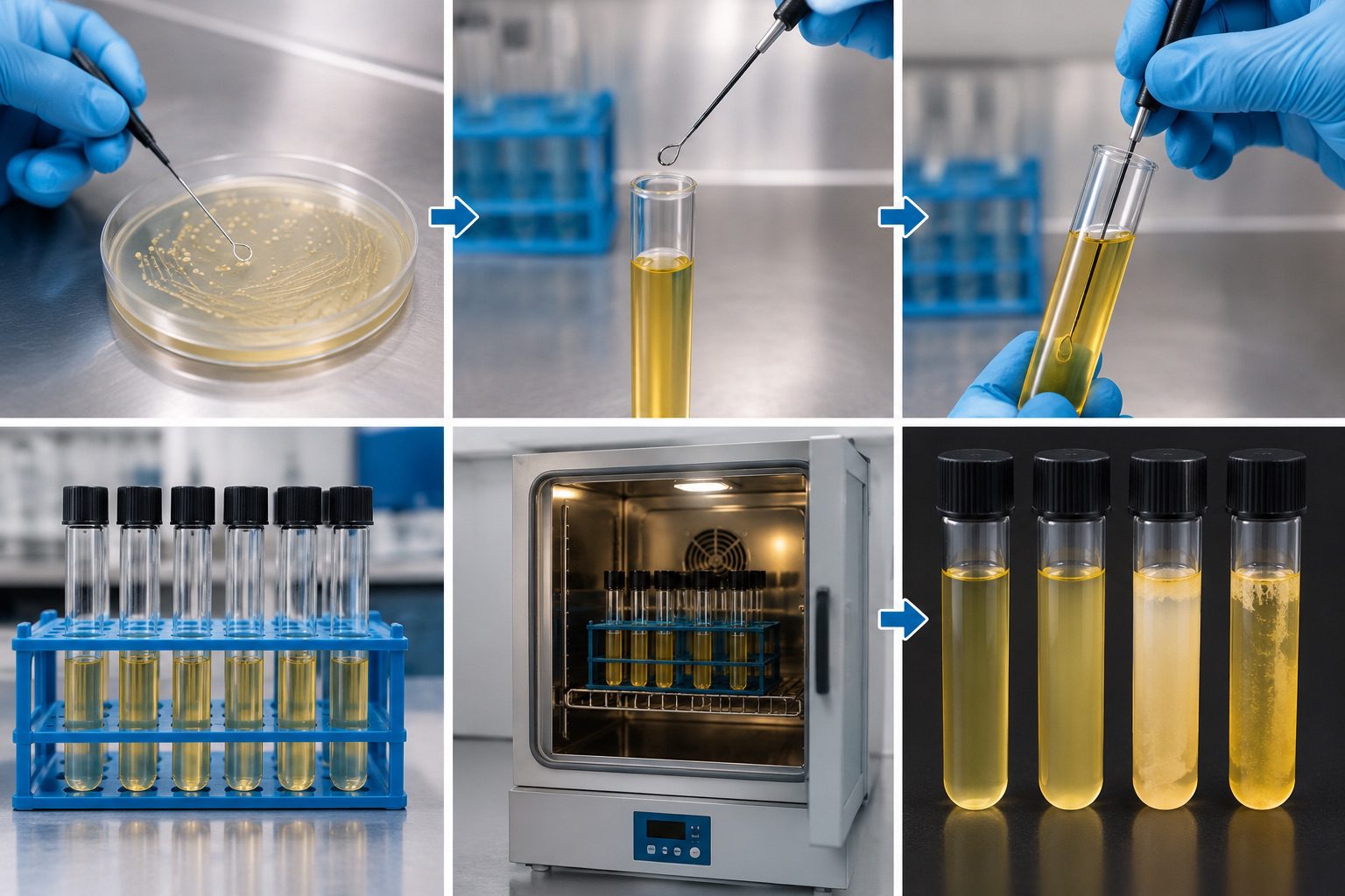
The container is then closed and incubated under conditions suitable for the microorganism. Growth may become evident through turbidity, sediment formation, surface films, colour changes in indicator-containing media, or other observable characteristics. However, the absence of visible turbidity does not necessarily indicate that growth has not occurred. Broth inoculation is commonly employed to prepare microbial suspensions, increase cell numbers, conduct biochemical tests, and support experiments involving microbial metabolism.
It is also useful in procedures that require relatively large quantities of actively growing microorganisms. Unlike solid media, broth generally does not provide direct separation of individual colonies, so additional isolation methods may be necessary when pure cultures are required. Reliable outcomes depend on suitable medium composition, inoculum quality, incubation conditions, and prevention of contamination. This technique remains fundamental to routine microbiological cultivation and laboratory investigations.
5. Stab inoculation technique
Stab inoculation is a microbiological technique used to introduce microorganisms into the deeper regions of a solid or semisolid culture medium. It is particularly useful for investigating bacterial motility, oxygen requirements, and certain physiological characteristics that can be observed along the path of microbial growth.
The procedure involves using a sterile straight inoculating needle to introduce a small quantity of culture into a deep agar medium (Figure 6). The needle is inserted vertically into the medium and withdrawn carefully along the same path to minimise disruption. Following incubation under appropriate conditions, the distribution of growth is examined. Motile organisms may produce diffuse growth extending away from the stab line, whereas non-motile organisms typically grow more narrowly along the inoculation track. In suitable media, differences in growth at various depths may also provide information about oxygen tolerance.
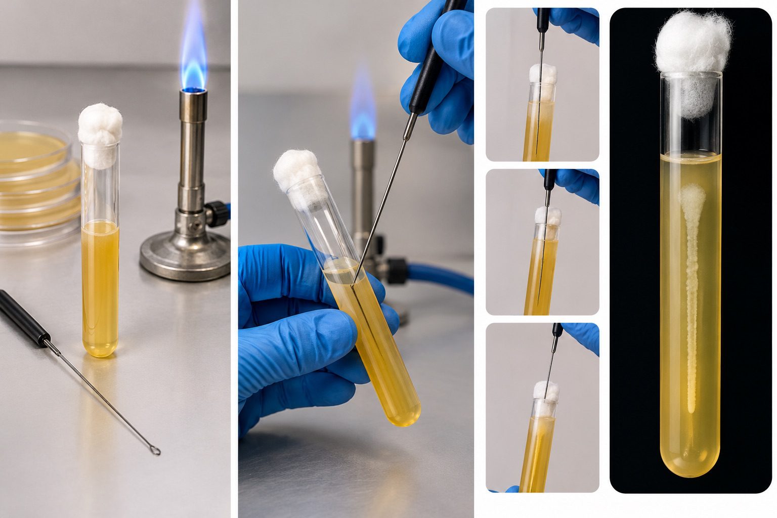
Stab inoculation is frequently used in diagnostic microbiology, particularly in motility testing and the investigation of selected biochemical reactions. The technique requires a medium with appropriate consistency, correct needle handling, and careful interpretation of growth patterns. Excessive movement during insertion or withdrawal may produce misleading results by mechanically spreading the inoculum. Because growth characteristics vary between organisms and media, findings should be interpreted alongside relevant control results and other identification tests.
6. Lawn culture technique
The lawn culture technique is a microbiological method designed to produce a dense, relatively uniform layer of microbial growth across the surface of an agar plate. Unlike the streak plate technique, which aims to separate individual colonies, lawn culture promotes continuous growth over the inoculated area. The method commonly involves preparing a suitable microbial suspension and distributing it evenly across the agar surface using a sterile swab or another appropriate spreading instrument (Figure 7).
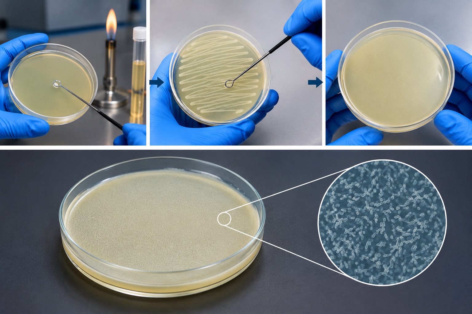
The inoculum is applied systematically to minimise untreated areas and achieve consistent coverage. After incubation under suitable conditions, a confluent or near-confluent layer of growth develops. The uniformity of this layer is important because uneven inoculation can affect the interpretation of experimental results. Lawn culture is particularly important in antimicrobial susceptibility testing, including the disc diffusion method, in which antimicrobial discs are placed on an inoculated agar surface.
Following incubation, zones of inhibited growth around the discs may be measured and interpreted according to the applicable testing standards. The technique is also used in bacteriophage assays and other experiments requiring broad surface coverage. Reliable lawn formation depends on standardised inoculum density, appropriate agar depth and composition, consistent application, and controlled incubation conditions. These factors help ensure reproducibility and meaningful comparison between experimental results.
Materials and equipment used in microbiological inoculation
The successful performance of microbiological inoculation depends on the appropriate selection and careful handling of laboratory materials and equipment. These instruments facilitate the aseptic transfer of microorganisms into suitable culture media while minimizing contamination and maintaining the reliability of experimental results.
1. Inoculating loop and inoculating needle: An inoculating loop is a laboratory instrument used to collect and transfer small quantities of microorganisms onto or into culture media. It is particularly useful in streak plate procedures for obtaining isolated colonies. An inoculating needle, which has a straight, pointed tip, is primarily employed in stab inoculation to introduce microorganisms into semisolid or deep agar media. These instruments must be sterile before use and handled according to established laboratory protocols.
2. Petri dishes containing solid agar media: Petri dishes are shallow, circular laboratory containers used to hold solidified agar, which provides a suitable surface for microbial growth. They support techniques such as streak plating and spread plating, enabling the observation, separation, and examination of microbial colonies. Proper labelling and careful handling are essential for accurate identification and prevention of contamination.
3. Test tubes containing broth or agar media: Test tubes serve as containers for liquid broth, agar slants, and deep agar media. Broth cultures facilitate microbial multiplication in liquid environments, whereas agar slants provide surfaces for maintaining appropriate laboratory cultures. Deep agar media are useful for stab inoculation and selected physiological investigations.
4. Sterilization equipment: Bunsen burners and other approved sterilization devices may be used in microbiology laboratories, depending on the facility’s safety procedures. Suitable equipment helps sterilize reusable instruments and supports contamination control. Where open flames are prohibited, validated alternatives, such as electric microincinerators or disposable sterile instruments, should be used.
5. Sterile pipettes and swabs: Sterile pipettes enable the measured transfer of liquid samples, suspensions, and culture media. Sterile swabs facilitate the collection and distribution of microorganisms across appropriate surfaces. Their sterility is essential for preserving sample integrity and reducing the introduction of unwanted organisms.
6. Personal protective equipment (PPE): Laboratory coats, appropriate gloves, and protective eyewear provide a barrier against potential exposure to biological materials. Their correct use, alongside hand hygiene and established biosafety practices, promotes a safer working environment during inoculation procedures.
Aseptic techniques during inoculation
Aseptic technique refers to a collection of laboratory practices designed to prevent the introduction, transfer, or spread of unwanted microorganisms during inoculation. In microbiology, these procedures are indispensable for preserving the purity of cultures, ensuring experimental accuracy, and protecting laboratory personnel from potential biological hazards. Effective aseptic practices create a controlled working environment in which microbial samples can be handled with minimal risk of contamination.
Prevention of microbial contamination: Contamination may occur when microorganisms from the surrounding environment, laboratory equipment, or the hands of personnel enter a culture medium. To minimize this risk, the work area should be disinfected before and after each procedure. Culture containers should remain open only for the time necessary to complete the transfer, and contact between sterile surfaces and non-sterile objects must be avoided.
Hand hygiene and personal protective equipment: Proper handwashing before and after laboratory activities reduces the transmission of microorganisms. Laboratory coats, appropriate gloves, and other required PPE provide additional barriers against exposure. Gloves should be changed when contaminated, and laboratory clothing should be handled according to institutional safety guidelines.
Sterilization of inoculating instruments: Reusable inoculating loops and needles must be sterilized using an approved method before and after use, following laboratory protocols. Instruments should be allowed to cool appropriately before contacting microbial specimens to prevent heat-related damage to the organisms and avoid splattering.
Proper handling of culture media: Culture plates, tubes, and other containers should be handled carefully to preserve their sterility. Media must be prepared and stored under suitable conditions, and containers should be labelled clearly to ensure accurate identification and traceability. Sterile instruments should be used whenever microorganisms are transferred between samples and culture media.
Safe disposal of contaminated materials: Used swabs, disposable pipettes, culture media, and other contaminated materials must be discarded into designated biohazard containers. Reusable equipment should undergo appropriate decontamination before cleaning or reuse. These measures reduce environmental contamination and support compliance with laboratory biosafety standards.
Procedure for microbial inoculation
Microbial inoculation is a fundamental laboratory procedure that involves introducing microorganisms into a suitable culture medium to facilitate their growth, isolation, and examination. The success of this process depends on meticulous preparation, appropriate instrument selection, aseptic handling, accurate labelling, and controlled incubation. Each stage must be performed carefully to preserve the integrity of the specimen and ensure the reliability of the resulting observations.
1. Preparation of the work area and culture media: The inoculation process begins with the preparation of a clean, organized laboratory workspace. The working surface is disinfected using an appropriate laboratory disinfectant, and unnecessary materials are removed to minimise contamination risks. Suitable culture media are selected according to the nutritional requirements of the target microorganism and the objective of the investigation. Before use, the media are examined for signs of contamination, dehydration, cracks, or other defects that could compromise microbial growth. Prepared media should be handled according to established laboratory protocols.
2. Selection and preparation of inoculating instruments: An appropriate inoculating instrument is selected based on the nature of the specimen and the intended culture technique. Common instruments include inoculating loops, sterile needles, swabs, and pipettes. Reusable instruments must be sterilised using an approved method, while disposable instruments should be sterile and used according to their intended purpose. Careful preparation of these tools helps prevent the introduction of extraneous microorganisms and supports the maintenance of culture purity throughout the procedure.
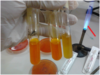
3. Transfer of the microorganism: The specimen is transferred into the designated culture medium using an appropriate aseptic technique (Figure 8). The mouth of culture tubes should also be flame-sterilized to reduce contamination during bacteria transfer. Culture containers should be opened only when necessary, and their openings should be protected from unnecessary exposure to the surrounding environment. The inoculating instrument must not contact non-sterile surfaces during the transfer process. These precautions reduce the likelihood of contamination and help maintain the original characteristics of the microbial sample.
4. Application of the appropriate inoculation method: The inoculation method is determined by the experimental objective. Streak plating facilitates the separation of individual colonies, whereas spread plating distributes a sample across the agar surface. Pour plating incorporates a specimen into molten medium under an approved protocol, while broth inoculation supports microbial propagation in liquid media. Stab inoculation is used when growth within a semisolid or deep medium is required. The selected technique should correspond to the purpose of the investigation and the characteristics of the organism.
5. Labelling of culture containers: Following inoculation, culture plates and tubes are labelled clearly and accurately. Essential information includes the specimen or organism identifier, culture medium, date of inoculation, and relevant experimental details. Appropriate labelling ensures traceability, prevents sample mix-ups, and facilitates the systematic recording and interpretation of results.
6. Incubation under appropriate conditions: Inoculated media are incubated under conditions specified for the organism and experimental purpose. Relevant parameters include temperature, duration, atmospheric requirements, and other organism-specific growth conditions. Cultures are subsequently examined according to the laboratory protocol for characteristics such as colony morphology, growth patterns, or changes in the medium. Throughout incubation and examination, appropriate biosafety precautions must be maintained to protect personnel and prevent environmental contamination.
Importance, applications, and precautions of inoculation techniques
Inoculation techniques are indispensable procedures in microbiology because they facilitate the controlled introduction of microorganisms into appropriate culture media for growth, examination, and analysis. Their application enables microbiologists to investigate microbial characteristics, assess biological activity, and obtain reliable experimental results under controlled laboratory conditions.
One major application of inoculation is the isolation and identification of microorganisms from mixed populations. Techniques such as streak plating promote the separation of individual colonies, allowing their morphological and biochemical characteristics to be examined. Inoculation also supports the cultivation of bacteria and fungi for physiological studies, antimicrobial susceptibility testing, and investigations into microbial growth patterns.
Quantitative methods, including spread plating and pour plating, assist in estimating viable microbial populations in samples. These procedures are particularly valuable in clinical microbiology, where they contribute to the investigation of infectious agents, and in food microbiology, where they help assess product quality and detect potential microbial contamination. In scientific research, inoculation provides a foundation for studying microbial metabolism, environmental interactions, and industrial applications.
Accurate inoculation requires strict adherence to aseptic principles to preserve culture purity and protect laboratory personnel. Hands must be cleaned appropriately, and suitable personal protective equipment must be worn throughout the procedure. Culture media, instruments, and containers should be handled carefully to minimize exposure to airborne contaminants and prevent the unintended transfer of microorganisms between samples. Reusable instruments must be sterilized using an appropriate validated method, while sterile disposable materials should be used as intended.
Culture vessels must be labelled clearly with relevant sample identification and experimental details to prevent mix-ups. Incubation conditions should be selected according to the organism and the approved laboratory protocol. Potentially hazardous cultures must be handled in accordance with the applicable biosafety requirements. Following completion of the procedure, contaminated materials must be decontaminated or discarded through approved waste-management systems.
References
Tomasello, F., Valero, A., Serraino, A., & Possas, A. (2023). Methods of inoculation and quantification for collecting data on microbial responses in foods. In V. O. Alvarenga (Ed.), Basic protocols in predictive food microbiology. Humana.
Fusaro, R. M. (1972). Inoculation technique for fungus cultures. Applied Microbiology, 23(1), 174–176.
Garcia L.S (2010). Clinical Microbiology Procedures Handbook. Third edition. American Society of Microbiology Press, USA.
Garcia L.S (2014). Clinical Laboratory Management. First edition. American Society of Microbiology Press, USA.
Ira R (1995). Bacteriology, Standard Operative procedure manual for microbiology laboratories, National Institute of Biologicals. Pp. 73-97.
Madigan M.T., Martinko J.M., Dunlap P.V and Clark D.P (2009). Brock Biology of Microorganisms, 12th edition. Pearson Benjamin Cummings Inc, USA.
Woods GL and Washington JA (1995). The Clinician and the Microbiology Laboratory. Mandell GL, Bennett JE, Dolin R (eds): Principles and Practice of Infectious Diseases. 4th ed. Churchill Livingstone, New York.
Discover more from Microbiology Class
Subscribe to get the latest posts sent to your email.