
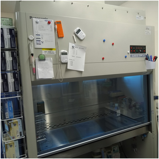
Laminar flow cabinet (hood)
The laminar flow biological safety cabinet which can also be called hood or cell culture hood is one of the most important equipment in the cell (tissue) culture laboratory. This piece of equipment helps to provide sterile environment to work with cells without any fear of contamination. It is a filtration system which protects a technician/scientist working in the cell culture laboratory from microorganisms or contamination by cells being handled within the hood.
The cell culture hood helps to prevent aerosols from coming in contact with the researcher. The constant and steady air filtration system of the hood which is distributed throughout the entire working surface of the laminar flow biological safety cabinet ensures that the whole working surface of the hood is adequately protected from extraneous contaminants and dust particles that might compromise the experimentation. Working in a clean environment (which the hood provides) is important to avoid personnel contamination and to keep everyone around safe.
The air filtration system in the hood is usually of two types: the vertical airflow and the horizontal airflow. This air filtration system is also used in classifying the hood as either a vertical airflow hood or a horizontal airflow hood. In the vertical airflow system, the air blows down from the top of the biosafety laminar flow cabinet to the entire work surface within the hood and re-circulated. But in the horizontal airflow system, the air is drawn from the sides of the hood facing the researcher.
This type of air filtration is not re-circulated as is obtainable in the vertical airflow system. While hoods with vertical airflow system gives more protection to the researcher, those with horizontal airflow systems gives lesser protection to the worker and more sterile protection to the reagents and culture media in the hood. The horizontal airflow hood gives the most stable airflow since the air it produces is not vented or re-circulated as is the case with the vertical airflow hood.
Inverted phase microscope
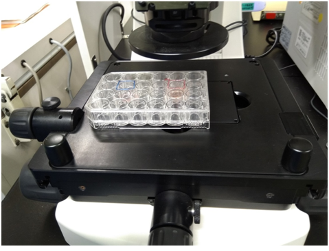
The inverted phase microscope unlike the normal light microscope does not require any staining of cells prior to its usage (Figure 2). It is inverted because the objective lenses are located below the stage; and condenser is located above the stage too. Cells or tissues in a cell culture flask or dish are viewed directly without being removed for staining (as is normally the case in light microscopy). Inverted microscopy allows scientists to view cells in a cell culture flask or bottle directly (Figure 3). Staining is not usually allowed in the cell culture lab because it can kill the cells being stained. The microscope is used to check for cell growth or viability, contamination and the health of the cells. It also helps the researcher to ensure that he or she is working with the correct cell line. Another type of microscope used in the cell culture laboratory is the contrast microscope. The inverted microscope is a basic piece of equipment required in the setting up of a cell (tissue) culture laboratory. This type of microscope allows scientists to routinely check cell culture bottles for possible signs of deterioration especially in the face of microbiological infection.
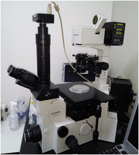
The inverted microscope is usually fitted with digital camera and phase-contrast optics which improves the magnification of the object. Unlike in the normal light microscopy, the condenser of the inverted microscope is above the stage while the objective lenses are below the stage as shown in Figure 3. Inverted microscopes can also be fitted with CCD (closed-circuit digital) cameras as aforementioned, and they are usually connected to computer monitors which enables digital viewing of cells in the cell culture bottle as well as allow the scientists to print an image of the cells.
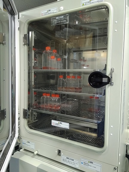
CO2 Incubator
The incubator used in the cell culture laboratory is a CO2 humidified incubator. The CO2 humidified incubator provides and maintains CO2 level (at 5-10%), humidity and temperature (at 37oC) to stimulate in vivo conditions necessary for the optimum growth of cells in the cell culture medium. Cell culture flasks should be evenly spaced when placed inside the CO2 incubator to avoid overcrowding and ensure even distribution of air or gases.
Overcrowding of cell culture flasks within the CO2 incubator should be avoided so as to ensure proper gas exchange within the incubator. Very tall stacks of culture flasks should be avoided within the incubator in order to avoid them falling over. The CO2 incubator for cell culture experiments should be routinely cleaned to remove fungal growths, traces of detergents, dust particles and dampness or wetness common with humidified incubators used for cell culture experimentations (Figure 4).
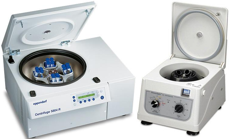
Centrifuge
The centrifuge is important in the cell culture laboratory because it is used to sediment particles or cells suspended in fluid (Figure 5). Centrifugation is a routine in the cell/tissue culture laboratory because suspensions of cells are often centrifuged to either wash-off a reagent or increase the concentration of cells required for a particular analysis.
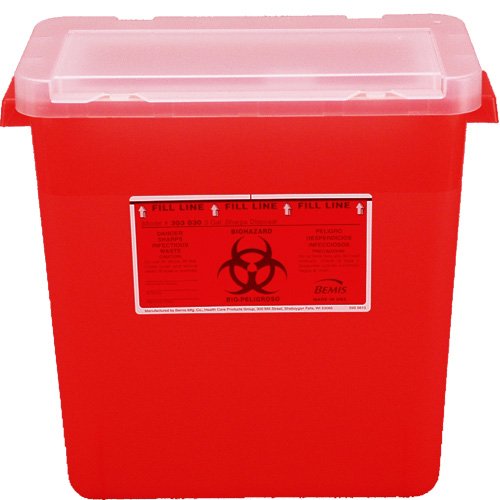
Waste container/bin
The waste container is used for collecting wastes including used pipettes, used cotton wools, broken dishes, used hand gloves and other wastes that arise during cell culture procedures (Figure 6). Old culture flasks/dishes and other wastes should be autoclaved first before their disposal.
Pipette
Pipette is used for collecting and transferring cells and media. Mouth pipetting is not allowed in the cell culture lab (Figure 7). Thus micropipettes or pipette is used for the collection and transferring of reagents or cell cultures in the cell culture laboratory. Plugged pipettes or disposable plastic pipettes are the pipettes routinely used for cell culture experimentations. Pipette pumps are used with plugged pipettes and this helps the scientist to draw certain concentration or amount of solution without using mouth pipetting method.

Cell counting machine (haemocytometer)
The haemocytometer is used for counting cells from cell cultures (Figure 8). It counts cells manually under the microscope. There are other automatic cells counting machines such as the countess machine which can be used to count cells in the cell culture lab. Cell growth can also be measured by using spectrophotometer.
Spectrophotometer is a device that measures the optical density of cells in suspension. The results of a spectrophotometer are extrapolated in order to determine the growth of the cells. A haemocytometer has an indention down the center where cells can be suspended in a liquid, and then the numbers of cells in a particular sized square are counted to calculate the overall cell count.

Refrigerator
The refrigerator is used for the storage of cell culture reagents and media. Reagents and media should always be stored according to the manufacturer’s instructions. All reagents for cell culture experimentation should always be stored at the appropriate temperature to avoid spoilage.
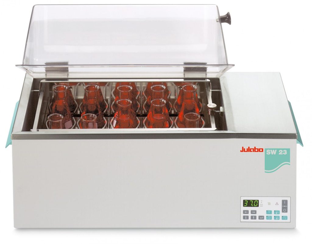
Water bath
Water bath is used to incubate bottles of cell culture media or other reagents and liquids in flasks in order to bring them to a particular or optimum temperature prior to their usage (Figure 9). Flasks should always be kept upright in the water bath, and the neck of the flasks should always be above the water in the water bath. Reagents in water baths should not be left more than is necessary because excessive warming could lead to the loss of activity in the media or reagents being warmed.
OTHER REAGENTS, EQUIPMENTS OR MATERIALS FOUND IN THE CELL CULTURE LABORATORY
Other reagents, equipment and materials found in the cell culture laboratory include:
- Automatic dispenser and water purifying system
- Colony counter
- Sterilizing oven
- Autoclave
- pH meter
- Plate reader
- Digital camera and monitor
- 70% Ethanol. Ethanol is used for disinfection
- Trolleys
- Osmometer
- Pipette washer and pipette drier
- Deep washing sink for wash-ups
- Low temperature freezer
- Cell culture lines and Growth media
- Trypan blue. This stain is used to determine the viability of cells. Living cells do not take up the colour of the dye/stain. Only dead cells take up the colour of the trypan blue stain.
- Cell culture flasks/dish
- Phosphate buffer saline (PBS)
- Sodium hypochlorite (bleach)
- Vacuum pump
- -20oC freezers
- Microplates (i.e. microtiter plates)
- Lyophilizers or lyophilizing equipment
- Disposable papers or salvage papers
- Hand gloves
References
Alberts B, Bray D, Johnson A, Lewis J, Raff M, Roberts K andWalter P (1998). Essential Cell Biology: An Introduction to the Molecular Biology of the Cell. Third edition. Garland Publishing Inc., New York.
Alberts B, Bray D, Lewis J, Raff M, Roberts K and Watson J.D (2002). The molecular Biology of the Cell. Fourth edition. New York, Garland, USA.
Ausubel, F.M., Brent, R., Kingston, R.E., Moore, D.D., Seidman, J.G., Smith, J.A., Struhl, K., eds (2002). Short Protocols in Molecular Biology, 5th edn. John Wiley & Sons, New York.
Caputo J.L (1996). Safety Procedures. In: Freshney, R.I., Freshney, M.G., eds., Culture of Immortalized Cells. New York, Wiley-Liss, Pp. 25-51.
Cooper G.M and Hausman R.E (2004). The cell: A Molecular Approach. Third edition. ASM Press.
Davis J.M (2002). Basic Cell Culture, A Practical Approach. Oxford University Press, Oxford, UK.
Freshney R.I (2005). Culture of Animal Cells, a Manual of Basic Technique, 5th Ed. Hoboken NJ, John Wiley and Sons Publishers.
Health Services Advisory Committee (HSAC) (2003). Safe Working and the Prevention of Infection in Clinical Laboratories. HSE Books: Sudbury
Lodish H, Berk A, Matsudaira P, Kaiser C.A, Kreiger M, Scott M.P, Zipursky S.L and Darnell J (2004). Molecular Cell Biology. Fifth edition. Scientific American Books, Freeman, New York, USA.
Marcovic O and Marcovic N (1998). Cell cross-contamination in cell cultures: the silent and neglected danger. In Vitro Cell Dev Biol. 34:108.
Mather J and Barnes D (1998). Animal cell culture methods, Methods in cell biology. 2rd eds, Academic press, San Diego.
Verma P.S and Agarwal V.K (2011). Cytology: Cell Biology and Molecular Biology. Fourth edition. S. Chand and Company Ltd, Ram Nagar, New Delhi, India.
Discover more from Microbiology Class
Subscribe to get the latest posts sent to your email.
