
The increasing prevalence of vancomycin-resistant enterococci (VRE) has created a persistent challenge for clinical microbiology, particularly in settings where rapid recognition of colonized patients can influence infection-control decisions. Conventional approaches to enterococcal detection may require several sequential procedures, increasing turnaround time and potentially delaying the identification of individuals who can serve as reservoirs for transmission. Chromatic VRE agar offers a selective and visually oriented approach that addresses this diagnostic gap by combining antimicrobial selection with chromogenic differentiation.
Rather than relying solely on biochemical reactions or subsequent identification procedures, chromatic media exploit enzyme-substrate interactions to generate characteristic colony colours when target organisms grow. In VRE screening, this principle is particularly valuable because vancomycin-resistant enterococci must be distinguished from other organisms that may be present in complex clinical specimens, especially rectal or perineal swabs. The medium therefore functions not simply as a growth surface, but as a selective ecological environment in which resistance and metabolic activity become directly observable.
The development of chromatic VRE agar also reflects a broader shift in diagnostic microbiology toward methods that integrate detection, discrimination, and workflow efficiency. Its application can support surveillance programmes by facilitating the recognition of presumptive VRE colonies without depending entirely on conventional culture characteristics. However, colony colour should be interpreted within an appropriate laboratory workflow because chromogenic reactions can vary among species and strains, while resistant organisms may occasionally display atypical appearances.
The significance of VRE agar lies in transforming a complex screening problem into a more readily interpretable culture-based process, potentially strengthening the speed and consistency with which VRE can be recognized in routine diagnostic and epidemiological investigations.
Vancomycin resistance in Enterococci and the importance of VRE surveillance
Vancomycin resistance in enterococci is primarily significant because of its clinical consequences and its capacity for dissemination. VRE emerged as an important healthcare-associated problem during the late twentieth century and subsequently became established in hospitals in many regions. The epidemiology is dominated by acquired resistance mechanisms, particularly those associated with the vanA and vanB gene clusters. These determinants alter the biochemical target of glycopeptide antibiotics, reducing their ability to interfere with bacterial cell-wall synthesis.
Enterococcus faecium is a major VRE species encountered in healthcare environments, although Enterococcus faecalis can also acquire vancomycin resistance. vanA-associated resistance generally produces high-level resistance to vancomycin and is commonly accompanied by resistance to teicoplanin. In contrast, vanB can produce variable levels of vancomycin resistance while retaining susceptibility to teicoplanin. These phenotypic differences are clinically and epidemiologically relevant, but they cannot be reliably inferred from colony colour on chromogenic agar alone.
Other resistance genotypes require a different epidemiological interpretation. vanC is intrinsically associated with species such as E. gallinarum and E. casseliflavus, and typically produces lower-level vancomycin resistance. These organisms differ from the more commonly encountered acquired vanA and vanB VRE in both their genetic organization and epidemiological behaviour. The presence of vanD, and less frequently other van-associated determinants, further demonstrates the genetic diversity underlying glycopeptide resistance in enterococci.
The public-health importance of VRE arises partly from the ability of enterococci to act as reservoirs of antimicrobial-resistance genes. The potential movement of resistance determinants between bacterial populations is a major concern, particularly in healthcare environments where multiple antimicrobial pressures and vulnerable patients coexist. The possibility of transfer of glycopeptide-resistance determinants to Staphylococcus aureus has historically intensified concern surrounding VRE surveillance, although such events remain uncommon.
For this reason, screening programmes may target patients or clinical areas considered to have an increased likelihood of VRE carriage. Detecting colonization is different from diagnosing infection: a patient can carry VRE without exhibiting disease. Nevertheless, colonized individuals can contribute to transmission, making surveillance microbiology an important component of infection prevention.
Chromatic VRE agar fits into this surveillance framework by providing a culture-based mechanism for identifying specimens that require further investigation. Rapid recognition of presumptive VRE allows laboratories and infection-prevention teams to respond before transmission chains become extensive. Its role is therefore epidemiological as much as diagnostic: the objective is not simply to name an organism but to identify a potential reservoir of clinically important antimicrobial resistance.
Purpose and diagnostic significance of VRE agar
Chromatic VRE agar is a selective and differential chromogenic culture medium designed for the qualitative and presumptive detection of vancomycin-resistant E. faecium and E. faecalis in clinical specimens (Figure 1). Its principal value lies in screening for intestinal or other carriage of VRE, particularly among patients who may contribute to healthcare-associated transmission. Unlike conventional culture media, chromogenic media incorporate substrates that interact with bacterial enzymes and produce characteristic pigmentation. This allows presumptive recognition of target organisms directly on the primary isolation plate, reducing dependence on a sequence of conventional biochemical tests during the initial screening stage.
The importance of such screening is closely associated with the epidemiology of enterococcal antimicrobial resistance. Enterococci are capable of surviving under diverse environmental conditions and can persist on hospital surfaces, medical equipment, and within the gastrointestinal tract. When strains acquire resistance to glycopeptide antibiotics such as vancomycin, therapeutic options may become substantially restricted. VRE are therefore important not only as individual clinical pathogens but also as reservoirs of transferable resistance determinants. Early recognition of colonization can support infection-prevention measures, including appropriate isolation strategies and enhanced environmental precautions, according to institutional policy.
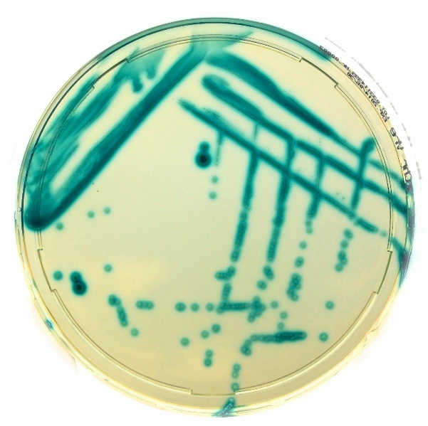
Chromatic VRE agar is primarily a screening medium, rather than a standalone diagnostic system. A coloured colony compatible with VRE should be regarded as presumptive because colony pigmentation alone does not establish species identity, vancomycin-resistance phenotype, or the underlying genetic mechanism. Following primary growth, presumptive colonies should be recovered by subculture onto a suitable non-selective medium and subjected to confirmatory identification and antimicrobial susceptibility testing. Where required, molecular or other epidemiological methods can subsequently be applied to characterize resistance determinants and establish relationships among isolates.
The ability to obtain an interpretable result after approximately 24 hours of incubation can make chromogenic screening particularly useful in surveillance laboratories. Earlier recognition of possible VRE carriage can shorten the interval between specimen collection and implementation of infection-control measures. Nevertheless, interpretation must remain linked to validated laboratory procedures, because atypical colony colours, mixed cultures, and non-target organisms can complicate visual assessment.
The significance of Chromatic VRE agar extends beyond its distinctive colony appearance. It represents a practical microbiological approach that integrates selective pressure with phenotypic differentiation, allowing laboratories to identify presumptive VRE more efficiently while retaining confirmatory testing as an essential second stage.
Constituents and functional roles of chromatic VRE agar
The composition of Chromatic VRE agar is specifically structured to create an environment in which resistant enterococci can grow while many competing microorganisms are suppressed. The principal constituents for the medium include peptones, sodium chloride, a chromogenic and selective mixture, and agar. The formulation is adjusted to a final pH of approximately 7.2 ± 0.2 at 25°C. Each component contributes a distinct physicochemical or microbiological function, and the combined formulation determines the medium’s selectivity and differential behaviour.
Peptones constitute the primary nutritional component and are supplied at approximately 30.0 g/L. They provide a complex source of peptides, amino acids, nitrogenous compounds, and other growth-supporting nutrients. Enterococci can utilize a wide range of nutritional substrates, allowing resistant strains to establish visible colonies under the conditions created by the medium. The nutritional base is therefore essential for recovering organisms from clinical specimens in which target bacteria may be present at relatively low abundance.
Sodium chloride, present at approximately 5.0 g/L, contributes to maintenance of the osmotic environment surrounding bacterial cells. Appropriate osmotic conditions are necessary for cellular integrity and metabolic activity. Although sodium chloride is not itself responsible for VRE selection, its concentration contributes to the overall physiological suitability of the formulation.
The chromogenic and selective mixture, approximately 6.5 g/L, represents the most functionally distinctive component. The selective portion incorporates antimicrobial compounds, including vancomycin, that impose pressure against susceptible competing organisms. Enterococci possessing relevant vancomycin-resistance mechanisms are able to grow under these selective conditions more readily than susceptible enterococci. However, selection should not be interpreted as absolute exclusivity; unusual resistant organisms or atypical strains may occasionally grow, which reinforces the need for confirmatory identification.
The chromogenic portion contains substrates designed to respond to enzymatic activity associated with the target organisms. When the appropriate bacterial enzymes act upon these substrates, coloured reaction products accumulate within or around colonies. Consequently, the laboratory can evaluate both growth and colony appearance. This differential characteristic distinguishes chromogenic media from conventional selective agar, where growth may indicate resistance but provide little immediate information about organism identity.
Agar, approximately 15.0 g/L, serves as the solidifying agent. It creates a stable surface on which individual microorganisms can develop discrete colonies, allowing their size, morphology, pigmentation, and distribution to be assessed.
These components create a controlled microbial environment rather than merely supplying nutrients. The formulation applies selective pressure, supports enterococcal growth, and generates a visual signal associated with characteristic metabolic activity. The effectiveness of the medium therefore depends on the interaction between its chemical constituents and the biological properties of the organisms being screened.
Microbiological principle of chromogenic VRE detection
The operating principle of Chromatic VRE agar is based on the simultaneous exploitation of antimicrobial resistance and bacterial enzymatic activity. These two properties provide complementary information: resistance permits the survival of the target organism under selective conditions, while enzyme-mediated chromogenic reactions contribute to presumptive differentiation. The resulting colony appearance can therefore provide a rapid visual indication that a specimen may contain VRE.
When a clinical specimen is inoculated onto the medium, it contains a mixture of microorganisms rather than a single bacterial population. Enterococci may coexist with members of the intestinal microbiota, Gram-negative bacteria, yeasts, and other organisms. The selective components of the medium are intended to restrict the growth of much of this background flora. Vancomycin is particularly important because susceptible enterococci are inhibited, whereas organisms carrying effective vancomycin-resistance mechanisms have a selective advantage.
The chromogenic component provides the second layer of discrimination. Target organisms possess enzymatic activities capable of interacting with specific chromogenic substrates incorporated into the medium. Enzymatic cleavage releases or modifies a chromophore, producing a visible colour associated with colony growth. The exact colour produced depends on the proprietary formulation and substrate system of the manufacturer; consequently, interpretation should always follow the manufacturer’s validated colour chart and laboratory instructions rather than relying on generalized descriptions.
This combination of selective growth and pigmentation makes the medium especially useful for direct screening. A conventional approach may require isolation followed by biochemical identification and separate susceptibility testing before a presumptive VRE result becomes available. Chromogenic media compress some of these early steps into a single culture-based procedure. The result is not equivalent to molecular detection, however, because the method depends on viable organisms growing under the conditions established by the medium.
The distinction between screening and confirmation is therefore fundamental. A presumptive coloured colony indicates that an organism compatible with the target phenotype has grown under selective conditions. It does not independently demonstrate the presence of E. faecium or E. faecalis, nor does it establish whether resistance is mediated by vanA, vanB, or another determinant. Confirmatory identification and susceptibility testing remain necessary, particularly because intrinsic low-level glycopeptide resistance in certain enterococcal species can produce findings that require careful interpretation.
The chromogenic approach is consequently best understood as a phenotypic screening strategy. It translates invisible biochemical activity into a visible colony signal while simultaneously applying antimicrobial selection. This design can improve the efficiency of VRE surveillance, but its interpretation remains dependent on controlled incubation, appropriate quality assurance, and confirmatory laboratory methods.
Interpretation, confirmation, and laboratory application of chromatic VRE agar
The practical application of Chromatic VRE agar begins with appropriate specimen selection and inoculation, followed by incubation under the validated conditions specified for the medium. After the recommended incubation period, commonly approximately 24 hours, plates are examined for growth and characteristic colony pigmentation. Colonies displaying the expected appearance are considered presumptive VRE and should be processed through a confirmatory workflow.
Visual interpretation is central to chromogenic culture, but it should not be treated as an independent identification method. The appearance of a colony is influenced by organism physiology, incubation conditions, inoculum density, medium preparation, and the specific chromogenic substrates used by the manufacturer. Mixed cultures can further complicate interpretation because neighbouring colonies may obscure colour or morphology. Laboratories should therefore apply standardized interpretation criteria and appropriate quality-control organisms when validating and routinely using the medium.
Presumptive colonies should be subcultured onto a non-selective medium to obtain a purified isolate. This step is important because downstream identification and antimicrobial susceptibility testing require an adequately isolated organism. Species confirmation can be performed using validated biochemical, mass-spectrometric, or molecular methods, depending on laboratory resources and diagnostic requirements. Antimicrobial susceptibility testing then establishes the phenotypic resistance profile and may provide information needed for clinical or epidemiological interpretation.
Where epidemiological investigation is required, molecular characterization can provide additional resolution. Detection of resistance determinants such as vanA or vanB can clarify the mechanism underlying the observed phenotype. Genotyping or other typing approaches may subsequently be used to investigate whether isolates recovered from different patients or locations are related. Such testing is particularly relevant during suspected healthcare-associated outbreaks.
A crucial limitation is that chromogenic VRE agar does not diagnose infection. Detection of VRE from a screening specimen generally indicates colonization or carriage and must be interpreted according to the specimen type and clinical context. A positive screening culture should therefore not automatically be equated with invasive enterococcal disease. Likewise, the medium should not be used alone to determine antimicrobial treatment.
The greatest value of Chromatic VRE agar is consequently achieved when it is incorporated into a broader laboratory pathway. Selective chromogenic culture provides an efficient first-line screen; subculture establishes a recoverable isolate; identification determines the organism; susceptibility testing characterizes the phenotype; and molecular or epidemiological methods can investigate the resistance mechanism and transmission pattern.
This staged approach preserves the principal advantage of chromogenic screening, which is rapid visual recognition without compromising diagnostic accuracy. In practice, Chromatic VRE agar is best regarded as an early-warning tool within antimicrobial-resistance surveillance. Its contribution is not the replacement of confirmatory microbiology, but the acceleration and prioritization of the investigations required to recognize, characterize, and control VRE in healthcare environments.
Further reading
D V, S V, S T S, M K Y, S M. (2014). Evaluation of chromogenic media in detection of vancomycin resistant enterococci. Journal of Clinical and Diagnostic Research, 8(11), DC25-DC27.
Stamper, P. D., Shulder, S., Bekalo, P., Manandhar, D., Ross, T. L., Speser, S., Kingery, J., & Carroll, K. C. (2010). Evaluation of BBL CHROMagar VanRE for detection of vancomycin-resistant enterococci in rectal swab specimens. Journal of Clinical Microbiology, 48(11), 4294-4297.
Zakaria, N. D., Hamzah, H. H., Salih, I. L., Balakrishnan, V., & Abdul Razak, K. (2023). A review of detection methods for vancomycin-resistant Enterococci (VRE) genes: From conventional approaches to potentially electrochemical DNA biosensors. Biosensors, 13(2), 294.
O’Driscoll, T., & Crank, C. W. (2015). Vancomycin-resistant enterococcal infections: Epidemiology, clinical manifestations, and optimal management. Infection and Drug Resistance, 8, 217-230.
Boschert, A. L., Arndt, F., Hamprecht, A., Wolke, M., & Walker, S. V. (2023). Comparison of five different selective agar for the detection of vancomycin-resistant Enterococcus faecium. Antibiotics, 12(4), 666.
Gouliouris, T., Blane, B., Brodrick, H. J., Raven, K. E., Ambridge, K. E., Kidney, A. D., Hadjirin, N. F., Török, M. E., Limmathurotsakul, D., & Peacock, S. J. (2016). Comparison of two chromogenic media for the detection of vancomycin-resistant enterococcal carriage by nursing home residents. Diagnostic Microbiology and Infectious Disease, 85(4), 409-412.
www.liofilchem.net/login/pd/ifu/11621_IFU.pdf
Discover more from Microbiology Class
Subscribe to get the latest posts sent to your email.