In the previous section on DEPARAFFINIZATION PRIOR TO IMMUNOHISTOCHEMISTRY STAINING, we looked at deparaffinization and rehydration of paraffin-embedded tissue. In this section, we look at epitope unmasking protocol. Deparaffinization is an important process in the immunohistochemistry staining process; and it must be done prior to immunohistochemistry, especially for paraffin-embedded tissue samples.
Deparaffinization is simply defined as the process of removing the paraffin from the tissue. Paraffin covers the tissue structures and may affect immunohistochemistry analysis, especially by masking or blocking the antigen of interest. Therefore, paraffin must be removed first before proceeding with the experiment. Deparaffinization is achieved by using a series of solvents including xylene, ethanol and water; and the sole aim is to remove the wax covering the tissue sample on the slide.
Antigen retrieval can be achieved using different protocol and reagents. However, all these steps are geared towards unmasking the covered or hidden antigen in the paraffin-embedded tissue sample on the slide. Antigen retrieval is done at a very high temperature (e.g., 95-100oC). The equipment used to achieve antigen retrieval includes microwave, autoclave or a heating flask / cooker or kettle. This equipment or instrument helps to provide the requisite temperature necessary to achieve antigen retrieval.
Why is antigen retrieval important in the immunohistochemistry staining technique? Fixation by paraffin leads to cross-linking of proteins on the embedded tissue on the slide. And this may result in the masking or covering of the epitope on the sample. The masking of the epitope may result in a weak staining or negative staining; and this can affect the integrity of the entire test result. To overcome the challenge presented by paraffinization process as aforesaid, antigen retrieval must be performed.
There are usually two methods of achieving epitope retrieval viz:,
- Heat-induced epitope retrieval (HIER)
- Proteolytic-induced epitope retrieval (PIER)
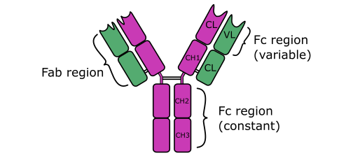
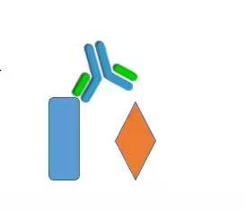
Note: The phrase epitope means antigenic determinant. Epitope is the particular place or site on an antigen, on which an antibody binds.
The choice of which type of antigen retrieval technique to use is largely dependent on the type of tissue sample to be processed and also on the type of antibody to be used. While HIER makes use of a citrate buffer and heating source via a microwave or autoclave, the PIER makes use of enzymes known as peptidases (e.g., trypsin, pepsin, proteinase K) – to unmask the hidden antigen or epitope.
Both HIER and PIER helps to unmask the hidden antigen as well as to increase the intensity and specificity of the test antibody. A high temperature (e.g., 100oC) is used for the HIER protocol while a much lower temperature (e.g., 37oC) is used in the PIER process. Only the HIER process will be explained in this section because it is mostly used. The difference between the HIER and PIER process is just on the solution used and on the temperature levels as aforesaid.
STEP BY STEP PROCESS OF PERFORMING HIER
- Place the entire slides in a slide rack. Ensure to allows maintain a high humidity for the sectioned tissues on the slides. At no point in time should the slides be allowed to dry out. Therefore, slides should always be kept and carried in a humidified container to prevent drying.
- Heat steam the slides harbouring the tissue samples in a solution of citrate buffer (pH 6.0) at 98-100°C for about 45 min. Sodium citrate buffer is a typical example of a citrate buffer solution that can be used for this step. Pour the citrate buffer solution in a heating vessel (e.g., cooker or autoclave) and heat it up to a high temperature.
- After the heating process in the presence of citrate buffer solution, remove the slides from the heating vessel.
- Allow the slides to cool in buffer at room temperature for 20 min. In some cases, this cooling process can be skipped; but it depends on the researcher.
- Rinse the slides in 1x TBS with Tween (TBST) 80 at room temperature for 1 min. In some quarters, slides are washed by immersing them in distilled water for about 5-10 min.
- Continue with the immunohistochemistry staining protocol after the above processes. The next step in the immunohistochemistry staining process is permeabilization and blocking non-specific binding. THIS WILL BE EXPLAINED IN THE NEXT SECTION ON THIS WEBSITE.
TBST is a mixture of tris-buffered saline (TBS) and Polysorbate 20 (also known as Tween 20)
Note:
At any point in time, every laboratory should maintain optimal conditions in carrying out this experiment.
Most importantly, it is also advisable to follow manufacturer’s instruction on the use of the reagents and other materials.
All experiments should be conducted in line with the specific information guideline and the laboratories protocol.
Discover more from Microbiology Class
Subscribe to get the latest posts sent to your email.