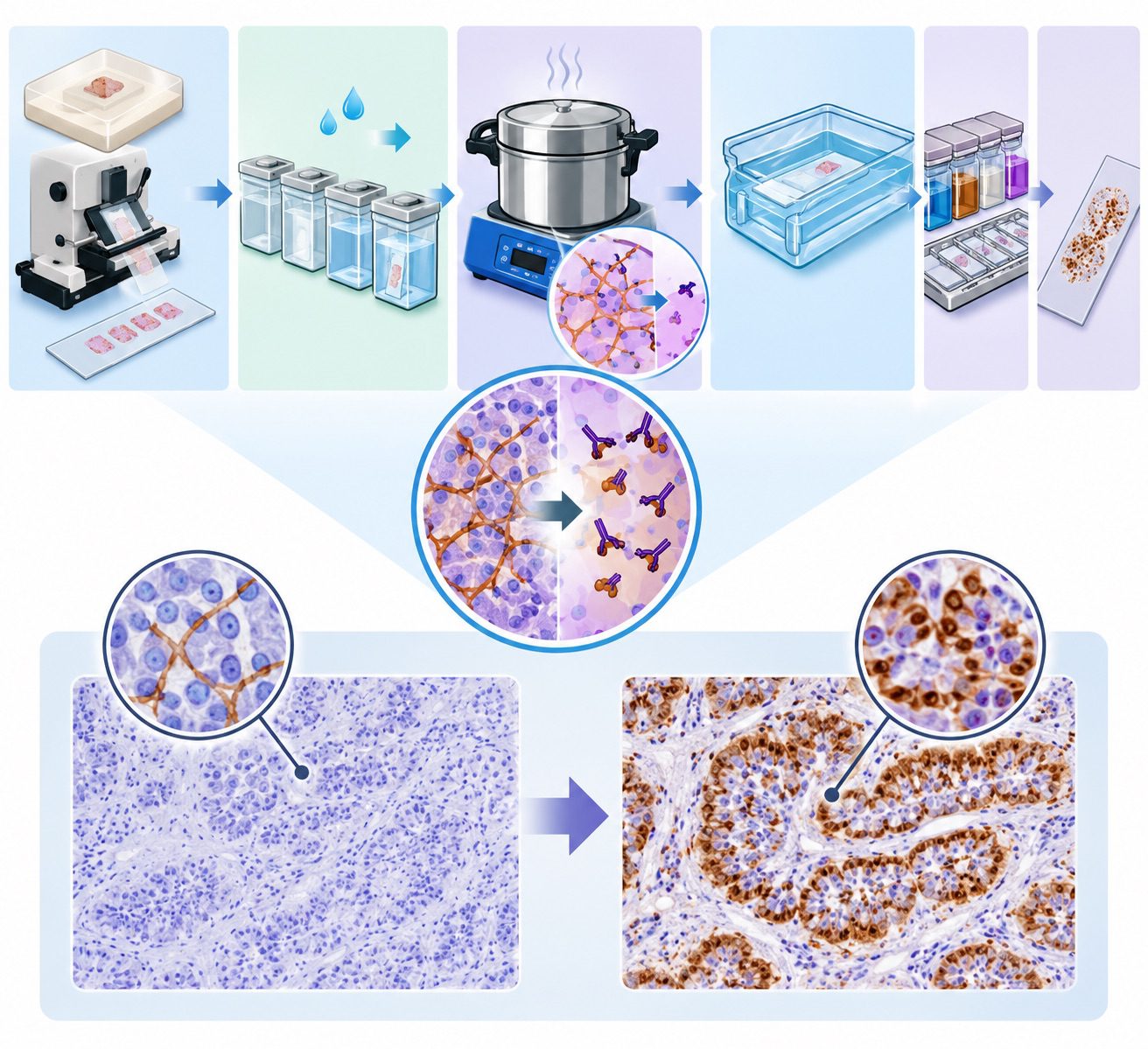

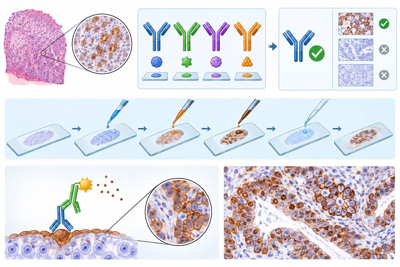
Immunohistochemistry (IHC) is a specialized laboratory technique that combines immunological recognition with histological examination to identify and localize specific antigens within tissue sections. It relies on the selective interaction between antibodies and their corresponding antigens, enabling the visualization of proteins and other molecular targets while preserving the structural organization of tissues. By integrating molecular detection with microscopic assessment, IHC provides valuable information about cellular composition, tissue architecture, and the distribution of biologically significant molecules.
Its effectiveness, however, depends substantially on the appropriate selection of antibodies and the careful execution of staining procedures. Antibodies are central to immunohistochemical analysis because they recognize particular molecular features, known as epitopes, on target antigens. The specificity of this recognition determines the reliability with which a particular protein or cellular marker can be identified within a tissue specimen. Antibody selection therefore represents a critical preliminary step in the development of an effective IHC protocol.
Factors such as antibody specificity, affinity, sensitivity, target-antigen accessibility, and compatibility with the selected detection system must be considered to obtain meaningful staining results. An unsuitable antibody may produce nonspecific reactions, weak signals, or misleading staining patterns, potentially compromising the interpretation of tissue findings. The staining process involves several coordinated procedures designed to facilitate antigen detection and produce a visible signal.
Tissue specimens are prepared through fixation, processing, and sectioning before antibodies are introduced to interact with the target antigens. Depending on the nature of the tissue and the antigen under investigation, additional procedures, including antigen retrieval and blocking of nonspecific binding, may be required. Following primary antibody binding, the antigen-antibody interaction is visualized using an appropriate detection system, such as an enzyme-linked chromogenic method or a fluorescent label. The resulting signal enables the identification of target molecules according to their staining intensity, distribution, and subcellular localization.
The diagnostic and investigative value of IHC extends across numerous areas of biomedical science. In diagnostic pathology, it assists in distinguishing morphologically similar lesions, classifying neoplasms, identifying the likely origin of metastatic tumors, and detecting disease-associated biomarkers. In research, the technique supports investigations into protein expression, cellular differentiation, tissue responses, and molecular changes associated with pathological processes. It is also useful in identifying immune-cell populations, characterizing tissue-specific markers, and evaluating the expression of therapeutic targets.
Despite its broad utility, immunohistochemical analysis is not entirely dependent on the presence of a target antigen alone. Tissue preservation, fixation conditions, antigen accessibility, antibody performance, staining optimization, and appropriate controls collectively influence the quality and interpretability of the final result. Antibody selection and staining should be regarded as interconnected components of a systematic analytical process rather than isolated laboratory tasks.
Types and characteristics of antibodies used in IHC
Antibodies are essential immunological reagents in immunohistochemistry (IHC), enabling the visualization of specific antigens within tissue sections while preserving their spatial relationships to surrounding cells and structural components. Their performance depends on molecular recognition, binding characteristics, and compatibility with the selected detection system.
Monoclonal and polyclonal antibodies
Monoclonal antibodies are antibodies that originate from a single B-cell clone and recognize a particular epitope, which is a specific molecular region of an antigen. Their restricted recognition profile provides a high degree of consistency between experiments and makes them useful for identifying defined cellular proteins, distinguishing closely related molecular targets, and evaluating disease-associated biomarkers. However, their narrow epitope recognition can make them vulnerable to alterations caused by tissue fixation, antigen retrieval, or protein modification. When the targeted epitope becomes inaccessible or structurally altered, staining intensity may decrease substantially.
Polyclonal antibodies comprise a mixture of immunoglobulins produced by different B-cell clones in response to the same antigen. They recognize multiple epitopes on that antigen, potentially generating stronger signals when the target is present at low concentrations. This broader recognition can be advantageous when tissue processing compromises some antigenic sites. Nevertheless, polyclonal preparations may exhibit greater batch-to-batch variability and an increased likelihood of cross-reactivity with unrelated tissue components. Antibody selection should reflect the biological characteristics of the target, the condition of the specimen, and the degree of specificity required.
Primary and secondary antibodies
Primary antibodies bind directly to the antigen of interest within the tissue section. They determine the molecular target being investigated and therefore exert considerable influence on the biological relevance of the final staining pattern. Their selection requires consideration of antigen identity, tissue distribution, host species, immunoglobulin class, and compatibility with the intended detection method. Secondary antibodies recognize immunoglobulins from the species in which the primary antibody was produced.
In indirect IHC, these reagents are frequently conjugated to enzymes or linked to detection systems that generate a visible signal. Multiple secondary antibodies may associate with a single primary antibody, amplifying signal intensity and improving the detection of relatively scarce antigens. Appropriate species matching and cross-adsorption are important for reducing unintended binding, particularly when examining tissues containing immunoglobulins or when multiple antibodies are used simultaneously.
Direct and indirect antibody detection methods
Direct detection involves a labelled primary antibody that binds to the target antigen and produces a detectable signal without requiring a secondary antibody. This approach offers procedural simplicity, reduces the number of incubation steps, and may limit background arising from secondary-reagent interactions. However, it generally provides less signal amplification and may require individual labelling of primary antibodies.
Indirect detection employs an unlabelled primary antibody followed by a labelled secondary antibody or another amplification system. Its principal advantage is enhanced signal generation, alongside flexibility because one labelled secondary reagent can be used with several compatible primary antibodies. The additional binding steps, however, may increase nonspecific staining if incubation conditions, reagent concentrations, and washing procedures are inadequately controlled.
Factors influencing antibody performance in IHC
Antibody specificity describes its ability to recognize the intended antigen without substantial binding to unrelated molecules. Sensitivity refers to its capacity to detect the target at low abundance, whereas affinity represents the strength of interaction between an individual antibody-binding site and its epitope. These properties are related but not interchangeable. Antibody performance is influenced by epitope accessibility, antigen abundance, tissue fixation, retrieval conditions, reagent concentration, incubation duration, temperature, and washing stringency.
Excessive antibody concentration can increase background staining, while insufficient concentration may produce weak signals. Careful titration, appropriate controls, and application-specific validation are therefore necessary to establish reliable staining conditions. Selecting an antibody with suitable recognition characteristics and detection compatibility is central to preserving the analytical accuracy and diagnostic value of immunohistochemical investigations.
Criteria for antibody selection in immunohistochemistry
Antibody selection is a fundamental determinant of the accuracy and reliability of IHC analysis. The quality of an immunohistochemical result depends not only on the staining procedure but also on the ability of the selected antibody to recognize its intended antigen within the tissue microenvironment. An appropriate antibody must demonstrate adequate specificity, sensitivity, binding performance, and compatibility with the analytical conditions under which it will be used. Careful consideration of these characteristics helps minimize misleading staining patterns and strengthens the validity of diagnostic and experimental findings.
Antigen specificity is one of the principal considerations during antibody selection. It describes the capacity of an antibody to recognize its intended molecular target without substantial interaction with unrelated tissue components. Insufficient specificity may produce cross-reactivity, resulting in staining that does not accurately represent the distribution of the target antigen. Antibodies should therefore be selected using evidence from validated applications, manufacturer documentation, peer-reviewed studies, and independent performance assessments where available. Particular attention should be given to the specificity of antibodies against structurally related proteins, especially when several antigens share similar epitopes. Where feasible, specificity can be investigated through genetic controls, such as target-deficient tissues, or through complementary analytical methods.
Sensitivity and binding affinity are equally important in determining antibody suitability. Sensitivity refers to the ability of an immunohistochemical assay to detect the target antigen when it is present, including at relatively low abundance. Affinity describes the strength of the interaction between an antibody-binding site and its corresponding epitope. Although high-affinity antibodies may improve antigen detection, affinity alone does not guarantee superior staining performance. Antibody concentration, incubation conditions, antigen accessibility, and the detection system can substantially influence the final signal. The selected antibody should provide sufficient signal intensity while maintaining a clear distinction between genuine antigen localization and nonspecific background staining.
Compatibility with tissue preparation and staining methodology must also guide antibody selection. Fixation, tissue processing, embedding, and antigen retrieval can alter epitope accessibility and influence antibody binding. Consequently, an antibody validated for frozen tissue may not perform equivalently in formalin-fixed, paraffin-embedded specimens. Selection should account for the intended specimen type, fixation conditions, retrieval method, and detection platform. Compatibility with enzyme-based or fluorescence-based detection systems is also essential to ensure that the antibody produces a measurable and interpretable signal.
Antibody validation using appropriate controls provides additional assurance of analytical reliability. Positive controls demonstrate whether the expected antigen can be detected under the selected conditions, whereas negative controls help identify nonspecific reactions and background signals. Appropriate controls should be incorporated during assay development and routine quality assessment. Validation should establish the expected staining location, intensity, and distribution rather than relying solely on the presence of a coloured reaction product.
The intended diagnostic or research application should determine the overall selection strategy. Antibodies used for diagnostic classification may require stringent specificity and reproducible staining patterns, whereas research applications may prioritize detection of subtle changes in antigen expression or distribution. In the end, antibody selection should be based on documented performance under defined experimental conditions, ensuring that the resulting immunohistochemical findings are technically sound, biologically meaningful, and suitable for their intended interpretation.
Tissue preparation and antigen retrieval in immunohistochemistry
Tissue preparation and antigen retrieval are fundamental stages in IHC, as they determine how effectively antibodies can recognize and bind to specific antigens within tissue sections. The quality of the final staining reaction depends not only on antibody selection but also on the preservation of tissue architecture, the chemical integrity of target molecules, and the accessibility of antigenic sites. Inadequate preparation may produce weak staining, nonspecific background reactions, or misleading results. Tissue processing and antigen retrieval must be carefully adapted to the biological characteristics of the specimen and the intended immunohistochemical analysis.
Tissue fixation and processing methods
Fixation stabilizes tissue components and prevents autolysis and microbial degradation after specimen collection. Neutral buffered formalin is widely used because it preserves cellular morphology and maintains tissue architecture sufficiently well for routine histopathological examination. However, prolonged fixation can alter antigenic structures and reduce antibody accessibility. Other fixatives, including alcohol-based solutions, may be appropriate for particular antigens but can produce different effects on tissue morphology and protein preservation. Following fixation, tissue samples are dehydrated through graded alcohol concentrations, cleared using a suitable solvent, and infiltrated with paraffin wax. Each stage requires appropriate control because excessive processing, inadequate dehydration, or unsuitable fixation conditions may compromise subsequent staining performance.
Preparation of paraffin-embedded and frozen tissue sections
Formalin-fixed, paraffin-embedded (FFPE) tissue is commonly used in diagnostic laboratories because it provides stable specimens that can be stored and sectioned consistently. After embedding, tissues are cut into thin sections using a microtome, mounted onto glass slides, and prepared for immunostaining (Figure 1). Paraffin must be removed before aqueous staining reagents can penetrate the tissue effectively. In contrast, frozen tissue sections are prepared from specimens rapidly frozen and cut using a cryostat. This approach may preserve certain antigens, lipids, and molecular features that are sensitive to routine processing. Nevertheless, frozen sections can exhibit ice-crystal artefacts, uneven morphology, or reduced structural stability. The choice between FFPE and frozen tissue depends on antigen sensitivity, diagnostic requirements, specimen availability, and the staining protocol.

Effects of formalin fixation on antigen preservation
Formalin fixation primarily preserves tissue through the formation of chemical cross-links between proteins and other cellular constituents. Although these interactions maintain structural integrity, they may conceal antibody-binding sites or modify the local chemical environment of an epitope. This phenomenon, commonly described as epitope masking, can diminish immunoreactivity even when the target protein remains present in the specimen. Fixation duration, formalin concentration, tissue thickness, and the interval between specimen collection and fixation may influence antigen preservation. Excessive fixation can be particularly problematic for certain nuclear and membrane-associated markers. Standardized fixation procedures are therefore essential for reducing variability and improving the comparability of immunohistochemical results across specimens.
Antigen retrieval techniques
Antigen retrieval is performed to restore antibody access to epitopes that have become inaccessible during fixation and tissue processing. Heat-induced epitope retrieval (HIER) involves heating tissue sections in an appropriate retrieval buffer, commonly citrate or Tris-based solutions, under controlled conditions. Heat can disrupt or modify fixation-associated interactions, thereby improving antigen recognition. Enzymatic-induced epitope retrieval uses proteolytic enzymes, such as proteinase K or trypsin, to partially digest tissue proteins and expose concealed antigenic sites. The effectiveness of either method depends on the target antigen, fixation history, buffer composition, temperature, incubation duration, and enzyme concentration. Excessive heating or digestion may damage tissue architecture, detach sections from slides, or destroy antigenic determinants. Retrieval conditions should be optimized for each antibody-tissue combination.
Prevention of tissue damage and preservation of morphology
Maintaining tissue integrity throughout preparation and retrieval is necessary for accurate interpretation. Tissue sections should be adequately secured to slides, handled gently, and exposed to controlled temperatures and reagent conditions. Overheating, excessive enzymatic digestion, prolonged incubation, and inappropriate buffer selection should be avoided. Positive and negative controls help establish whether observed staining reflects genuine antigen recognition rather than technical artefacts. Careful optimization ensures that antigen accessibility is improved without compromising cellular boundaries, nuclear details, or the spatial distribution of tissue components. Effective tissue preparation and antigen retrieval establish the foundation for specific, reproducible, and diagnostically meaningful immunohistochemical staining.
Materials and equipment used for immunohistochemistry (IHC)
The following materials and equipment are commonly used in IHC to prepare tissue sections, preserve tissue morphology, detect specific antigens, and visualize antibody-antigen interactions under a microscope. Each material and piece of equipment plays an important role in ensuring accurate, reliable, and reproducible IHC staining results.
Materials and reagents
Frozen or paraffin-embedded tissue: Tissue samples serve as the biological specimens for IHC analysis, with frozen sections preserving certain antigens and paraffin embedding providing long-term tissue preservation and structural integrity.
Cryo-embedding medium (e.g., OCT): OCT (Optimal Cutting Temperature) compound supports and stabilizes tissue during freezing and facilitates the preparation of thin frozen tissue sections.
Sucrose: Sucrose is used as a cryoprotective agent to prevent ice crystal formation and minimize tissue damage during freezing.
4% Paraformaldehyde (PFA): PFA is a tissue fixative that preserves cellular architecture and immobilizes proteins by forming cross-links between cellular molecules.
Xylene: Xylene is an organic solvent used to remove paraffin wax from tissue sections before rehydration and subsequent staining.
Ethanol (different concentrations): Graded ethanol solutions are used to rehydrate deparaffinized tissue sections through a series of decreasing alcohol concentrations before aqueous staining procedures.
Primary and secondary antibodies: Primary antibodies specifically recognize and bind to the target antigen, while secondary antibodies bind to the primary antibodies and carry or facilitate the detection signal.
Hematoxylin: Hematoxylin is a nuclear counterstain that stains cell nuclei blue to purple, providing morphological contrast against the brown IHC staining.
Serum: Normal serum, typically obtained from the species in which the secondary antibody was raised, is used as a blocking reagent to reduce nonspecific antibody binding and background staining.
Citrate buffer (pH 6): Citrate buffer is commonly used for heat-induced epitope retrieval (HIER) to unmask antigenic sites that may have been concealed during tissue fixation.
Tris-EDTA buffer (pH 9): Tris-EDTA buffer is an alkaline antigen retrieval solution used to expose certain masked epitopes and improve antibody accessibility in tissue sections.
3,3′-Diaminobenzidine tetrahydrochloride (DAB): DAB is a chromogenic substrate that reacts with horseradish peroxidase (HRP) to produce an insoluble brown precipitate at sites of antibody–antigen localization.
Mounting medium: Mounting medium is used to preserve stained tissue sections and secure coverslips for microscopic examination.
Equipment and laboratory apparatus
Microtome: A microtome is an instrument used to cut thin, uniform sections of paraffin-embedded tissue for mounting onto glass slides.
Cryostat: A cryostat is used to cut thin sections of frozen tissue embedded in OCT medium while maintaining low temperatures to preserve tissue structure.
Glass slides: Glass slides provide a flat supporting surface for mounting and processing tissue sections during IHC staining.
Coverslips: Coverslips are placed over stained tissue sections to protect the specimen, prevent physical damage, and improve optical clarity during microscopic examination.
Refrigerator: A refrigerator is used to store antibodies, buffers, tissue samples, and other temperature-sensitive reagents under appropriate conditions to maintain their stability.
Incubator: An incubator provides controlled temperature conditions to facilitate antibody-antigen binding and other incubation steps during IHC staining.
Boiling source (e.g., water bath or microwave): A boiling source is used to heat tissue sections in antigen retrieval buffers, helping to reverse formalin-induced protein cross-links and expose masked antigenic sites.
Forceps: Forceps are used to handle tissue specimens, glass slides, and coverslips during tissue preparation and staining procedures.
Staining jars or Coplin jars: These containers are used to hold glass slides during fixation, washing, dehydration, rehydration, and staining procedures.
Pipettes and micropipette tips: These instruments are used to accurately measure and dispense antibodies, buffers, and other reagents onto tissue sections.
Humidified chamber: A humidified chamber prevents tissue sections from drying out during antibody incubation and helps maintain optimal reaction conditions.
Fume hood: A fume hood provides ventilation and protection when handling volatile or hazardous chemicals, such as xylene and paraformaldehyde.
Microscope: A light microscope is used to examine stained tissue sections, identify the distribution and localization of target antigens, and evaluate staining intensity and tissue morphology.
The choice of antigen retrieval buffer (citrate buffer at pH 6 or Tris-EDTA buffer at pH 9), antibody concentration, and incubation conditions should be optimized for the specific antibody and target antigen. Frozen and paraffin-embedded tissues also require different preparation workflows. DAB, paraformaldehyde, and xylene should be handled using appropriate laboratory safety procedures and personal protective equipment.
Main steps of IHC using a Chromogenic-labeled secondary antibody on paraffin-embedded tissue sections
IHC involves several steps to detect specific antigens in paraffin-embedded tissue sections using chromogenic detection. First, tissue sections are deparaffinized with xylene and rehydrated through graded ethanol concentrations. Antigen retrieval is then performed using an appropriate heated buffer, followed by blocking to minimize nonspecific binding. The primary antibody is applied to bind the target antigen, followed by a chromogen-labeled secondary antibody for detection. DAB is added to produce a brown reaction product, and hematoxylin is used for nuclear counterstaining. Finally, slides are mounted and examined microscopically (Figure 2).
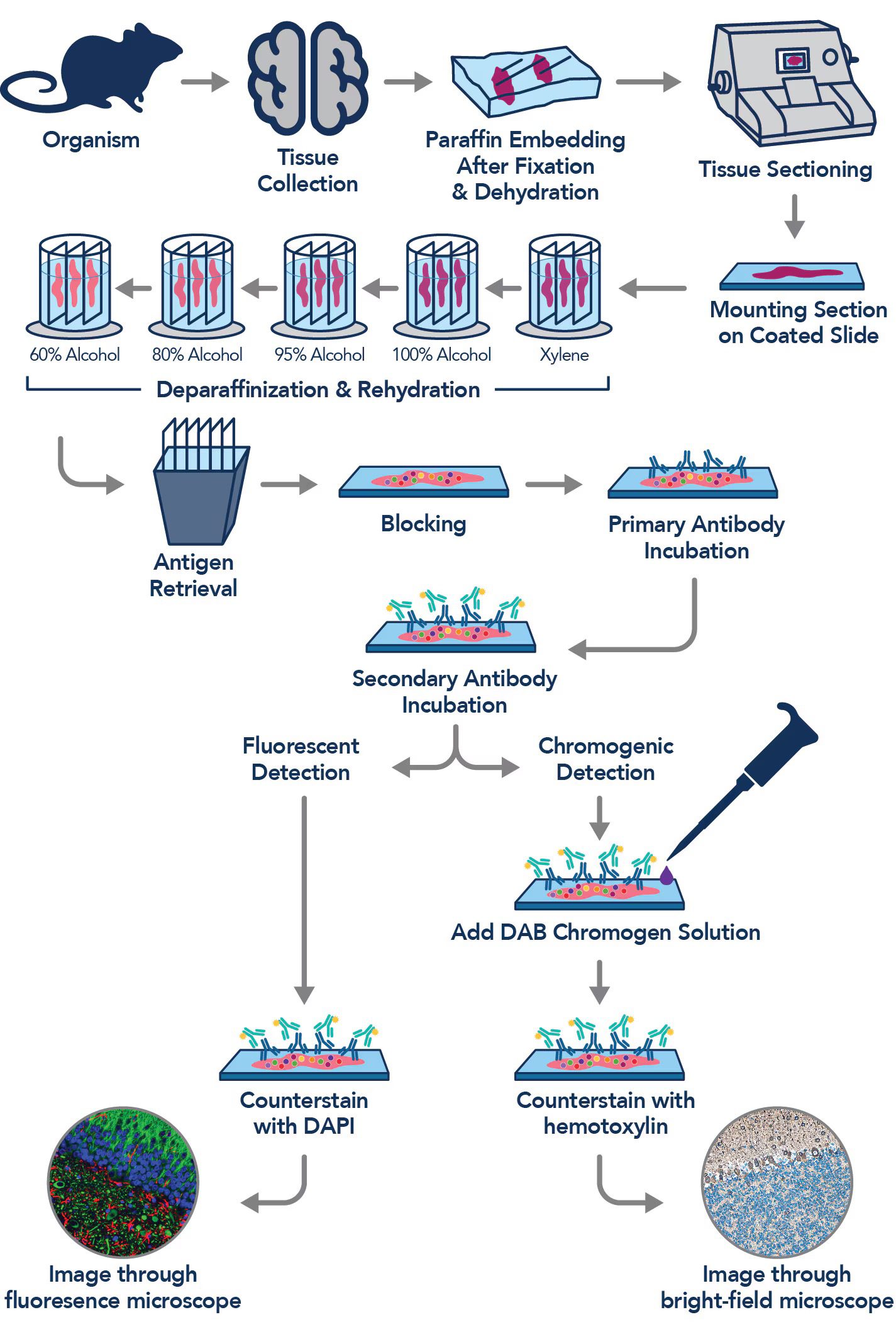
As illustrated in Figure 2, the process begins with the collection of tissue from an organism, followed by fixation to preserve tissue architecture and prevent degradation. The tissue is dehydrated through graded alcohol concentrations, cleared with xylene, and embedded in paraffin wax to provide structural support. Thin tissue sections are then prepared using a microtome and mounted onto coated glass slides.
Next, tissue sections undergo deparaffinization with xylene and rehydration through descending concentrations of ethanol, followed by antigen retrieval using an appropriate heated buffer to expose masked antigenic sites. Blocking is subsequently performed to minimize nonspecific antibody binding and reduce background staining. The sections are then incubated with a primary antibody that specifically recognizes and binds to the target antigen. Following incubation and washing, a chromogen-labeled secondary antibody is applied to bind the primary antibody and facilitate antigen detection.
For chromogenic detection, 3,3′-diaminobenzidine (DAB) is added as a substrate, producing an insoluble brown precipitate at sites of target antigen expression. The tissue sections are subsequently counterstained with hematoxylin, which stains cell nuclei blue and provides morphological contrast. Finally, the stained slides are examined under a bright-field microscope to evaluate antigen presence, distribution, localization, and staining intensity, while assessing the overall tissue and cellular morphology.
Antibody staining procedures in immunohistochemistry
Antibody staining in IHC is a carefully coordinated laboratory procedure designed to visualize specific antigens within tissue sections while preserving their structural integrity. The accuracy of the final staining result depends on the quality of tissue preparation, the effectiveness of antibody binding, the suitability of the detection system, and the precision of each experimental step. A well-controlled staining procedure produces distinct, interpretable signals that support the identification and localization of target proteins in their natural tissue environment.
1. Deparaffinization and rehydration of tissue sections: For formalin-fixed, paraffin-embedded tissues, staining begins with the removal of paraffin wax to expose the tissue to aqueous reagents. Sections are immersed in xylene or a suitable substitute, followed by graded concentrations of ethanol to remove residual solvent and progressively reintroduce water. Final rinsing in distilled water or an appropriate buffer prepares the tissue for subsequent reactions. Inadequate deparaffinization may obstruct antibody penetration, resulting in uneven staining or reduced antigen accessibility. Frozen tissue sections generally require a different preparation procedure, depending on the fixation method and tissue characteristics.
2. Blocking endogenous enzyme activity and nonspecific binding
Blocking procedures minimize unwanted signals that could compromise the specificity of immunostaining. When an enzyme-based detection system is used, endogenous peroxidase activity is commonly suppressed with hydrogen peroxide, particularly in tissues containing blood cells or other peroxidase-rich components. Endogenous alkaline phosphatase may also require inhibition when relevant to the selected detection system. Nonspecific antibody interactions are reduced using appropriate blocking solutions containing proteins or other validated blocking agents. These reagents occupy potential nonspecific binding sites, thereby improving the distinction between genuine antigen-associated staining and background coloration.
3. Application and incubation of primary antibodies
The primary antibody is selected according to its ability to recognize the target antigen with suitable specificity and sensitivity. Before application, the antibody is prepared at an experimentally validated concentration using an appropriate diluent. The reagent is distributed evenly across the tissue section and incubated under conditions optimized for the antibody, antigen, and staining platform. Incubation may occur at room temperature or under refrigerated conditions, depending on the validated protocol. Insufficient incubation or an unsuitable antibody concentration may produce weak signals, whereas excessive concentration can increase nonspecific staining. Careful optimization is therefore essential for reliable antigen detection.
4. Application of secondary antibodies or detection reagents: Following primary antibody incubation, tissue sections are washed with an appropriate buffer to remove unbound antibodies. In indirect IHC, a labelled secondary antibody recognizes the primary antibody and facilitates signal detection. Alternatively, polymer-based detection systems employ multiple enzyme molecules associated with antibody-binding components to enhance signal intensity. The detection reagent must be compatible with the primary antibody’s host species and immunoglobulin class. Appropriate incubation and washing conditions help maximize target-associated signal while limiting background staining.
5. Chromogen development, counterstaining, and mounting: For chromogenic IHC, an enzyme substrate is introduced to generate a visible, localized reaction product at antigen-binding sites. In horseradish peroxidase-based systems, 3,3′-diaminobenzidine (DAB) commonly produces a brown precipitate. Chromogen development must be monitored carefully because excessive reaction time can obscure tissue details, while insufficient development may yield weak staining. Counterstaining, commonly with hematoxylin, provides nuclear contrast and facilitates morphological assessment. Finally, sections are prepared for mounting using a method compatible with the chromogen and counterstain. Proper mounting preserves tissue architecture, supports microscopic examination, and contributes to the long-term stability of the prepared slides.
Detection systems and staining techniques in immunohistochemistry
Detection systems and staining techniques constitute essential components of IHC, enabling the visualization and localization of specific antigens within tissue sections. Their effectiveness depends on the interaction between antigen recognition, antibody binding, signal amplification, and the generation of a detectable reaction product. Selecting an appropriate detection strategy is therefore critical to obtaining reliable, interpretable, and reproducible staining results. The choice of technique is influenced by antigen abundance, tissue characteristics, background activity, and the intended diagnostic or experimental application.
Enzyme-based detection systems: Enzyme-based detection remains a widely established approach in routine IHC. Horseradish peroxidase (HRP) and alkaline phosphatase (AP) are commonly employed enzymes that catalyze reactions producing localized, visible staining. HRP is frequently paired with chromogenic substrates that generate insoluble colored deposits at sites of antigen expression. AP provides an alternative enzymatic pathway, offering distinct substrate options that may be advantageous when endogenous peroxidase activity interferes with interpretation. Appropriate blocking procedures and optimized reaction conditions are necessary to minimize nonspecific signals and preserve staining specificity.
Chromogenic substrates and signal development: Chromogens convert enzymatic activity into visible deposits that reveal the distribution of target antigens. 3,3′-Diaminobenzidine (DAB) is a commonly used HRP substrate that produces a brown precipitate at antigen-positive sites. Its compatibility with hematoxylin counterstaining facilitates the simultaneous assessment of tissue architecture and cellular morphology. Other chromogens can produce contrasting colors, supporting the differentiation of multiple markers when suitable detection systems are used. Chromogen selection, substrate concentration, and development duration influence signal intensity and background appearance, making careful optimization necessary for consistent results.
Direct and indirect staining methods: Direct immunohistochemical staining employs a labeled primary antibody that binds directly to the target antigen. This approach reduces procedural complexity and may shorten staining time, although its signal strength can be limited when antigen expression is low. Indirect staining uses an unlabeled primary antibody followed by a labeled secondary antibody directed against the primary antibody. Because multiple secondary antibody molecules may associate with a primary antibody, this method can improve signal amplification and detection sensitivity. The choice between these approaches depends on the required sensitivity, available reagents, and experimental objectives.
Polymer-based detection systems: Polymer-based systems enhance detection by coupling multiple enzyme molecules to a polymer backbone associated with antibody-binding components. This arrangement can increase signal intensity while reducing the need for multiple incubation steps. Polymer systems may also limit nonspecific background associated with endogenous biotin in methods that rely on biotin-avidin interactions. Their performance, however, remains dependent on antibody compatibility, reagent quality, and appropriate optimization.
Fluorescence-based detection and multiplex staining: Fluorescence-based IHC uses fluorophore-labeled antibodies or fluorescent detection reagents to identify antigens through emitted light following excitation. Unlike conventional chromogenic staining, fluorescence can facilitate the simultaneous examination of several molecular targets within the same tissue section. Multiplex staining combines multiple markers using distinguishable fluorophores, enabling the assessment of cellular populations, spatial relationships, and co-expression patterns. Careful fluorophore selection, spectral separation, and controls for autofluorescence and signal overlap are essential for accurate interpretation.
Optimization, controls, and troubleshooting in immunohistochemical staining
Optimization, controls, and troubleshooting are fundamental to achieving accurate, specific, and reproducible results in IHC. Effective optimization ensures that the selected antibody interacts appropriately with its target antigen while minimizing nonspecific reactions and background interference. Antibody concentration must be carefully titrated to determine the optimal dilution that produces distinct staining without excessive background. Incubation time, temperature, antigen retrieval conditions, and detection system compatibility should also be adjusted according to the antibody characteristics and tissue requirements.
Appropriate controls are essential for verifying staining reliability. Positive controls contain tissues known to express the target antigen and confirm that the staining procedure is functioning correctly. Negative controls help identify nonspecific staining and assess the possibility of unintended antibody interactions. Reagent controls, including procedures that omit the primary antibody, can reveal background signals arising from secondary antibodies, detection reagents, or endogenous tissue components. Using suitable controls allows researchers to distinguish genuine antigen localization from technical artefacts.
Background staining may result from inadequate blocking, excessive antibody concentration, insufficient washing, endogenous peroxidase activity, or nonspecific binding to tissue components. Weak or absent staining may indicate poor antigen preservation, inappropriate antigen retrieval, unsuitable antibody dilution, or inadequate incubation. Excessively intense staining may reflect antibody overconcentration, prolonged chromogen development, or excessive detection sensitivity. Troubleshooting requires systematic evaluation of these variables, changing one parameter at a time whenever possible to identify the underlying cause.
Standardization strengthens the consistency of immunohistochemical procedures across experiments, tissue samples, and laboratory personnel. Maintaining detailed records of antibody clones, lot numbers, working dilutions, incubation conditions, retrieval methods, and detection reagents facilitates protocol replication. Routine quality-control assessments, documented acceptance criteria, and periodic revalidation are equally important. Through systematic optimization, appropriate control selection, and structured troubleshooting, laboratories can improve staining specificity, reduce technical variability, and generate dependable results for diagnostic interpretation and scientific investigation.
Interpretation of results and quality assurance in immunohistochemistry
Interpretation of IHC results requires a systematic assessment of staining intensity, cellular localization, distribution, and specificity to ensure reliable diagnostic conclusions. Staining intensity is commonly classified as weak, moderate, or strong, while distribution may be focal, multifocal, or diffuse. These characteristics should be evaluated alongside the proportion of positively stained cells and the expected expression profile of the target antigen. Importantly, staining intensity alone does not establish diagnostic significance, as expression levels may vary according to tissue type, disease progression, and technical conditions.
The subcellular localization of immunoreactivity provides an additional interpretive dimension. Nuclear staining may indicate the presence of transcription factors or proliferation-associated proteins, whereas cytoplasmic staining frequently reflects intracellular enzymes, structural proteins, or other cytoplasmic constituents. Membranous staining is particularly relevant when evaluating cell-surface receptors and adhesion molecules. Recognition of these distinct patterns helps differentiate specific antigen expression from nonspecific background reactions and staining artefacts.
Analytical validity is strengthened through the appropriate application of positive and negative controls, which demonstrate whether the staining procedure performs as expected and whether unintended reactivity is present. Antibody specificity must also be established through appropriate validation procedures, including assessment of expected tissue reactivity and, where necessary, comparison with complementary analytical methods. Inadequate fixation, insufficient antigen retrieval, excessive antibody concentration, and endogenous enzyme activity may compromise the reliability of observed staining patterns.
Quality assurance encompasses standardized protocols, reagent traceability, equipment maintenance, personnel competency, and systematic documentation of staining conditions and results. Reproducibility should be assessed through consistent evaluation criteria and periodic verification of assay performance. Where quantitative or semiquantitative scoring systems are employed, clearly defined thresholds and interpretation guidelines are essential for minimizing observer variability.
Despite its diagnostic utility, immunohistochemistry has inherent limitations, including cross-reactivity, heterogeneous antigen expression, technical variability, and subjective interpretation. Immunohistochemical findings should be integrated with histomorphological features, clinical history, and relevant laboratory investigations. This comprehensive approach promotes analytical consistency, strengthens diagnostic confidence, and supports the responsible application of IHC in clinical pathology and biomedical research.
References
Kim, S. W., Roh, J., & Park, C. S. (2016). Immunohistochemistry for pathologists: Protocols, pitfalls, and tips. Journal of Pathology and Translational Medicine, 50(6), 411–418.
Daneshtalab, N., Doré, J. J., & Smeda, J. S. (2010). Troubleshooting tissue specificity and antibody selection: Procedures in immunohistochemical studies. Journal of Pharmacological and Toxicological Methods, 61(2), 127–135.
Ivell, R., Teerds, K., & Hoffman, G. E. (2014). Proper application of antibodies for immunohistochemical detection: Antibody crimes and how to prevent them. Endocrinology, 155(3), 676–687.
De Matos, L. L., Trufelli, D. C., de Matos, M. G. L., & da Silva Pinhal, M. A. (2010). Immunohistochemistry as an important tool in biomarkers detection and clinical practice. Biomarker Insights, 5, 9–20.
Grizzle, W. E., Fredenburgh, J. L., & Myers, R. B. (2008). Fixation of tissues. In J. D. Bancroft & M. Gamble (Eds.), Theory and practice of histological techniques (6th ed., pp. 53–74). Churchill Livingstone.
Discover more from Microbiology Class
Subscribe to get the latest posts sent to your email.