
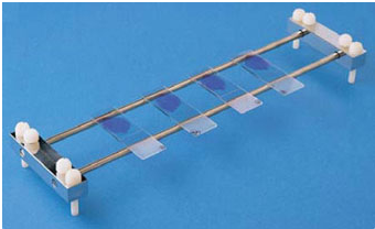
Staining is an essential technique in microbiology that involves applying dyes or stains to microorganisms to enhance contrast and improve their visibility under a microscope. It facilitates the observation, identification, and differentiation of microorganisms based on their staining reactions, morphology, and cellular structures. The procedure typically involves preparing a microbial smear on a glass slide, followed by the application of appropriate staining reagents.
Depending on the purpose of the examination, staining techniques may employ a single dye or a combination of dyes and reagents to highlight specific microbial features. Commonly used stains include crystal violet, methylene blue, carbol fuchsin, Giemsa stain, and lactophenol cotton blue. Major staining techniques include Gram staining, acid-fast staining, and negative staining, each of which serves a specific purpose in microbial examination. Staining is fundamental to the microscopic examination, identification, and classification of microorganisms in the microbiology laboratory.
Lactophenol cotton blue (LPCB)
LPCB is a commonly used fungal staining technique for examining the morphology of moulds, including their hyphae, spores, and conidia, under a light microscope. Principle: Cotton blue stains chitin in fungal cell walls, imparting a blue coloration to fungal structures. Phenol acts as a fungicidal agent, lactic acid helps preserve fungal morphology, and glycerol prevents the preparation from drying out rapidly. Procedure: A small amount of fungal growth is transferred to a drop of LPCB on a clean glass slide, covered with a coverslip, and examined microscopically.
Microscopic appearance: Fungal hyphae, conidiophores, and spores appear blue against a relatively clear background, allowing their structural features to be observed. LPCB is used to identify moulds based on their characteristic morphology, particularly Aspergillus, Penicillium, and dermatophytes. It is especially useful for examining hyphal branching, septation, and spore-bearing structures.
Potassium hydroxide (KOH) preparation
KOH preparation is a direct microscopic examination technique used to detect fungal elements in clinical specimens. It is a clearing method rather than a true staining technique. Principle: Potassium hydroxide (KOH) dissolves keratin, epithelial cells, and other organic debris, clearing the specimen while leaving fungal structures sufficiently intact for microscopic examination. Procedure: Skin scrapings, hair, or nail clippings are placed on a clean glass slide. One or two drops of 10-20% KOH solution are added, and the preparation is covered with a coverslip.
It is allowed to stand until the specimen clears; gentle warming may be used when appropriate, according to laboratory protocol. Microscopic appearance: Fungal hyphae, yeast cells, and pseudohyphae appear as refractile structures against a cleared background. KOH preparation is commonly used to detect fungal elements in skin, hair, and nail specimens and supports the diagnosis of dermatophytosis, cutaneous candidiasis, and other superficial fungal infections. However, it does not reliably identify the fungal species.
Periodic acid-Schiff (PAS) stain
PAS is a histochemical staining technique used to demonstrate fungal cell walls in tissue sections. Principle: Periodic acid oxidizes certain carbohydrate groups to form aldehydes. Schiff reagent reacts with these aldehydes, producing a magenta or pink color. Procedure: Tissue sections are treated with periodic acid, followed by Schiff reagent, and then counterstained as required.
Microscopic appearance: Fungal cell walls appear magenta or pink, while surrounding tissue structures take contrasting colors. PAS is used for the detection of fungal organisms in tissue biopsies, including Candida, Aspergillus, and dermatophytes. PAS is useful for demonstrating fungal invasion in infected tissues. Nonetheless, PAS may highlight other carbohydrate-rich tissue components, so fungal identification depends on morphology and clinical findings.
Gomori methenamine silver (GMS) stain
Gomori methenamine silver (GMS) is a special histological staining technique widely used to detect fungal organisms in tissue specimens. Principle: Periodic acid oxidation of fungal cell-wall carbohydrates generates aldehydes, which reduce silver ions to metallic silver, producing a dark stain. Procedure: Tissue sections are oxidized, treated with methenamine silver solution, and counterstained to enhance contrast between fungal structures and surrounding tissues.
Microscopic appearance: Fungal cell walls appear black to dark brown against a contrasting background, which may be pale green or light-colored depending on the counterstain. Applications: GMS is used to detect fungi in infected tissues, including Aspergillus, Candida, and Pneumocystis jirovecii. It is particularly useful when fungal organisms are sparse or difficult to visualize with routine hematoxylin and eosin (H&E) staining. Although GMS highlights fungal structures effectively, it does not independently identify the fungal species or establish whether the detected organisms are responsible for active infection.
Calcofluor white stain
Calcofluor white is a fluorescent stain used for rapid detection of fungal elements in clinical specimens. Principle: The dye binds to chitin and other polysaccharide-containing structures in fungal cell walls. Under suitable fluorescence illumination, the bound dye emits a bright fluorescent signal. Procedure: A clinical specimen is mixed with calcofluor white reagent, sometimes combined with KOH, and examined using a fluorescence microscope.
Microscopic appearance: Fungal hyphae, yeast cells, and other fungal structures fluoresce bright blue-white or greenish-white, depending on the optical setup. Calcofluor white stain is used for the rapid screening of skin scrapings, nail clippings, and other specimens for fungal elements. But it requires a fluorescence microscope and may also stain some nonfungal structures, so interpretation requires appropriate expertise.
Chicago sky blue stain
Chicago Sky Blue is a dye used in certain fungal diagnostic preparations to improve visualization of fungal structures. Principle: The dye provides contrast between fungal elements and the surrounding specimen. It may be used with other reagents, depending on the specific diagnostic method. Procedure: The specimen is treated with the appropriate validated staining or clearing reagent and examined microscopically.
Microscopic appearance: Fungal structures become more distinguishable from the background, with coloration depending on the specific formulation. Certain methods use Chicago Sky Blue in the direct examination of suspected superficial fungal infections. It is not as universally standardized as LPCB, KOH, PAS, or GMS. The exact procedure and expected appearance should therefore be taken from the laboratory’s validated protocol.
Gram staining for yeasts cell
Gram staining is a differential staining technique that can demonstrate yeasts in clinical specimens. Principle: Gram-positive organisms retain the crystal violet-iodine complex during decolorization. Many medically important yeasts, including Candida species, stain purple. Procedure: A fixed smear is treated with crystal violet, iodine, decolorizer, and safranin or another counterstain. Microscopic appearance: Yeast cells commonly appear purple, oval, or round.
Budding cells and pseudohyphae may also be visible. Rapid examination of specimens such as vaginal discharge, sputum, and other clinical materials when yeast infection is suspected. Gram staining can support the detection of yeast but is not sufficient for definitive species identification. Culture, antigen testing, or molecular methods may be needed.
India ink preparation for Cryptococcus
India ink is a negative staining technique historically used to demonstrate the prominent capsule of Cryptococcus neoformans in cerebrospinal fluid. Principle: India ink darkens the background but does not penetrate the polysaccharide capsule. The capsule therefore appears as a clear halo around the yeast cell. Procedure: A drop of specimen is mixed with India ink on a glass slide, covered with a coverslip, and examined microscopically.
Microscopic appearance: Round or oval yeast cells appear against a dark background, often surrounded by a clear halo. It is used for the rapid presumptive detection of encapsulated Cryptococcus in cerebrospinal fluid, particularly in suspected cryptococcal meningitis. Sensitivity is lower when the fungal burden is low. Cryptococcal antigen testing is generally more sensitive and is preferred where available.
Mucicarmine stain
Mucicarmine is a special histological stain used to demonstrate the polysaccharide capsule of Cryptococcus in tissue sections. Principle: The carmine-containing staining complex binds to acidic mucopolysaccharide material in the cryptococcal capsule. Procedure: Tissue sections are treated with mucicarmine reagent and counterstained to distinguish the organisms from surrounding tissue.
Microscopic appearance: The cryptococcal capsule stains rose-red to deep red, while the yeast cells and background show contrasting colors. Mucicarmine stain is used for the histopathological diagnosis of cryptococcosis, especially when examining tissue biopsies. Mucicarmine is particularly useful for capsular demonstration but is not a stand-alone method for identifying every fungal species. It is often interpreted alongside H&E, GMS, and other tests.
Fontana-Masson stain
Fontana-Masson is a silver impregnation stain used to demonstrate melanin and melanin-like pigments in certain fungi. Principle: Melanin can reduce silver ions to visible metallic silver, producing a dark deposit at pigment-containing sites. Procedure: Tissue sections or suitable preparations undergo silver impregnation according to the laboratory’s validated protocol.
Microscopic appearance: Melanin-containing fungal structures appear brown to black. Demonstration of fungal melanin, particularly in pigmented fungi such as Cryptococcus and certain dematiaceous fungi. Fontana-Masson stain may help support the identification of melanized fungal organisms in tissue. The reaction is not absolutely specific for fungal melanin, and results must be interpreted with morphology and other diagnostic findings.
Fluorescent antibody staining for fungal antigens
Fluorescent antibody staining uses antibodies labelled with fluorescent dyes to detect specific fungal antigens in specimens. Principle: Labelled antibodies bind to target antigens on or within fungal cells. When illuminated at the appropriate wavelength, the fluorescent label emits light that reveals the target.
The different types of fluorescent antibody staining techniques used in fungal investigations include direct immunofluorescence -in which a fluorescently labelled primary antibody binds directly to the antigen, and indirect immunofluorescence – in which an unlabelled primary antibody binds the antigen, followed by a fluorescently labelled secondary antibody. Microscopic appearance: Target organisms or antigens display characteristic fluorescence.
They are used for the detection of selected fungal organisms or antigens, depending on the validated antibody test. Applications vary by fungal pathogen and specimen type. However, the specificity of fluorescent antibody staining in fungal diagnosis is usually dependent on the antibody and assay. Not every fungal infection has a routinely available fluorescent antibody test, and results may require confirmation by other methods.
Direct immunofluorescence (DIF)
Direct immunofluorescence (DIF) is a diagnostic staining technique used to detect specific antigens in a specimen or demonstrate tissue-bound immunoglobulins and complement components using fluorescently labelled antibodies. The resulting fluorescence is observed under a fluorescence microscope. Principle: DIF is based on the specific binding of an antibody to its corresponding antigen or target.
A fluorescent dye, such as fluorescein isothiocyanate (FITC), emits visible light when exposed to light of an appropriate excitation wavelength, allowing the target to be visualized. Procedure: A specimen is placed on a microscope slide and treated with a fluorescently labelled antibody. After incubation, the slide is washed to remove unbound antibodies and examined under a fluorescence microscope.
Applications: DIF is used to detect selected microbial antigens and diagnose certain infections. It is also widely used to demonstrate immune deposits in tissue biopsies, particularly in dermatology and renal pathology. DIF provides rapid and specific results and requires fewer steps than indirect immunofluorescence. However, it generally offers less signal amplification and requires a separately labelled antibody specific to each target.
Indirect immunofluorescence (IIF)
Indirect immunofluorescence is a staining technique used to detect specific antibodies in a patient’s serum or antigens in a specimen using a fluorescently labelled secondary antibody. Principle: The primary antibody binds to the target antigen, while a fluorescently labelled secondary antibody binds to the primary antibody. This produces fluorescence that can be observed under a fluorescence microscope.
Procedure: A specimen containing the target antigen is placed on a slide and incubated with the patient’s serum or an appropriate primary antibody. After washing, a fluorescently labelled secondary antibody is added. The slide is washed again and examined under a fluorescence microscope.
Applications: It is used in serological diagnosis, detection of autoantibodies, and identification of certain infectious diseases. IIF provides signal amplification, is sensitive, and allows one labelled secondary antibody to be used with multiple compatible primary antibodies. However, it requires additional incubation and washing steps and may produce nonspecific background fluorescence.
Immunohistochemical staining (IHC)
Immunohistochemical (IHC) staining is a laboratory technique used to detect and localize specific antigens in tissue sections using antigen-specific antibodies and a detectable label. It enables the visualization of antigen distribution within cells and tissues under a light microscope. Principle: Specific antibodies bind to target antigens in tissue. This binding is detected using a labelled detection system, commonly an enzyme that reacts with a chromogenic substrate to produce a coloured precipitate at the antigen site.
Procedure: Tissue is fixed, embedded, sectioned, and mounted on a glass slide. Antigen retrieval may be performed, followed by blocking and incubation with primary antibodies. A suitable detection system and chromogenic substrate are then applied. The section is counterstained, commonly with hematoxylin, and examined microscopically.
Applications: IHC is used to detect microbial antigens in tissues, identify tumour markers, classify cancers, and investigate the distribution of specific proteins in cells and tissues. IHC preserves tissue architecture while revealing the precise localization of target antigens. But results depend on antibody specificity, fixation, antigen preservation, and appropriate controls. Cross-reactivity, nonspecific binding, and background staining may affect interpretation.
Immunoperoxidase staining
Immunoperoxidase staining is an antibody-based technique that uses the enzyme horseradish peroxidase (HRP) to detect specific antigens in cells or tissues. The enzyme produces a coloured reaction product that can be examined using a light microscope. Principle: An antibody binds specifically to the target antigen. HRP is linked to the antibody or to a secondary detection system.
When a suitable substrate, commonly 3,3′-diaminobenzidine (DAB), is added with hydrogen peroxide, an insoluble coloured product forms at the antigen’s location. Procedure: The specimen is fixed and prepared on a slide. Blocking and antigen retrieval may be performed as needed. The primary antibody and HRP-based detection system are applied, followed by the chromogenic substrate. The slide may then be counterstained and examined under a light microscope.
Applications: Immunoperoxidase staining is used to detect microbial antigens, viral proteins, and specific cellular markers in tissue sections. Immunoperoxidase provides permanent, easily documented staining and does not require a fluorescence microscope. But endogenous peroxidase activity, nonspecific antibody binding, and inadequate controls may produce false-positive results.
Negative staining for transmission electron microscopy (TEM)
Negative staining is a preparation technique used in transmission electron microscopy to visualize the size, shape, and surface features of small biological particles, including viruses and certain microorganisms. Principle: An electron-dense stain surrounds the specimen rather than primarily staining its internal structures. The stain scatters electrons strongly, producing a dark background, while the specimen appears relatively light.
Procedure: A small amount of specimen is placed on a carbon-coated or suitable TEM grid. Excess liquid is removed, and a drop of negative stain is applied. The grid is allowed to dry before examination in a transmission electron microscope. Applications: Negative staining is used to examine viral particles, bacteriophages, bacterial appendages, and macromolecular structures. It is particularly useful for assessing viral morphology in suitable specimens.
Negative stating for TEM is rapid, requires relatively simple preparation, and can reveal surface morphology with good contrast. But it provides limited information about internal structure, and drying can distort delicate specimens. The technique does not establish the identity or infectivity of a virus on its own.
Uranyl acetate negative staining
Uranyl acetate negative staining is a technique used in transmission electron microscopy to visualize small biological particles by creating contrast between the specimen and its surrounding background. Principle: Uranyl acetate is an electron-dense heavy-metal stain that scatters electrons strongly. It accumulates around the specimen, making the background appear dark while the specimen appears comparatively light.
Procedure: A specimen is applied to a suitable TEM grid. Excess liquid is removed, and uranyl acetate solution is added for a short period. Excess stain is removed, and the grid is dried before examination using a transmission electron microscope. Exact conditions depend on the specimen and laboratory protocol. Applications: This method is used to study viral particles, bacteriophages, bacterial surface structures, and macromolecular assemblies.
It produces strong electron contrast and can reveal particle shape, size, and surface details. Uranyl acetate is toxic and contains radioactive uranium, so it requires appropriate handling, storage, and hazardous-waste disposal according to laboratory regulations. Staining and drying may also introduce structural artefacts.
Phosphotungstic acid negative staining
Phosphotungstic acid (PTA) negative staining is a transmission electron microscopy technique used to visualize biological particles against a contrasting background. Principle: Phosphotungstic acid contains heavy tungsten atoms that scatter electrons. The stain surrounds the specimen and creates a dark background, while the specimen appears relatively light because it excludes much of the stain.
Procedure: A small quantity of specimen is placed on a suitable TEM grid. Excess liquid is removed, and phosphotungstic acid solution is applied. The excess stain is drained, and the grid is dried before examination under a transmission electron microscope. The concentration and pH are adjusted according to the specimen and protocol. Applications: PTA negative staining is used to examine viruses, bacteriophages, bacterial appendages, and protein complexes.
It is useful for studying particle morphology and surface features. It provides good electron contrast and is an alternative to uranyl acetate for many specimens. The stain’s pH may affect sensitive biological structures, and drying may cause distortion. The resulting image may not accurately represent the specimen’s native hydrated state.
Trypan blue exclusion
Trypan blue exclusion is a dye-based method used to distinguish viable cells from non-viable cells based on plasma membrane integrity. Viable cells with intact membranes exclude the dye and remain unstained, whereas non-viable cells with compromised membranes take up the dye and appear blue under a microscope. This method is widely used to assess cell viability and estimate viable cell numbers in cultured cell populations. It is simple, rapid, and inexpensive. However, it does not directly measure metabolic activity and may fail to detect early stages of cell death before significant membrane damage occurs.
Neutral red staining
Neutral red staining is a vital dye-based assay used to assess cell viability based on the ability of viable cells to take up and retain the dye within their lysosomes. The dye accumulates in acidic lysosomal compartments, producing a red coloration. Cells with impaired viability or lysosomal function generally exhibit reduced dye uptake or retention. This method is commonly used in cytotoxicity testing, cell culture studies, and toxicological research. Although it provides a useful indirect measure of cell viability, the results may be influenced by changes in lysosomal activity and cellular physiology.
Janus green B staining
Janus green B staining is a supravital staining technique traditionally used to visualize mitochondria in living cells. The dye functions as a redox indicator and produces a characteristic blue-green coloration under appropriate staining conditions. Its staining pattern is influenced by mitochondrial distribution and cellular redox activity. The technique has historically been used to investigate mitochondrial morphology and activity in living cells. However, staining intensity and distribution depend on cell type, dye concentration, incubation conditions, and mitochondrial physiological state; therefore, staining alone does not provide a definitive quantitative measure of cell viability.
Methylene blue viability staining
Methylene blue is a redox-sensitive dye used in certain methods to assess cellular or microbial viability. Metabolically active cells may reduce the blue dye to its colorless form, whereas cells with lower reducing activity may retain the blue coloration. The method has been applied to microbial viability assessments, yeast studies, and selected cell-counting procedures. Its interpretation depends on the organism, dye concentration, incubation conditions, and metabolic state of the cells. Although inexpensive and relatively simple, methylene blue staining primarily reflects cellular reducing activity under the conditions used and does not definitively distinguish all living cells from dead cells.
Acridine orange staining
Acridine orange is a fluorescent nucleic acid stain that binds to DNA and RNA. Under appropriate fluorescence microscopy conditions, DNA generally produces green fluorescence, whereas RNA and certain acidic intracellular compartments may exhibit orange-red fluorescence. The technique is used to examine cellular morphology, nucleic acid distribution, and microbial populations. In viability studies, acridine orange is often combined with other fluorescent dyes to differentiate cells according to membrane integrity or other physiological characteristics. However, because acridine orange can enter both viable and non-viable cells, it cannot reliably distinguish living from dead cells when used alone.
Fluorescein diacetate staining
Fluorescein diacetate (FDA) staining is a viability assessment method based on intracellular esterase activity and, in part, membrane integrity. The non-fluorescent FDA molecule enters cells and is hydrolyzed by intracellular esterases to produce fluorescein, which emits green fluorescence under suitable illumination. Viable cells with sufficient esterase activity and the ability to retain fluorescein generally exhibit green fluorescence. This technique is widely used in microbial ecology, plant cell research, and cell culture studies. However, fluorescence intensity depends on esterase activity, membrane permeability, dye loading, and experimental conditions; therefore, it should not be interpreted as an absolute measure of viability.
Propidium iodide taining
Propidium iodide (PI) is a fluorescent nucleic acid stain used to identify cells with compromised plasma membranes. It is generally excluded by cells with intact membranes but enters membrane-damaged cells, binds to DNA and RNA, and produces red fluorescence. PI staining is widely used in flow cytometry, fluorescence microscopy, and cell death studies. It is frequently combined with other fluorescent dyes, such as fluorescein diacetate or SYTO 9, to distinguish membrane-intact from membrane-compromised cells. However, PI primarily indicates loss of membrane integrity and cannot independently identify every stage or mechanism of cell death.
LIVE/DEAD fluorescent staining systems
LIVE/DEAD fluorescent staining systems use combinations of fluorescent dyes to distinguish cells according to membrane integrity and, in some systems, metabolic activity. For example, the LIVE/DEAD BacLight bacterial viability kit commonly uses SYTO 9, which stains many bacterial cells green, and propidium iodide, which preferentially enters cells with damaged membranes and produces red fluorescence.
These systems are used to assess bacterial populations in laboratory cultures, biofilms, and environmental samples. They provide a convenient means of visualizing differences in cellular membrane integrity. Nevertheless, fluorescence patterns should be interpreted cautiously because membrane integrity does not always correspond directly to reproductive viability or the ability of a cell to grow and divide.
Resazurin-based viability assays
Resazurin-based viability assays evaluate cellular metabolic activity using resazurin. Resazurin is a blue, weakly fluorescent dye that can be reduced by metabolically active cells to resorufin, a pink compound with strong fluorescence. The extent of dye conversion generally reflects cellular reducing activity and may correlate with the number of viable cells under validated experimental conditions.
These assays are widely used to assess microbial growth, antimicrobial susceptibility, and cell culture viability. They are relatively simple, sensitive, and suitable for high-throughput screening. However, results can be affected by metabolic differences, cell density, incubation time, and chemical interference. Appropriate controls and assay-specific validation are therefore essential for reliable interpretation.
Neisser-Albert staining for metachromatic granules
Neisser-Albert staining is a differential staining technique used to demonstrate metachromatic granules, which are intracellular phosphate-storage inclusions found in certain bacteria, particularly Corynebacterium diphtheriae. The granules typically appear dark blue or bluish-black, while the bacterial cytoplasm stains yellowish or greenish-blue, depending on the staining method. The technique employs specific dyes that produce contrasting colors in the granules and surrounding cytoplasm. It is useful for the presumptive identification of C. diphtheriae, although confirmation requires appropriate culture, biochemical, or molecular tests.
Pappenheim staining
Pappenheim staining, commonly associated with the May-Grünwald-Giemsa (MGG) method, is a differential staining technique primarily used to examine peripheral blood films and bone marrow smears. It combines acidic and basic dyes to differentiate cellular components according to their chemical properties. Nuclei typically stain purple, while the cytoplasm and granules exhibit varying shades of blue, pink, and red. In microbiology and parasitology, this technique is useful for detecting blood parasites, such as Plasmodium species, and for demonstrating certain intracellular microorganisms in appropriate specimens.
Hucker’s modification of Gram staining
Hucker’s modification is a widely used variation of the Gram staining technique for differentiating bacteria according to their cell-wall characteristics. The procedure uses crystal violet as the primary stain, Gram’s iodine as the mordant, alcohol or an alcohol-acetone solution as the decolorizer, and safranin as the counterstain. Gram-positive bacteria retain the crystal violet–iodine complex and appear purple, whereas Gram-negative bacteria are decolorized and subsequently stain pink or red. This method is essential for preliminary bacterial classification and helps guide the selection of subsequent identification tests.
Kopeloff-Beerman Gram staining
Kopeloff-Beerman staining is a historical modification of the Gram staining technique developed to improve the differentiation of Gram-positive and Gram-negative bacteria. It employs a modified staining and decolorization procedure, with reagents that differ from those used in conventional Gram staining. Gram-positive bacteria generally retain the primary stain and appear purple, whereas Gram-negative bacteria take up the counterstain and appear pink or red. Although historically important, its use is less common than that of standard Gram staining, and the results should be interpreted according to the specific laboratory protocol.
Bartholomew-Mittwer endospore staining
Bartholomew-Mittwer staining is a differential technique used to demonstrate bacterial endospores and distinguish them from vegetative cells. The method exploits differences in staining behavior between these structures. Endospores typically appear green, while vegetative cells stain red or pink, depending on the protocol. The technique is useful for examining spore-forming bacteria, particularly species of Bacillus and Clostridium. It helps determine the presence, position, and morphology of endospores within bacterial cells.
Dyar’s method for bacterial flagella
Dyar’s method is a specialized staining technique used to visualize bacterial flagella, which are slender, filamentous structures responsible for motility in many bacteria. Because flagella are generally too thin to be resolved clearly by conventional light microscopy, the technique uses staining reagents that deposit material on their surfaces, increasing their apparent thickness. The stained flagella can then be examined microscopically to determine their number and arrangement. This information can assist in bacterial characterization and the study of motility. Results depend on careful specimen preparation and adherence to the prescribed staining procedure.
Ryu’s flagella staining
Ryu’s flagella staining is a specialized method for demonstrating bacterial flagella by coating them with staining material to increase their visibility under a light microscope. The flagella appear as fine filaments extending from bacterial cells, allowing their number and arrangement to be assessed. Common flagellar arrangements include monotrichous, lophotrichous, amphitrichous, and peritrichous. The technique is useful in bacterial characterization and motility studies. Accurate interpretation requires careful preparation, as improper handling may damage flagella or produce staining artifacts.
Loeffler’s Methylene blue staining
Loeffler’s methylene blue staining is a simple staining technique that uses methylene blue to enhance the visibility of bacterial cells. It facilitates the examination of bacterial morphology, including cell shape, size, and arrangement. Bacteria generally appear blue against a relatively clear background. The method is particularly useful for examining Corynebacterium diphtheriae, in which characteristic pleomorphic cells and arrangements may be observed. However, it does not differentiate Gram-positive from Gram-negative bacteria and cannot independently confirm the identity of a bacterial species.
McCallum-Goodpasture staining
McCallum-Goodpasture staining is a historical staining method associated with the demonstration of microorganisms in histological specimens. The precise application and reagents may vary among historical references and laboratory protocols; therefore, the specific method should be verified before describing its diagnostic use. In general, histological staining techniques of this type enhance the visibility of microorganisms against surrounding tissue structures and support the microscopic investigation of infectious diseases. Interpretation should consider the staining characteristics, specimen type, and potential for nonspecific reactions.
Gimenez staining for selected intracellular bacteria
Gimenez staining is a differential staining technique historically used to demonstrate certain bacteria that are difficult to visualize using routine staining methods, particularly Rickettsia species. The method typically employs basic fuchsin as the primary stain and a contrasting green dye for the background. The organisms generally appear red or magenta against a green background. Gimenez staining has historical significance in the microscopic investigation of rickettsial organisms and may be used in selected preparations. However, molecular and immunological methods generally provide more reliable evidence for specific identification.
Steiner-Steiner staining
Steiner-Steiner staining is a silver impregnation technique used to demonstrate delicate microorganisms in tissue sections. Silver-based reagents deposit metallic silver on or around microbial structures, enhancing their visibility under a microscope. The method has been used in the investigation of spirochetes and certain other bacteria in tissue specimens. Its application and staining characteristics depend on the specimen and the specific protocol employed. Appropriate controls and confirmatory methods are important because silver impregnation can produce nonspecific deposits that may complicate interpretation.
Levaditi silver impregnation
Levaditi silver impregnation is a histological technique used to demonstrate spirochetes in tissue specimens, particularly Treponema pallidum, the causative agent of syphilis. The method uses silver salts to impregnate delicate organisms, which typically appear dark brown or black against a lighter tissue background. It has historical significance in the histopathological investigation of syphilis and other spirochetal infections. Although the technique can demonstrate spirochetal structures in tissue, interpretation may be challenging because of nonspecific staining and the morphological similarity of different organisms. Modern immunohistochemical and molecular methods may provide more specific evidence of infection.
Giemsa staining for selected intracellular bacteria
Giemsa staining is a Romanowsky-type staining technique widely used to examine blood cells, detect blood parasites, and demonstrate selected intracellular microorganisms. It is particularly useful for identifying Plasmodium species in blood films and may help demonstrate certain intracellular bacterial pathogens, including Chlamydia in appropriate specimens. Cellular nuclei typically appear purple, while cytoplasm and microbial structures exhibit varying shades of blue, violet, and pink. The diagnostic value of the method depends on specimen quality, staining conditions, and the organism being investigated. Specific identification may require additional laboratory tests.
Modified Kinyoun staining
Modified Kinyoun staining is a variation of the acid-fast staining technique that uses carbol fuchsin without heating. Depending on the formulation and protocol, it can be used to detect selected acid-fast or partially acid-fast organisms. In diagnostic parasitology, modified acid-fast staining of stool specimens is commonly used to detect coccidian parasites, including Cryptosporidium and Cyclospora, which typically appear red to pink against a blue or green background.
The specific reagents, decolorization procedures, and counterstains vary according to the intended application. This technique is particularly useful in investigating selected intestinal parasitic infections; however, staining findings should be interpreted in conjunction with clinical information and, when indicated, appropriate confirmatory tests.
References
Atlas R.M (2010). Handbook of Microbiological Media. Fourth edition. American Society of Microbiology Press, USA.
Balows A, Hausler W, Herrmann K.L, Isenberg H.D and Shadomy H.J (1991). Manual of clinical microbiology. 5th ed. American Society of Microbiology Press, USA.
Basic laboratory procedures in clinical bacteriology. World Health Organization (WHO), 1991. Available from WHO publications, 1211 Geneva, 27-Switzerland.
Beers M.H., Porter R.S., Jones T.V., Kaplan J.L and Berkwits M (2006). The Merck Manual of Diagnosis and Therapy. Eighteenth edition. Merck & Co., Inc, USA.
Black, J.G. (2008). Microbiology: Principles and Explorations (7th ed.). Hoboken, NJ: J. Wiley & Sons.
Garcia L.S (2010). Clinical Microbiology Procedures Handbook. Third edition. American Society of Microbiology Press, USA.
Garcia L.S (2014). Clinical Laboratory Management. First edition. American Society of Microbiology Press, USA.
Ira R (1995). Bacteriology, Standard Operative procedure manual for microbiology laboratories, National Institute of Biologicals. Pp. 73-97.
Madigan M.T., Martinko J.M., Dunlap P.V and Clark D.P (2009). Brock Biology of Microorganisms, 12th edition. Pearson Benjamin Cummings Inc, USA.
Woods GL and Washington JA (1995). The Clinician and the Microbiology Laboratory. Mandell GL, Bennett JE, Dolin R (eds): Principles and Practice of Infectious Diseases. 4th ed. Churchill Livingstone, New York.
Discover more from Microbiology Class
Subscribe to get the latest posts sent to your email.