
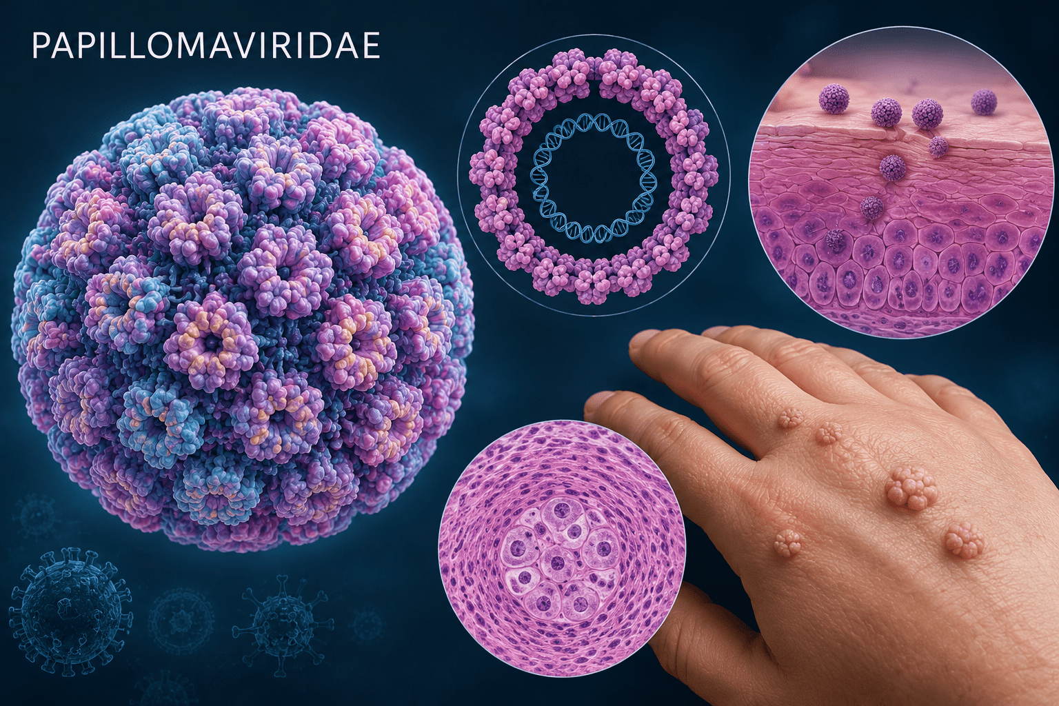
The Papillomaviridae family comprises a large and diverse group of small, non-enveloped DNA viruses collectively referred to as papillomaviruses (PVs). These viruses are widely distributed among vertebrates and are recognized for their strong affinity for epithelial tissues, where they establish localized infections that may remain asymptomatic or progress to proliferative and neoplastic diseases. Papillomaviruses are of considerable medical and veterinary importance because of their ability to induce both benign lesions and malignant transformation in infected hosts.
Historically, papillomaviruses were classified together with polyomaviruses under the now-obsolete family Papovaviridae, a term derived from papilloma, polyoma, and vacuolating viruses. However, advances in molecular virology, structural biology, and genome analysis demonstrated that papillomaviruses and polyomaviruses differ substantially in their genomic organization, replication strategies, evolutionary relationships, and biological properties. As a result, the family Papovaviridae was discontinued, and papillomaviruses are currently classified independently within the family Papillomaviridae.
Papillomaviruses infect a broad range of vertebrate hosts, including humans, cattle, horses, rabbits, dogs, birds, reptiles, and several other animal species. Despite their broad host distribution, these viruses exhibit a high degree of host specificity and tissue tropism, meaning that individual papillomavirus types typically infect particular host species and preferentially replicate within specific epithelial tissues. Most papillomaviruses target stratified squamous epithelial cells of the skin and mucosal membranes.
To date, hundreds of papillomavirus types have been identified, with more than 200 types characterized in humans alone. Among these, human papillomaviruses (HPVs) are the most extensively studied due to their major public health significance and established association with a range of benign and malignant conditions.
Members of the Papillomaviridae family are among the most biologically specialized DNA viruses infecting vertebrates. They possess a unique life cycle that is closely linked to epithelial differentiation and tissue maturation. Unlike many viruses that cause acute systemic infections, papillomaviruses generally establish localized infections confined to epithelial surfaces. These viruses are characterized as small, non-enveloped virions containing a circular double-stranded DNA (dsDNA) genome enclosed within an icosahedral protein capsid. Virions measure approximately 50-60 nm in diameter, with an average size of about 55 nm.
The absence of a lipid envelope contributes to the environmental stability of papillomaviruses and enhances resistance to environmental degradation. Consequently, papillomaviruses are relatively resistant to ether and certain physical stressors but remain susceptible to ultraviolet (UV) radiation and chemical disinfectants such as formalin. Replication occurs entirely within the nucleus of infected epithelial cells, and newly assembled virions are released primarily through epithelial shedding and desquamation rather than active budding.
Although many papillomavirus infections are transient and self-limiting, persistent infection with specific viral types may lead to uncontrolled epithelial proliferation and, in some cases, cancer development. For this reason, papillomaviruses are classified as oncogenic viruses, meaning they possess the capacity to contribute to malignant transformation. Human papillomavirus infection commonly produces papillomas (warts) affecting the skin, lips, and mucosal surfaces and is strongly associated with cancers of the cervix, anus, vulva, vagina, penis, and oropharyngeal region. Among human papillomaviruses, HPV-16 and HPV-18 are regarded as high-risk oncogenic types and are major causative agents of cervical cancer globally.
Biology of papillomaviruses
Papillomaviruses exhibit a highly specialized biological life cycle that is closely linked to the differentiation program of epithelial cells. Their replication strategy enables persistent infection while minimizing immune detection and maximizing viral production within stratified epithelial tissues. Infection typically begins when virions gain access to the basal layer of the epithelium through microscopic abrasions or disruptions in epithelial integrity that expose basal keratinocytes, the only epithelial cells capable of supporting long-term viral persistence.
After attachment to host cell surface receptors, the virus enters the cell through endocytic pathways. The viral capsid subsequently undergoes disassembly, allowing delivery of the viral genome into the nucleus. Once inside the nucleus, papillomaviral DNA is maintained as an episomal molecule, remaining physically separate from the host chromosome during most stages of infection. This episomal persistence enables the virus to replicate efficiently without immediate integration into the host genome.
During the initial phase of infection, expression of early viral genes supports establishment and maintenance of the viral genome at low copy numbers within proliferating basal cells. Viral proteins regulate DNA replication and manipulate host cellular pathways to maintain an environment favorable for viral persistence. As infected basal cells divide and migrate upward through the epithelial layers, the virus adapts its replication strategy to match the host cell differentiation process.
In suprabasal and differentiated epithelial cells, viral genome amplification occurs alongside increased expression of late genes encoding structural proteins required for virion assembly. Newly synthesized viral particles accumulate in the upper epithelial layers and are ultimately released as terminally differentiated cells are naturally shed from the epithelial surface, generally without causing cell lysis or significant inflammation. This differentiation-dependent replication cycle allows papillomaviruses to maintain persistent infections and, in certain high-risk viral types, contributes to pathological changes that may progress to neoplastic transformation and cancer development.
The papillomavirus replication cycle consists of three major phases:
1. Establishment phase
During the initial stage of infection, viral genomes undergo limited replication to establish a stable reservoir of viral DNA within dividing basal epithelial cells.
2. Maintenance phase
During maintenance, viral DNA replicates at low copy numbers and is distributed to daughter cells during normal epithelial turnover, enabling long-term persistence of infection.
3. Productive replication phase
As infected epithelial cells migrate toward the upper differentiated layers of the epithelium, viral replication intensifies. Large quantities of viral DNA are synthesized, structural proteins are expressed, and new virions are assembled.
This differentiation-dependent replication strategy allows papillomaviruses to maximize viral production while reducing exposure to host immune surveillance. Papillomaviruses also possess mechanisms for modifying host cellular pathways to support replication. Viral proteins alter normal cell cycle regulation, enabling continued cellular proliferation even in epithelial cells that would normally stop dividing. In oncogenic HPV infections, prolonged expression of viral proteins can interfere with cellular tumor suppressor pathways, promoting genomic instability and increasing the risk of malignant transformation.
Structure of papillomaviruses
Members of the Papillomaviridae family possess a relatively simple yet highly organized structure optimized for efficient infection and genome protection. Papillomaviruses are non-enveloped viruses with an approximately spherical morphology and icosahedral symmetry (Figure 1). The viral capsid serves as a protective shell surrounding the viral genome and facilitating attachment to host cells.
The capsid of papillomaviruses consists primarily of two structural proteins:
1. Major capsid protein (L1)
The L1 protein constitutes the majority of the viral capsid and self-assembles into pentameric units known as capsomeres. These capsomeres collectively form the characteristic icosahedral architecture of papillomaviruses.
L1 is essential for:
- Viral capsid formation
- Viral host cell attachment
- Maintenance of virion stability
- Induction of protective immune responses
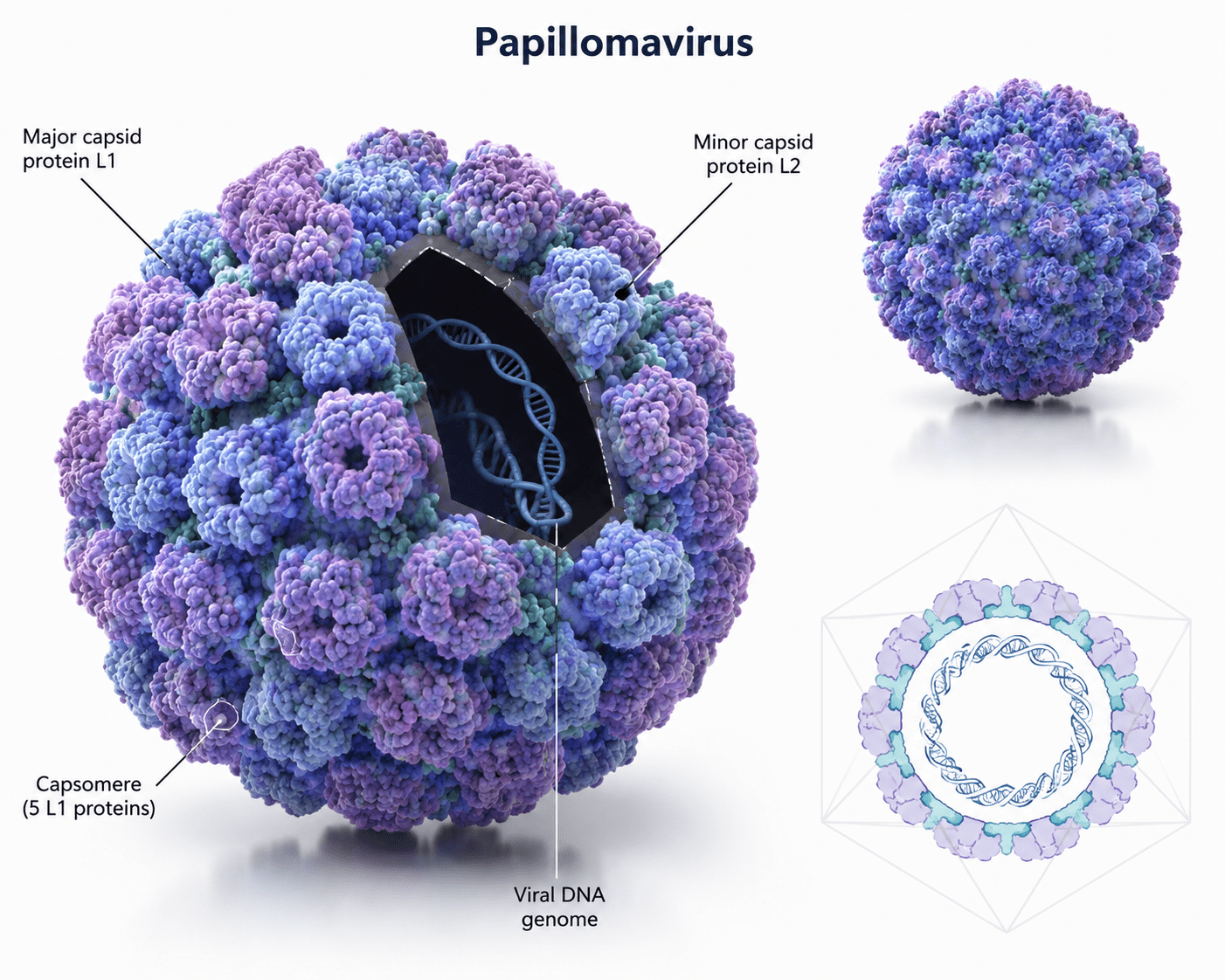
2. Minor capsid protein (L2)
The L2 protein is present in smaller amounts and performs several important functions during infection.
The roles of L2 protein include:
- Viral genome packaging
- Facilitation of viral entry
- Intracellular transport
- Delivery of viral DNA into the host nucleus
Because papillomaviruses lack an envelope, attachment and entry rely primarily on interactions between capsid proteins and receptors located on epithelial cell surfaces. The compact and durable structural organization of papillomaviruses supports effective transmission while ensuring preservation of the viral genome under external environmental conditions.
Genome organization of papillomaviruses
Papillomaviruses possess a compact yet highly efficient circular double-stranded DNA genome measuring approximately 7,000-8,000 base pairs (7-8 kb) in length. Despite this relatively small genome size, papillomaviruses exhibit a remarkably organized genetic architecture that supports precise regulation of viral replication, persistence, and host interaction.
The viral genome is functionally divided into three major regions:
- the early region (E region),
- the late region (L region), and
- the long control region (LCR).
Each of these regions contribute distinct but coordinated roles throughout the viral life cycle of the virus. The early region contains genes primarily responsible for establishing and maintaining infection within host epithelial cells. Proteins encoded in this region regulate viral DNA replication, transcriptional control, interaction with host cellular machinery, and modulation of cellular proliferation pathways.
Among these proteins, E1 functions as the viral helicase and plays a central role in initiating and sustaining viral DNA replication by unwinding the viral genome and recruiting host replication factors. E2 acts as a multifunctional regulatory protein involved in controlling viral transcription, coordinating replication, and ensuring stable maintenance and partitioning of the viral genome during cell division. E4 contributes predominantly to the productive stage of infection by facilitating viral maturation and promoting efficient release of viral particles from infected epithelial cells. E5 participates in altering host cellular signaling pathways, enhancing conditions favorable for viral persistence and replication.
Particularly important within the early region are the E6 and E7 proteins, especially in high-risk human papillomavirus (HPV) types associated with malignant transformation. These viral oncoproteins interfere with normal cellular regulatory mechanisms that govern cell proliferation and survival. E6 promotes degradation or functional inhibition of tumor suppressor pathways, while E7 disrupts cell cycle checkpoints by interacting with regulatory proteins that control progression through the cell cycle. Through these mechanisms, E6 and E7 drive sustained cellular proliferation, facilitate accumulation of genetic abnormalities, and contribute significantly to oncogenesis. Their activities are central to the development of HPV-associated cancers and represent major determinants of viral pathogenicity.
The late region comprises genes expressed primarily during the final stages of the viral life cycle and is responsible for viral particle assembly. This region contains two structural genes, L1 and L2, which encode the major and minor capsid proteins, respectively. The L1 protein forms the bulk of the viral capsid and is highly conserved across papillomavirus species, while L2 contributes to virion assembly, genome encapsidation, and infection of new host cells. Expression of late genes occurs predominantly in differentiated epithelial cells during productive infection, reflecting the close dependence of papillomaviruses on host epithelial differentiation for successful completion of their replication cycle.
Separating and coordinating these coding regions is the long control region (LCR), also referred to as the upstream regulatory region (URR), a non-coding but functionally critical segment of the viral genome. The LCR contains multiple regulatory elements that govern initiation of viral DNA replication, transcriptional activation and repression, promoter activity, and overall coordination of the viral life cycle. Through interactions with both viral and host transcription factors, this region ensures precise temporal and spatial regulation of gene expression, allowing papillomaviruses to synchronize replication and virion production with epithelial cell differentiation.
The Papillomaviridae family represents a highly specialized group of DNA viruses characterized by strong epithelial tropism, marked host specificity, tightly regulated replication strategies, conserved genomic organization, and notable oncogenic potential. Their capacity to establish persistent infections and contribute to cancer development makes papillomaviruses among the most clinically significant viral families in both human and veterinary medicine.
Host range of papillomaviruses
Papillomaviruses exhibit one of the broadest host distributions among DNA viruses while maintaining a remarkable degree of host specificity and tissue specificity. Each papillomavirus type is generally adapted to a particular host species and preferentially infects specific epithelial tissues.
Human papillomaviruses (HPVs) are the most extensively studied because of their medical importance; however, papillomavirus infections occur naturally in a wide variety of animals. Documented hosts for papillomaviruses include cattle, rabbits, horses, elephants, dogs, birds, primates, cats, deer, and other mammals. Although papillomaviruses infect many vertebrate species, cross-species transmission is uncommon because viral evolution has occurred alongside host adaptation.
In cattle, bovine papillomaviruses cause cutaneous and mucosal papillomas and have been associated with lesions affecting the skin, digestive tract, and urinary bladder. Equine papillomaviruses infect horses and may produce papillomas and sarcoids, which are among the most common skin tumors in horses. Rabbits infected with papillomaviruses may develop epithelial growths that occasionally progress to malignant lesions under favorable conditions. Canine papillomaviruses are commonly associated with oral papillomatosis and cutaneous warts in dogs. Birds may also harbor papillomavirus infections affecting feathered and non-feathered epithelial regions.
Pathogenesis of papillomavirus infection
The pathogenesis of papillomavirus infection is closely linked to the biology of epithelial cell growth and differentiation. Infection is initiated when viral particles gain access to the basal epithelial layer through microscopic abrasions or disruptions in the skin or mucosal surfaces.
After entering basal epithelial cells, the viral capsid disassembles and delivers the viral DNA into the host cell nucleus. Unlike many cytolytic viruses that rapidly destroy infected cells, papillomaviruses establish localized and highly regulated infections. The viral genome persists as an episome within the nucleus and replicates alongside host cellular DNA.
Papillomavirus replication is synchronized with epithelial differentiation. Infected basal cells continue to divide and migrate toward the epithelial surface while carrying viral genomes. As cells differentiate and move upward through epithelial layers, expression of viral genes changes, leading to increased viral DNA replication and production of structural proteins required for assembly of new virions.
An important feature of papillomavirus pathogenesis is the induction of cellular hyperproliferation. Viral proteins interfere with normal mechanisms controlling cell division, allowing infected cells to continue proliferating beyond their normal limits. This abnormal proliferation leads to visible epithelial lesions.
In high-risk human papillomavirus infections, prolonged viral persistence may result in integration of viral DNA into the host genome. Viral proteins particularly E6 and E7 can interfere with host tumor suppressor pathways, disrupt regulation of apoptosis, promote genomic instability, and increase the likelihood of malignant transformation. The progression from infection to cancer is typically slow and occurs over many years, requiring persistent infection and accumulation of additional genetic and cellular alterations.
Clinical manifestations and symptoms of papillomavirus infection
Clinical manifestations of papillomavirus infection vary depending on the host species, anatomical site affected, viral type, and immune status of the infected individual. Many papillomavirus infections remain subclinical, meaning infected individuals show no visible symptoms despite harboring the virus. When lesions occur, they generally result from localized epithelial overgrowth.
The most common clinical manifestation is the development of papillomas, commonly referred to as warts. These lesions are benign epithelial proliferations that appear on the skin and may vary in size, texture, and location. Cutaneous warts may appear as raised, rough, flattened, or cauliflower-like growths.
When papillomavirus infects mucosal epithelial surfaces, the resulting lesions are called condylomas. Condylomas typically develop in moist mucosal regions including the oral cavity, respiratory tract, and genital tract.
Symptoms associated with papillomavirus infection may include:
- Raised or thickened skin lesions (warts)
- Painless growths on mucosal surfaces
- Itching or irritation around affected areas
- Discomfort during movement or contact
- Oral lesions causing feeding or swallowing difficulties
- Genital lesions causing irritation or discomfort
- Abnormal bleeding in advanced cervical disease
- Persistent ulcers or masses in malignant progression
Genital warts represent one of the most recognized manifestations of human papillomavirus infection. These lesions occur on the external genitalia and surrounding mucosal tissues of both males and females and may appear singly or in clusters. Many high-risk HPV infections remain asymptomatic for years before progression to precancerous changes or invasive malignancy.
Diseases caused by papillomaviruses
Papillomaviruses are medically and veterinary important pathogens because of their broad host range, epithelial tropism, and ability to produce outcomes ranging from transient benign lesions to persistent infections and malignant disease. Their role in cancer development makes them one of the most significant viral families in both human and animal health. Papillomaviruses cause a broad spectrum of diseases ranging from benign epithelial proliferations to invasive cancers. The diseases caused by papillomavirus include:
Cutaneous warts
Cutaneous warts are among the most common outcomes of papillomavirus infection. These benign lesions affect the skin of the hands, feet, face, and other body regions. Although usually harmless, they may cause discomfort, cosmetic concerns, or secondary infection.
Condylomas (genital warts)
Genital warts are sexually transmitted epithelial lesions caused primarily by low-risk human papillomavirus types. They occur on the external genitalia, perineal region, and adjacent mucosal surfaces. While generally benign, they may recur following treatment.
Cervical cancer
Cervical cancer is one of the most clinically significant diseases associated with papillomavirus infection. Persistent infection with high-risk HPV types can induce progressive cellular abnormalities within the cervical epithelium that advance from dysplasia to invasive carcinoma.
Oral and oropharyngeal cancer
Certain papillomavirus types contribute to malignant lesions of the mouth and throat. Persistent infection of oral mucosal tissues may lead to transformation of epithelial cells and development of head and neck cancers.
Skin cancer
Although most cutaneous papillomavirus infections remain benign, some viral types have been implicated in the development of skin malignancies, particularly under conditions involving ultraviolet exposure and immune compromise.
Urogenital tract cancer
Papillomavirus infection may contribute to cancers affecting the penis, vulva, vagina, and anal canal through persistent epithelial infection and progressive cellular transformation.
Gastrointestinal and ocular lesions
Papillomaviruses have also been associated with lesions involving portions of the gastrointestinal tract and ocular tissues, although these associations are less common than genital and cutaneous disease.
Animal papillomatosis and tumor formation
In animals, papillomaviruses cause a variety of proliferative diseases including bovine papillomatosis, equine sarcoids, canine oral papillomatosis, and epithelial tumors in several wildlife species.
Laboratory detection of papillomavirus infection
Laboratory diagnosis of infections caused by members of the Papillomaviridae family is essential for confirming infection, identifying viral types, monitoring disease progression, and detecting premalignant or malignant changes at an early stage. Since many papillomavirus infections remain asymptomatic or produce non-specific lesions, laboratory methods provide reliable tools for accurate diagnosis and clinical management.
One of the most commonly employed approaches for laboratory detection is cytological examination, especially in the diagnosis of human papillomavirus (HPV)-associated cervical disease. Cytology involves the microscopic examination of exfoliated epithelial cells collected from affected tissues to identify morphological changes associated with viral infection. The Papanicolaou (Pap) smear test remains one of the most widely used screening methods for cervical abnormalities caused by HPV. Cytological examination may reveal characteristic cellular changes such as koilocytosis, enlargement of nuclei, abnormal nuclear staining, and varying degrees of epithelial dysplasia. These abnormalities provide early evidence of HPV-associated lesions before progression to invasive disease.
Another important diagnostic approach is histopathological examination, which involves microscopic evaluation of tissue biopsy samples obtained from suspicious lesions. Histopathology allows detailed assessment of tissue architecture and cellular changes associated with papillomavirus infection. Characteristic findings may include epithelial hyperplasia, papillomatosis, hyperkeratosis, koilocytic alterations, and varying degrees of cellular atypia. Histopathological examination is particularly valuable in distinguishing benign papillomas from premalignant and malignant lesions and plays a critical role in cancer diagnosis.
Modern diagnosis increasingly relies on molecular detection techniques, which provide high sensitivity and specificity. Among these techniques, the polymerase chain reaction (PCR) is considered one of the most effective methods for detecting papillomavirus DNA. PCR amplifies viral genetic material from clinical specimens and enables detection even when viral concentrations are low. This method also permits viral genotyping, allowing differentiation between low-risk and high-risk HPV strains. Identification of high-risk types is clinically important because persistent infection with these variants is associated with cancer development. Advanced molecular methods such as real-time PCR further enhance diagnostic capability by enabling quantification of viral DNA and monitoring viral persistence over time. In addition, nucleic acid hybridization assays are used in screening programs to identify high-risk HPV types and support early intervention strategies.
Immunological methods, including enzyme-linked immunosorbent assay (ELISA) and immunohistochemistry, may also contribute to diagnosis by detecting viral proteins or host immune responses. However, these approaches are generally supplementary and are used less frequently than molecular diagnostic methods.
Treatment of infections caused by papillomaviruses
Treatment of infections caused by papillomaviruses primarily focuses on eliminating visible lesions, reducing symptoms, preventing recurrence, and minimizing progression to malignant disease. At present, there is no specific antiviral therapy capable of completely eradicating papillomavirus infection from the body. In many cases, host immune responses eventually clear the infection naturally; therefore, therapeutic interventions are usually directed toward management of clinical manifestations.
Medical treatment commonly involves the use of topical therapeutic agents for benign lesions and genital warts. These medications act through different mechanisms to destroy infected tissue or stimulate local immune responses. Agents such as podophyllotoxin inhibit cellular division and promote regression of wart tissue, while imiquimod enhances immune-mediated clearance of infected cells. Chemical treatments such as trichloroacetic acid (TCA) are also used to remove superficial lesions through controlled tissue destruction.
When lesions are persistent, extensive, recurrent, or located in areas that complicate topical therapy, physical and surgical interventions may be required. Procedures such as cryotherapy, which destroys lesions through freezing, are commonly employed in clinical practice. Other treatment modalities include electrocautery, laser ablation, and surgical excision, depending on lesion size, location, and severity.
For patients who develop papillomavirus-associated malignancies, treatment becomes more complex and depends on cancer type and stage. Management may involve a combination of surgery, chemotherapy, radiation therapy, and targeted therapeutic approaches. Early diagnosis remains one of the most important factors influencing successful treatment outcomes and survival.
Prevention and control of papillomavirus infections
Prevention and control remain the most effective approaches for reducing the global burden of diseases caused by papillomaviruses. Because these viruses can establish persistent infections and contribute to cancer development, preventive strategies emphasize reducing transmission, enhancing immunity, and detecting disease before progression occurs.
The most important preventive measure is vaccination. The development of prophylactic HPV vaccines has significantly transformed the prevention of papillomavirus-associated diseases. These vaccines are designed to stimulate protective immunity against the most clinically important HPV types, particularly those responsible for cervical cancer and genital warts. Vaccination is most effective when administered prior to exposure to the virus, typically during early adolescence. Widespread immunization programs have demonstrated substantial reductions in the incidence of genital warts, precancerous cervical lesions, and HPV-related cancers.
Another major component of prevention is routine screening and early detection. Screening programs enable identification of infected individuals and detection of precancerous lesions before they progress to invasive cancer. Regular Pap smear testing and HPV DNA testing are widely implemented in many healthcare systems to monitor individuals at risk and guide early treatment decisions.
Behavioral and public health interventions also contribute significantly to disease control. Adoption of safe sexual practices can reduce exposure and transmission of HPV. Public health education programs increase awareness regarding transmission routes, vaccination benefits, and the importance of regular screening. Prompt evaluation and treatment of suspicious lesions further reduce disease progression and limit spread within populations.
In veterinary medicine, prevention of papillomavirus infections in animals relies on good husbandry practices, surveillance, early identification of lesions, and appropriate management of infected animals to minimize transmission. Effective control of infections caused by the Papillomaviridae family depends on a combination of accurate laboratory diagnosis, appropriate clinical management, vaccination programs, routine screening, and sustained public health interventions. These integrated approaches continue to play a critical role in reducing the incidence of papillomavirus-associated diseases and improving health outcomes worldwide.
References
Acheson N.H (2011). Fundamentals of Molecular Virology. Second edition. John Wiley and Sons Limited, West Sussex, United Kingdom.
Alan J. Cann (2005). Principles of Molecular Virology. 4th edition. Elsevier Academic Press, Burlington, MA, USA.
Alberts B, Bray D, Johnson A, Lewis J, Raff M, Roberts K and Walter P (1998). Essential Cell Biology: An Introduction to the Molecular Biology of the Cell. Third edition. Garland Publishing Inc., New York.
Barrett J.T (1998). Microbiology and Immunology Concepts. Philadelphia, PA: Lippincott-Raven Publishers. USA.
Black, J.G. (2008). Microbiology: Principles and Explorations (7th ed.). Hoboken, NJ: J. Wiley & Sons.
Brian W.J Mahy and Mark H.C van Regenmortel (2010). Desk Encyclopedia of Human and Medical Virology. Elsevier Academic Press, San Diego, USA.
Brooks G.F., Butel J.S and Morse S.A (2004). Medical Microbiology, 23rd edition. McGraw Hill Publishers. USA.
Cann A.J (2011). Principles of Molecular Virology. Fifth edition. Academic Press, San Diego, United States.
Carter J and Saunders V (2013). Virology: Principles and Applications. Second edition. Wiley-Blackwell, New Jersey, United States.
Champoux J.J, Neidhardt F.C, Drew W.L and Plorde J.J (2004). Sherris Medical Microbiology: An Introduction to Infectious Diseases. 4th edition. McGraw Hill Companies Inc, USA.
Dimmock N (2015). Introduction to Modern Virology. Seventh edition. Wiley-Blackwell, New Jersey, United States.
Dimmock N.J, Easton A.J and Leppard K.N (2001). Introduction to modern virology. 5th edition. Blackwell Science publishers. Oxford, UK.
Discover more from Microbiology Class
Subscribe to get the latest posts sent to your email.