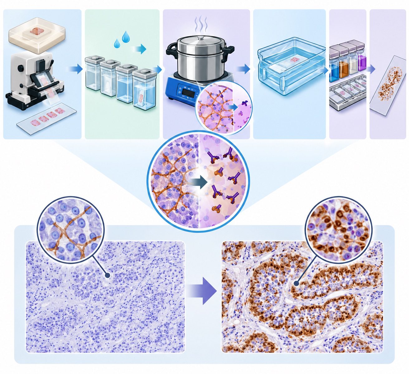

Immunoglobulin A (IgA) is a specialized glycoprotein antibody that plays a fundamental role in the body’s immune defence, particularly at surfaces exposed to the external environment. It is an essential component of the adaptive immune system, serving as an immunological barrier against invading microorganisms and foreign substances. Unlike immune components that primarily operate within the bloodstream and internal tissues, IgA is strategically positioned at mucosal surfaces, where continuous exposure to bacteria, viruses, allergens and environmental antigens occurs.
Its distinctive distribution and protective properties make it one of the most important immunoglobulins involved in maintaining the body’s first line of immunological defense. Antibodies, also known as immunoglobulins, are specialised proteins produced by plasma cells following the activation and differentiation of B lymphocytes. They recognise and bind to specific antigens, enabling the immune system to identify potentially harmful substances and initiate appropriate protective responses.
Through antigen recognition, antibodies contribute to pathogen neutralization, immune regulation and the elimination of foreign materials. Based on differences in their heavy-chain composition and biological functions, human immunoglobulins are classified into five major classes: IgG, IgA, IgM, IgE and IgD. Each class possesses distinct structural features and immunological responsibilities, allowing the immune system to respond effectively to different forms of biological threats.
Among these five classes, IgA is particularly associated with mucosal immunity. It is predominantly found in secretions such as saliva, tears, intestinal fluid, respiratory mucus and breast milk, although it is also present in the blood. This widespread distribution enables IgA to provide continuous immunological surveillance across several anatomical regions. In humans, IgA exists mainly in two structural forms: monomeric IgA, which is commonly found in serum, and dimeric IgA, which predominates in mucosal secretions.
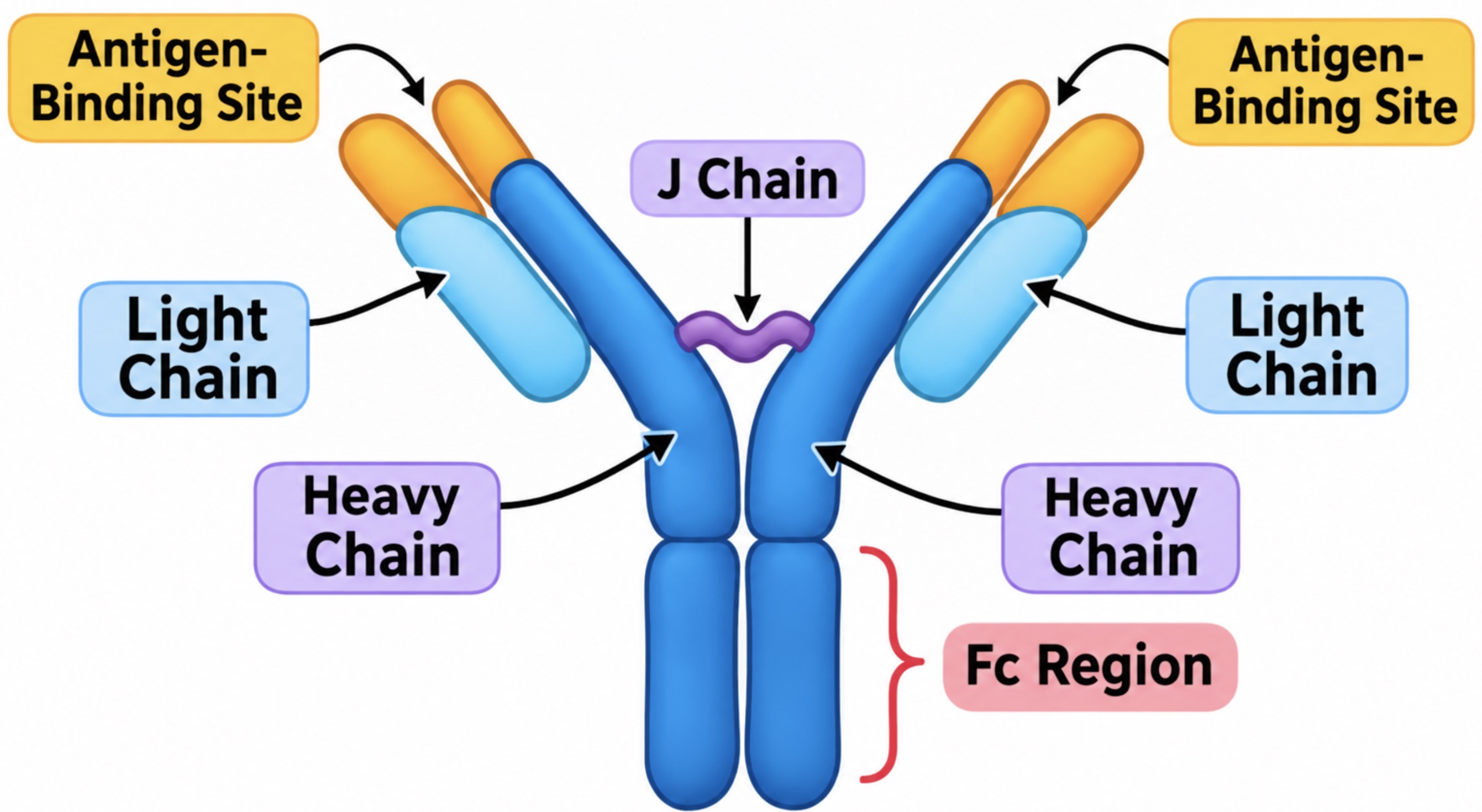
The dimeric form consists of two antibody units connected by a joining chain (J chain) and is associated with a secretory component that facilitates its transport across epithelial cells and enhances its stability in external secretions. Structurally, IgA consists of two identical heavy chains and two identical light chains arranged in a characteristic Y-shaped configuration. These chains form antigen-binding regions that enable the antibody to recognise specific molecular targets.
Its structural organisation is closely related to its biological versatility, particularly its ability to function effectively within mucosal environments where enzymes and other potentially destructive substances are present. The significance of IgA extends beyond simple pathogen recognition. It helps prevent microorganisms from attaching to and penetrating epithelial surfaces, neutralises toxins and viruses.
IgA also supports a balanced relationship between the host immune system and its resident microbial populations. Through these protective activities, IgA contributes to infection prevention while limiting unnecessary inflammatory reactions that could damage healthy tissues. Immunoglobulin A represents a vital component of human immune defence, linking antibody-mediated protection with the preservation of mucosal integrity and overall physiological health.
Structure and types of immunoglobulin A
IgA possesses a distinctive molecular architecture that enables it to perform its protective functions across mucosal surfaces and within the circulatory system. Like other antibody classes, IgA is a glycoprotein composed of four polypeptide chains: two identical heavy chains and two identical light chains. These chains are held together by disulfide bonds, which provide molecular stability and maintain the characteristic Y-shaped configuration of the antibody.
Each heavy chain is associated with a light chain, forming two antigen-binding arms that recognize and attach to specific antigens. The constant regions of the heavy chains contribute to the biological properties of IgA, while its variable regions determine antigen specificity. This structural organization allows IgA to recognize a wide range of foreign substances, including bacterial antigens, viral particles and environmental allergens.
Structurally, IgA occurs predominantly in two molecular forms: monomeric and dimeric IgA (Figure 1). Monomeric IgA consists of a single antibody unit and is the principal form found in blood serum. It circulates through the bloodstream, where it participates in systemic immune surveillance and antigen neutralization. In contrast, dimeric IgA consists of two antibody monomers joined together, creating a larger molecular complex. This form is particularly abundant in mucosal secretions, including saliva, tears, intestinal fluids, respiratory secretions and breast milk.

Its polymeric arrangement enhances its suitability for mucosal defense, allowing it to interact effectively with pathogens before they penetrate epithelial barriers. Another important structural characteristic of IgA is the existence of two subclasses, IgA1 and IgA2, which differ in their molecular composition and distribution. IgA1 contains a relatively extended hinge region between its antigen-binding arms and constant region. This hinge provides flexibility, enabling the antibody to interact with antigens at different orientations.
However, its extended structure makes it more susceptible to cleavage by certain bacterial proteases. IgA2, on the other hand, possesses a shorter hinge region and greater resistance to enzymatic degradation. This characteristic makes it particularly advantageous in environments with high microbial activity, especially the gastrointestinal and other mucosal compartments. Although both subclasses contribute to immune protection, their structural differences influence their tissue distribution and resistance to microbial enzymes.
The J chain (joining chain) is another essential component of polymeric IgA (Figure 2). The joining chain is a small polypeptide that facilitates the association of two IgA monomers during dimer formation. Beyond its structural contribution, the J chain plays a crucial role in enabling dimeric IgA to bind to the polymeric immunoglobulin receptor (pIgR) expressed on epithelial cells. Following receptor-mediated transport across the epithelial layer, the receptor undergoes cleavage, leaving a portion known as the secretory component attached to the antibody.
This component protects secretory IgA against enzymatic degradation and supports its stability within mucosal secretions. Collectively, the structural variations of IgA demonstrate how its molecular design is adapted to different immunological environments. Its monomeric form supports systemic immunity, while its polymeric and secretory forms provide specialized protection at mucosal surfaces. These distinctive features establish IgA as an essential component of both systemic and mucosal immune defense.
Synthesis and production of immunoglobulin A (IgA)
The synthesis and production of IgA constitute a highly coordinated immunological process through which the body establishes and maintains protective immunity at mucosal surfaces. Unlike antibodies that predominantly function within the bloodstream, IgA is strategically manufactured in association with mucosa-associated lymphoid tissues and distributed across epithelial barriers. Its production involves the concerted activities of B lymphocytes, antibody-secreting plasma cells, regulatory cytokines, and specialized epithelial transport mechanisms. These interconnected processes ensure that IgA reaches locations where microorganisms are most likely to enter the body.
Sites of IgA production in the body: IgA is predominantly synthesized in mucosa-associated lymphoid tissues (MALT), which include the gastrointestinal, respiratory, and genitourinary immune systems. Important production sites include the Peyer’s patches of the small intestine, the tonsils, the appendix, and other lymphoid structures distributed throughout mucosal surfaces. The intestinal lamina propria is particularly rich in IgA-producing plasma cells, making the gastrointestinal tract one of the most significant centres of IgA synthesis. Additional production occurs in the respiratory and salivary gland-associated lymphoid tissues. These anatomical locations provide an extensive network for localized antibody generation, allowing IgA to protect vulnerable mucosal surfaces against invading pathogens.
Role of B lymphocytes and plasma cells: B lymphocytes are specialized immune cells responsible for recognizing specific antigens and initiating antibody production. Following antigen recognition, these cells become activated through interactions with antigen-presenting cells and other immune components. Some activated B lymphocytes undergo proliferation and subsequently differentiate into plasma cells. Plasma cells are the terminally differentiated antibody-producing cells that synthesize and release substantial quantities of IgA. Their concentration within mucosal tissues enables continuous antibody secretion, even in the absence of an immediate infectious challenge. Consequently, B lymphocytes initiate the immune response, while plasma cells serve as the principal cellular factories responsible for sustained IgA production.
Differentiation of B cells into IgA-producing plasma cells: The transformation of B lymphocytes into IgA-secreting plasma cells involves a process known as immunoglobulin class-switch recombination. Initially, activated B cells may express IgM or IgD on their surfaces. Following appropriate immunological stimulation, they undergo genetic rearrangement that changes their antibody class to IgA without altering the antigen-binding specificity. This process involves the activation-induced cytidine deaminase (AID) enzyme, which facilitates the genetic modifications required for switching to the IgA heavy-chain constant region. After class switching, the B cells undergo further differentiation into plasma cells capable of producing either IgA1 or IgA2, depending on the surrounding tissue environment and regulatory signals.
Role of cytokines in IgA production: Cytokines function as essential biochemical messengers that regulate the development and specialization of IgA-producing cells. Transforming growth factor-beta (TGF-β) is particularly important because it promotes immunoglobulin class switching towards IgA. Other regulatory molecules, including interleukin-5 (IL-5), interleukin-6 (IL-6), and interleukin-10 (IL-10), contribute to B-cell differentiation, plasma-cell development, and antibody secretion. Additionally, B-cell activating factor (BAFF) and a proliferation-inducing ligand (APRIL) support IgA responses, particularly within mucosal tissues. Through these coordinated signalling pathways, cytokines establish an appropriate immunological environment for efficient IgA synthesis and sustained mucosal protection.
Transport of IgA across epithelial cells: Following synthesis, polymeric IgA, particularly its dimeric form containing a joining (J) chain, must cross epithelial barriers to reach mucosal secretions. This process occurs through receptor-mediated transcytosis involving the polymeric immunoglobulin receptor (pIgR), which is expressed on the basolateral surfaces of epithelial cells. The receptor binds dimeric IgA and transports it through the epithelial cell towards the apical surface. Upon reaching the outer membrane, the receptor undergoes cleavage, releasing IgA into the mucosal environment while retaining a portion known as the secretory component. This association produces secretory IgA (sIgA), a form that is relatively resistant to enzymatic degradation. Once released, secretory IgA provides a protective barrier in saliva, tears, intestinal mucus, respiratory secretions, and breast milk, thereby reinforcing the body’s first line of immune defence.
Distribution and location of immunoglobulin A (IgA)
IgA is widely distributed throughout the human body, with a particularly prominent presence in body fluids and mucosal tissues that are continuously exposed to environmental microorganisms. Its distribution reflects its specialised role in immune defence, especially at the surfaces through which pathogens may enter the body. IgA occurs in two principal forms: serum IgA, which circulates in the bloodstream predominantly as a monomer, and secretory IgA (sIgA), which is mainly found in external secretions as a dimer. These different forms enable IgA to provide immune protection in both internal and external compartments of the body.
Presence of IgA in blood serum: In blood serum, IgA constitutes the second most abundant immunoglobulin isotype after IgG. It is produced by plasma cells and released into circulation, where it contributes to systemic immune surveillance. Serum IgA exists mainly in a monomeric form and is transported through the bloodstream to different tissues. Although its concentration in serum is lower than that of IgG, it plays an important role in identifying and neutralising foreign antigens. Its presence in circulation also makes serum IgA a valuable laboratory marker in the assessment of certain immunological and inflammatory conditions.
Distribution in mucosal surfaces: Mucosal surfaces represent the principal sites of IgA activity. These surfaces line organs and body passages that communicate with the external environment, including the intestines, respiratory tract and reproductive system. IgA-producing plasma cells are abundant within the underlying mucosal tissues, particularly the lamina propria. From these locations, IgA is transported across epithelial cells into the mucosal lumen. This strategic positioning allows it to intercept invading microorganisms before they penetrate deeper tissues, thereby establishing an important protective barrier without necessarily triggering excessive inflammatory reactions.
Presence in saliva and tears: IgA is an essential constituent of saliva and tears, where it contributes to the protection of the oral cavity and ocular surfaces. In saliva, secretory IgA helps prevent microorganisms from adhering to the teeth, gums and oral mucosa. It also assists in neutralising microbial toxins and maintaining a balanced oral microbial environment. In tears, IgA provides local immune protection by binding to potentially harmful microorganisms that come into contact with the conjunctiva and corneal surfaces. Its presence in these fluids helps safeguard delicate tissues that are frequently exposed to environmental contaminants.
Occurrence in breast milk and colostrum: Breast milk, particularly colostrum, contains substantial amounts of secretory IgA. Colostrum is the early milk produced during the first days following childbirth and is especially rich in immunological components. Maternal IgA is transferred to the infant through breastfeeding, providing passive immune protection during the early stages of life when the infant’s immune system is still developing. Within the gastrointestinal tract, this antibody helps prevent pathogen attachment, supports intestinal barrier integrity and contributes to the establishment of a healthy microbial community. Breast milk IgA represents an important component of maternal-to-infant immune protection.
Presence in respiratory, gastrointestinal and genitourinary secretions: Secretory IgA is also extensively distributed throughout respiratory, gastrointestinal and genitourinary secretions. In the respiratory tract, it occurs in nasal mucus, bronchial secretions and other airway fluids, where it helps restrict the entry and colonisation of inhaled pathogens. Within the gastrointestinal tract, it is present in intestinal mucus and digestive secretions, where it protects the intestinal epithelium against bacteria, viruses and other foreign antigens. In the genitourinary tract, IgA contributes to mucosal defense within reproductive and urinary passages by limiting microbial attachment and penetration. These anatomical distributions demonstrate how IgA provides continuous, site-specific immune protection across multiple body systems.
Functions and mechanism of action of IgA
IgA plays a fundamental role in the body’s immune defence, particularly at mucosal surfaces that are constantly exposed to environmental microorganisms and foreign substances. It serves as a frontline antibody, safeguarding delicate epithelial barriers against potentially harmful pathogens while maintaining a balanced relationship between the immune system and the normal microbial population. Unlike other immunoglobulins that primarily operate within the bloodstream and tissues, IgA is strategically concentrated in mucosal secretions, including saliva, tears, intestinal fluids, respiratory mucus and breast milk. Its protective activity involves several coordinated mechanisms that prevent microbial invasion without necessarily triggering excessive inflammatory reactions.
Protection of mucosal surfaces against pathogens: One of the principal functions of IgA is to defend mucosal membranes against invading microorganisms. These membranes line the gastrointestinal, respiratory and genitourinary tracts, making them important entry points for infectious agents. Secretory IgA provides localized protection by binding to pathogens at these surfaces before they penetrate the underlying epithelial tissues. This protective activity helps preserve epithelial integrity and reduces the likelihood of infections developing. Through its continuous presence in mucosal secretions, IgA establishes an immunological barrier that responds to microbial exposure.
Neutralization of bacteria, viruses and toxins: IgA neutralizes harmful microorganisms and their toxic products through specific antigen recognition. It binds to viral particles, bacterial structures and soluble toxins, interfering with their biological activities. In viral infections, IgA can attach to viral surface proteins, preventing the virus from interacting with receptors on host cells. Similarly, its interaction with bacterial toxins can obstruct their attachment to target tissues and limit their harmful effects. This neutralizing capacity is particularly important in the intestinal and respiratory tracts, where exposure to infectious agents occurs frequently.
Prevention of microbial attachment to epithelial cells: Another important mechanism of IgA-mediated protection is the inhibition of microbial adhesion. Many pathogens initiate infection by attaching to receptors located on epithelial cell surfaces. IgA interferes with this process by binding to specific microbial adhesion molecules, thereby preventing pathogens from establishing contact with host tissues. This mechanism reduces microbial colonization and limits the opportunity for pathogens to multiply or penetrate epithelial barriers. IgA helps interrupt the early stages of infection before tissue invasion occurs.
Immune exclusion and trapping of pathogens in mucus: IgA contributes to a protective process known as immune exclusion, through which microorganisms and antigens are retained within the mucosal environment rather than gaining access to underlying tissues. By binding to pathogens, secretory IgA facilitates their entrapment within mucus layers covering epithelial surfaces. These antibody-antigen complexes can subsequently be removed through natural clearance mechanisms, including intestinal movement, coughing, sneezing and the flow of secretions. This process reduces microbial penetration while limiting direct tissue exposure to potentially harmful substances.
Regulation of immune responses and prevention of excessive inflammation: Beyond its antimicrobial activities, IgA plays an essential role in maintaining immunological balance. Its protective mechanisms generally operate with minimal activation of inflammatory pathways, helping to prevent unnecessary tissue damage. Unlike certain other antibody classes, IgA does not efficiently activate the classical complement pathway. This characteristic allows it to neutralize pathogens while reducing the possibility of excessive inflammatory reactions at sensitive mucosal surfaces. IgA also contributes to immune tolerance by helping the body coexist with beneficial intestinal microorganisms and harmless environmental antigens. Through these regulatory activities, IgA supports mucosal homeostasis, preserves tissue integrity and strengthens the overall effectiveness of the immune system.
Clinical significance and disorders associated with IgA
IgA plays a fundamental role in immune protection, particularly at mucosal surfaces, where it serves as a frontline defence against invading microorganisms. Beyond its protective function, IgA has considerable clinical significance because abnormalities in its production, regulation, distribution, or deposition can contribute to several pathological conditions. Alterations in IgA concentrations may reflect underlying immunological disturbances, renal complications, inflammatory processes, or systemic diseases. Some of the most notable conditions associated with IgA dysregulation include selective IgA deficiency, IgA nephropathy, IgA vasculitis, and certain autoimmune disorders.
Selective IgA deficiency:
Selective IgA deficiency is one of the most common primary immunodeficiency disorders. It is characterized by markedly reduced or undetectable serum IgA levels despite normal concentrations of other major immunoglobulin classes, particularly IgG and IgM. The condition results from impaired differentiation or maturation of B lymphocytes into IgA-producing plasma cells. Individuals with this deficiency may experience recurrent respiratory and gastrointestinal infections because of inadequate antibody-mediated protection at mucosal surfaces. Common manifestations include sinusitis, bronchitis, pneumonia, and persistent gastrointestinal infections. Some affected individuals also develop allergies, autoimmune conditions, or adverse reactions to blood products containing IgA. However, many remain asymptomatic and are diagnosed incidentally during routine immunological investigations.
IgA nephropathy (Berger’s disease)
IgA nephropathy, also known as Berger’s disease, is a kidney disorder characterized by the accumulation of IgA-containing immune complexes within the glomerular mesangium. These deposits trigger inflammatory responses that progressively damage the kidney’s filtration structures. The disease may present with recurrent episodes of visible haematuria, microscopic blood in the urine, proteinuria, and, in some cases, hypertension or declining renal function. Although its precise pathogenesis is multifactorial, abnormalities in the production and glycosylation of IgA1 are considered important contributors. Persistent inflammation may eventually result in glomerulosclerosis and chronic kidney disease, with some patients progressing to end-stage renal disease.
IgA vasculitis (Henoch–Schönlein Purpura)
IgA vasculitis is a systemic small-vessel inflammatory disorder associated with IgA-containing immune complex deposition in affected blood vessels. It commonly affects children but can also occur in adults. Its characteristic clinical features include palpable purpura, joint pain, abdominal discomfort, and renal involvement. The skin manifestations typically appear as raised, reddish-purple lesions, particularly on the lower limbs and buttocks. Gastrointestinal complications may include intestinal bleeding, while renal involvement can produce haematuria and proteinuria. The condition demonstrates how abnormal IgA-mediated immune responses can extend beyond mucosal immunity and affect multiple organ systems.
Association between IgA and autoimmune diseases
Dysregulated IgA responses have been associated with several autoimmune and immune-mediated disorders. In conditions such as coeliac disease, IgA antibodies directed against tissue transglutaminase are important diagnostic markers. IgA may also participate in inflammatory processes involving the gastrointestinal tract, liver, and other tissues. These associations demonstrate that disturbances in antibody recognition and immune tolerance can contribute to tissue injury. However, elevated IgA does not independently establish the presence of an autoimmune disease; clinical findings and disease-specific investigations are required for accurate diagnosis.
Clinical significance of increased and decreased IgA levels
Abnormal serum IgA concentrations can provide valuable information about an individual’s immunological status. Elevated IgA levels may occur in chronic infections, persistent inflammation, liver diseases, and certain autoimmune or plasma-cell disorders. Conversely, reduced IgA concentrations may indicate selective IgA deficiency, broader immunodeficiency, or secondary immunological disturbances. Laboratory assessment of IgA, usually alongside IgG and IgM measurements, helps clinicians investigate recurrent infections, suspected immune disorders, and certain inflammatory conditions. Nevertheless, IgA levels must be interpreted in conjunction with the patient’s clinical presentation and other laboratory findings. The clinical significance of IgA lies not only in its protective capacity but also in the consequences of its deficiency, excessive production, and abnormal tissue deposition.
Laboratory diagnosis, testing and therapeutic importance of IgA
Quantitative assessment of serum IgA
Measurement of serum Immunoglobulin A is an essential laboratory investigation used to evaluate humoral immune function. It determines the concentration of IgA circulating in the blood and helps identify abnormalities in antibody production. Serum IgA levels are commonly measured alongside Immunoglobulin G (IgG) and Immunoglobulin M (IgM) to provide a broader assessment of immune competence. Results are interpreted according to age-specific reference intervals because IgA concentrations vary throughout human development.
Laboratory techniques for IgA detection
Several analytical techniques are employed to determine IgA concentrations and detect its presence in biological specimens. Nephelometry and turbidimetry are widely used quantitative methods that measure changes in light scattering caused by antigen–antibody interactions. Enzyme-linked immunosorbent assay (ELISA) is another technique used to identify and quantify IgA in serum and mucosal secretions. Immunofixation electrophoresis may also help characterize abnormal immunoglobulin patterns, particularly when a monoclonal protein is suspected.
Interpretation of IgA concentrations
Abnormal IgA concentrations may provide important clues about underlying physiological or pathological conditions. Reduced IgA levels may indicate selective IgA deficiency, impaired antibody synthesis or certain immunodeficiency disorders. Conversely, elevated concentrations may occur in chronic infections, inflammatory conditions, autoimmune diseases and some liver disorders. Significantly increased monoclonal IgA may also be associated with plasma cell malignancies, including multiple myeloma. However, IgA results should never be interpreted in isolation; clinical findings and other laboratory investigations are necessary for accurate diagnosis.
Diagnostic relevance in immune disorders and infections
IgA testing contributes significantly to the investigation of recurrent respiratory and gastrointestinal infections, suspected immunodeficiency and autoimmune conditions. It is particularly useful in screening for coeliac disease, where IgA antibodies against tissue transglutaminase (tTG-IgA) are commonly measured. Total serum IgA is assessed alongside this test because IgA deficiency may produce falsely negative results. Additional IgA-based investigations may assist in evaluating selected inflammatory and immune-mediated disorders.
Therapeutic implications and clinical management
Management of IgA-related disorders depends on the underlying cause, severity and clinical presentation. Patients with selective IgA deficiency may require monitoring, prompt treatment of infections and appropriate vaccination strategies. In IgA nephropathy, treatment focuses on controlling blood pressure, reducing proteinuria and preserving renal function. IgA vasculitis may require supportive care or immunosuppressive treatment in severe cases. Although no routine IgA replacement therapy is established for selective IgA deficiency, individualized clinical management and regular follow-up remain important in preventing complications and improving patient outcomes.
References
Abbas A.K, Lichtman A.H and Pillai S (2010). Cellular and Molecular Immunology. Sixth edition. Saunders Elsevier Inc, USA.
Actor J (2014). Introductory Immunology. First edition. Academic Press, USA.
Alberts B, Bray D, Johnson A, Lewis J, Raff M, Roberts K and Walter P (1998). Essential Cell Biology: An Introduction to the Molecular Biology of the Cell. Third edition. Garland Publishing Inc., New York.
Bach F and Sachs D (1987). Transplantation immunology. N. Engl. J. Med. 317(8):402-409.
Barrett J.T (1998). Microbiology and Immunology Concepts. Philadelphia, PA: Lippincott-Raven Publishers. USA.
Jaypal V (2007). Fundamentals of Medical Immunology. First edition. Jaypee Brothers Medical Publishers (P) Ltd, New Delhi, India.
Levinson W (2010). Review of Medical Microbiology and Immunology. Twelfth edition. The McGraw-Hill Companies, USA.
Roitt I, Brostoff J and Male D (2001). Immunology. Sixth edition. Harcourt Publishers Limited, Spain.
Zon LI (1995). Developmental biology of hematopoiesis. Blood, 86(8): 2876–91.
Discover more from Microbiology Class
Subscribe to get the latest posts sent to your email.