
Endospore staining is a specialized differential staining technique employed in microbiology to detect and distinguish bacterial endospores from their vegetative cells. Endospores are dormant, highly resistant structures produced by certain bacteria in response to environmental stress, including nutrient depletion, desiccation, elevated temperatures, and exposure to harmful chemicals. Unlike reproductive spores, endospores do not increase bacterial numbers; instead, they serve as survival mechanisms that enable bacterial cells to withstand adverse conditions and resume metabolic activity when the environment becomes favorable.
The remarkable resistance of endospores arises from their distinctive structural organization, which includes multiple protective layers, a dehydrated core, and specialized chemical components such as calcium dipicolinate. These features contribute to their ability to endure conditions that would ordinarily destroy vegetative bacterial cells. Endospores are difficult to stain using conventional staining procedures because their protective coats restrict the penetration and retention of ordinary dyes.
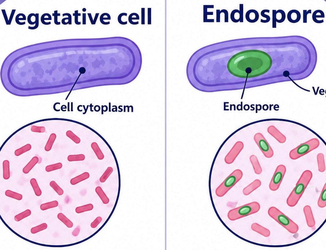
Endospore staining overcomes this limitation through the application of heat to facilitate the entry of a primary stain, typically malachite green, into the endospore. Following treatment with water to remove the stain from vegetative cells, safranin is applied as a counterstain to impart a contrasting colour to the surrounding bacterial cells. Under microscopic examination, endospores generally appear green, whereas vegetative cells appear pink or red, allowing their differentiation based on staining characteristics.
The genera Bacillus and Clostridium contain well-known endospore-forming bacteria of considerable medical, industrial, and environmental significance. Identifying endospores is important in bacterial characterization, laboratory diagnosis, food microbiology, and the evaluation of sterilization procedures. Endospore staining provides a valuable method for examining bacterial survival structures and understanding the adaptive mechanisms that enable certain microorganisms to persist under unfavorable environmental conditions.
Principle of endospore staining
The underlying principle of endospore staining is the differential penetration and retention of dyes by endospores and vegetative bacterial cells, enabling their microscopic differentiation based on contrasting colours. The technique commonly employs malachite green as the primary stain and safranin as the counterstain, with heat facilitating the penetration of the primary stain into the resistant endospore structure.
Endospores possess a complex, multilayered architecture consisting of a dehydrated core, inner membrane, cortex, and protective protein-rich coat (Figure 1). These structural components confer exceptional resistance to heat, desiccation, chemical disinfectants, and other adverse environmental conditions. The compact organization and limited permeability of the spore envelope impede the penetration of conventional aqueous dyes under ordinary staining conditions. A specialized staining procedure is required to visualize endospores within bacterial cells.

In the endospore staining procedure, malachite green is applied to the bacterial smear while heat is carefully introduced. Heating facilitates the penetration of the primary stain through the protective spore layers. The stain is maintained in contact with the specimen during heating to promote adequate dye uptake. Once malachite green penetrates the endospore, it is retained sufficiently to resist removal during subsequent rinsing with water.
Water serves as the decolorizing agent in this technique, readily removing malachite green from vegetative cells while leaving the stain retained within properly stained endospores. Following decolorization, safranin is applied as a counterstain to colour the vegetative cells. These cells readily take up safranin and appear pink or red, whereas endospores retain their green coloration. This differential staining produces a distinct contrast between endospores and vegetative bacterial cells.
The resulting colour contrast facilitates the identification of endospores and the assessment of their morphology and intracellular position. Depending on the bacterial species, endospores may be located centrally, subterminally, or terminally within the cell and may exhibit spherical or oval shapes. The principle of endospore staining relies on the combined effects of the resistant spore structure, heat-assisted dye penetration, differential dye retention during decolorization, and counterstaining. These processes enable the microscopic differentiation of endospores from vegetative cells and support the preliminary characterization of endospore-forming bacteria.
Aim of the experiment: Detection and characterization of bacterial endospores
The primary aim of endospore staining is to detect and examine the morphological characteristics of bacterial endospores using a specialized differential staining technique. Endospores are dormant, highly resistant structures produced by certain bacteria under unfavorable environmental conditions, such as nutrient depletion and other forms of stress. Their distinctive structural composition enables them to survive conditions that may be lethal to vegetative bacterial cells (Figure 2).
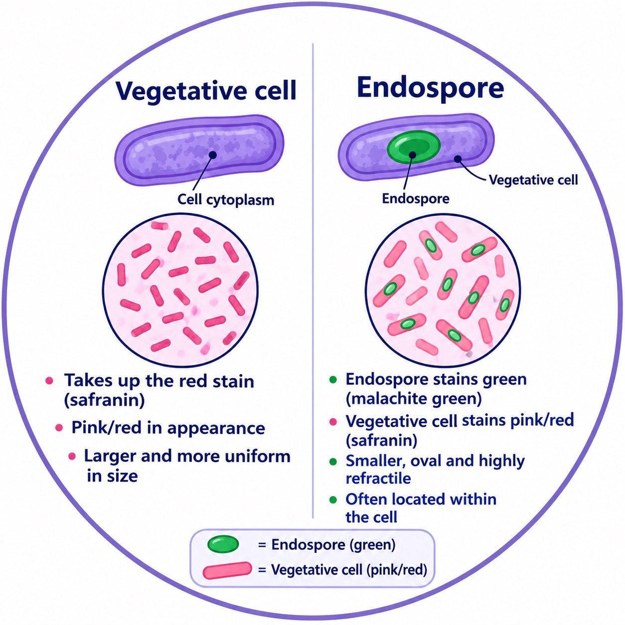
This experiment aims to differentiate endospores from vegetative cells based on their staining properties. Malachite green serves as the primary stain This stain penetrates the resistant endospore structure with the aid of heating. Safranin acts as the counterstain, coloring vegetative cells to provide contrast. This differential staining pattern facilitates the microscopic identification of endospores and vegetative cells within a prepared bacterial smear.
Another objective is to examine the morphological characteristics of endospores, particularly their shape, size, and position within bacterial cells. Depending on the bacterial species, endospores may be centrally located, subterminally positioned, or situated at the terminal end of the cell. These features provide valuable preliminary information for differentiating among endospore-forming bacteria.
The experiment also aims to develop practical skills in bacterial smear preparation, heat fixation, application of staining reagents, and microscopic examination using the oil-immersion objective. Careful observation of the stained preparation enables the detection of endospores and supports the preliminary morphological characterization of bacterial isolates.
Endospore staining is an important microbiological technique for investigating bacterial survival structures and distinguishing endospore-forming bacteria from non-spore-forming organisms. Although the staining pattern provides useful morphological evidence for bacterial characterization, it should be interpreted alongside other laboratory findings to support accurate bacterial identification.
Materials and reagents required for endospore staining
Endospore staining requires an appropriate combination of microbiological specimens, staining reagents, laboratory equipment, and microscopic examination tools. Each material serves a specific purpose in preparing the bacterial smear, facilitating stain penetration, differentiating endospores from vegetative cells, and enabling clear microscopic observation. The proper selection, preparation, and handling of these materials are essential for ensuring the accuracy and reliability of the staining procedure.
1. Bacterial culture: A bacterial culture containing suspected endospore-forming organisms, such as Bacillus species, serves as the biological specimen. A culture of appropriate age and grown under suitable conditions is important because endospore formation varies with nutrient availability, growth phase, and environmental stress.
2. Clean glass microscope slides: Microscope slides provide a smooth, transparent surface for preparing a thin bacterial smear. They must be free from grease, dust, and residual chemicals that could interfere with smear adhesion, staining quality, or microscopic observation.
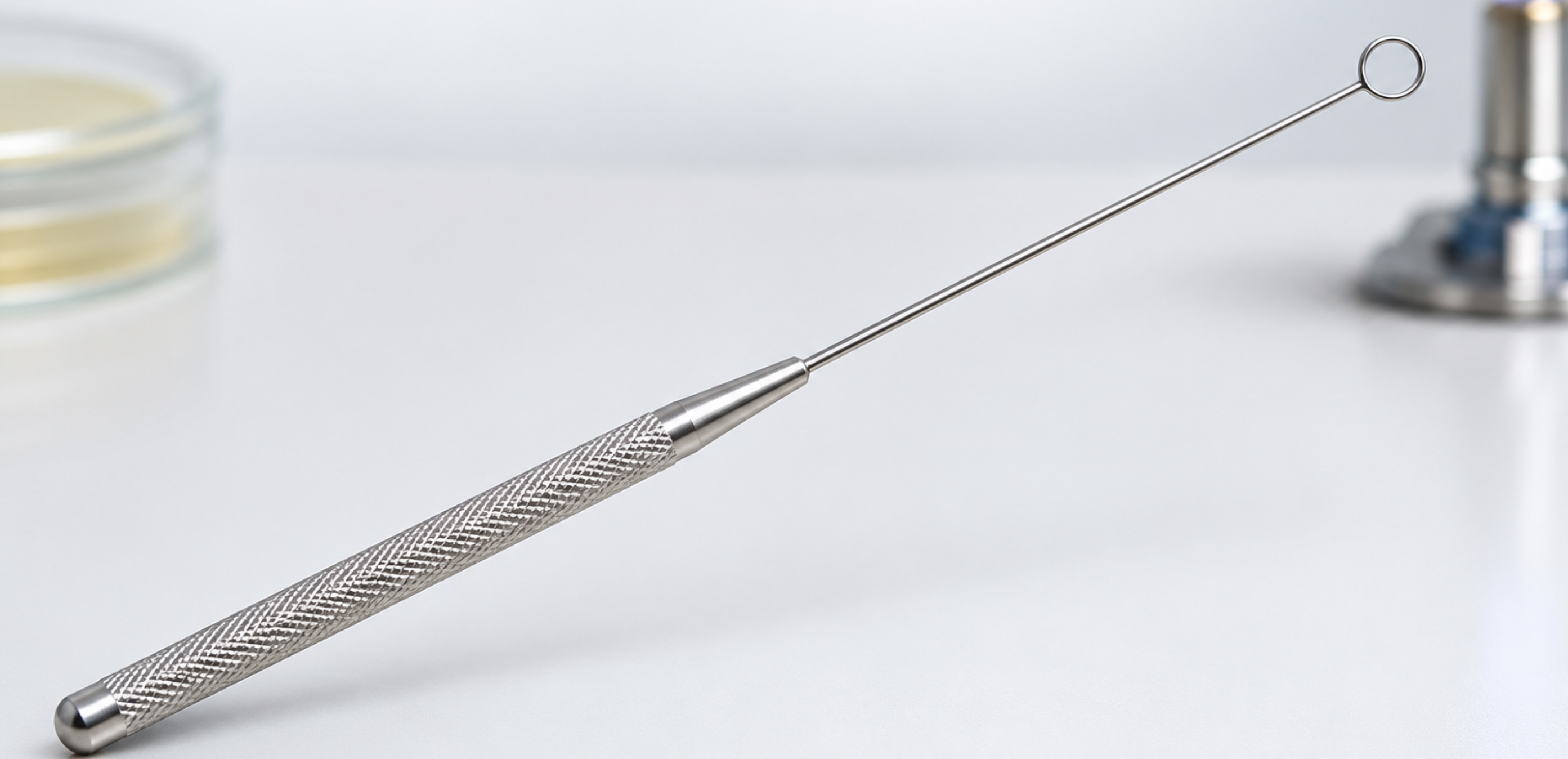
3. Inoculating loop: An inoculating loop is used to aseptically transfer bacterial material from the culture medium to the microscope slide. Proper sterilization and handling help prevent contamination and maintain the integrity of the specimen during smear preparation.
4. Bunsen burner or suitable heating apparatus: A controlled heat source is used for heat-fixing the bacterial smear and, in the Schaeffer–Fulton endospore staining method, facilitating the penetration of malachite green into the resistant endospore layers. Appropriate heating techniques are necessary to avoid overheating, distortion, or damage to the specimen.
5. Malachite green solution: Malachite green serves as the primary stain. When applied with heat, it penetrates the resistant endospore layers and imparts a green coloration to the endospores, allowing them to be distinguished from the surrounding vegetative cells.
6. Safranin solution: Safranin functions as the counterstain, imparting a pink or red coloration to vegetative bacterial cells. This contrasting coloration facilitates the differentiation of endospores from vegetative cells during microscopic examination.
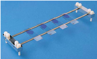
7. Distilled water and staining rack: Distilled water is used to rinse the smear and remove excess staining reagents at appropriate stages of the procedure. The staining rack supports the microscope slide during reagent application and rinsing, helping to maintain an organized and efficient staining process.
8. Compound microscope and immersion oil: A compound microscope is used to examine the stained specimen and assess the morphology, distribution, and position of endospores within bacterial cells. Immersion oil is applied when using the 100× oil-immersion objective to improve light transmission and optical resolution, thereby facilitating detailed observation of the stained bacterial structures.
Procedure for endospore staining
Preparation of the bacterial smear
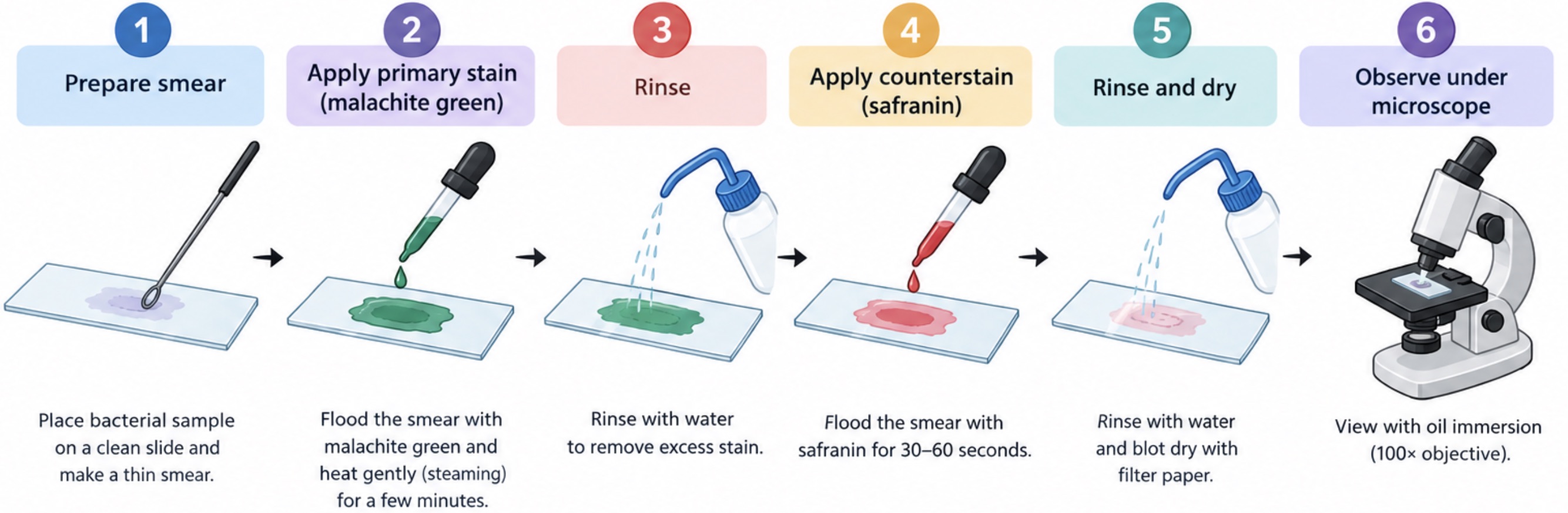
A clean, grease-free glass slide is selected to provide a suitable surface for specimen preparation. Using a sterile inoculating loop, a small quantity of the bacterial culture is transferred onto the centre of the slide (Figure 3). If the culture is grown on solid medium, a drop of sterile distilled water is added to facilitate the even dispersion of the bacterial cells. The specimen is then spread gently into a thin, uniform film. Proper smear preparation is essential because excessive bacterial material may obscure individual cells and hinder the accurate visualization of endospores under the microscope.
Air-drying and heat fixation
The prepared smear is allowed to air-dry completely at room temperature. Once dry, the slide is heat-fixed by passing it briefly through a flame or using an appropriate laboratory heat-fixation method. This process helps attach the bacterial cells to the glass surface, minimizing their loss during subsequent staining and rinsing. Excessive heating should be avoided because it may distort the cellular morphology and compromise the quality of microscopic observations.
Application of malachite green
The heat-fixed smear is placed on a staining rack, and the entire specimen is flooded with malachite green solution, which serves as the primary stain. This dye is used to impart colour to the endospores. Since the protective layers surrounding endospores restrict the penetration of ordinary staining solutions, an additional heating step is required to facilitate dye entry into these resistant structures.
Heating during primary staining
The slide is gently heated while the malachite green remains in contact with the smear, following the laboratory’s approved staining protocol. Heating facilitates the penetration of the primary stain through the resilient endospore covering. The preparation must be monitored carefully to prevent the staining solution from boiling, splattering, or evaporating completely. If necessary, additional stain is applied to keep the specimen moist throughout the heating period. Appropriate protective equipment and a safe heating method should be used to prevent accidental burns and exposure to stain aerosols.
Cooling and rinsing
After the heating stage, the slide is removed from the heat source and allowed to cool naturally. The preparation is then rinsed gently with water to remove excess malachite green from the smear. During this stage, vegetative cells generally lose most of the primary stain, whereas endospores retain the green dye because of its penetration and retention within their protective structures. Gentle rinsing is important to preserve the smear and prevent the unnecessary detachment of bacterial material from the slide.
Counterstaining with safranin
The rinsed preparation is covered with safranin solution, which acts as the counterstain. It is generally allowed to remain on the smear for approximately 30-60 seconds, or according to the laboratory’s validated staining procedure. Safranin colours the vegetative bacterial cells pink or red, thereby creating a distinct visual contrast between the cells and the endospores. This differential coloration enables the observer to distinguish spore-containing cells from their surrounding cellular structures more readily.
Final rinsing and drying
Following counterstaining, the slide is rinsed gently with clean water to eliminate excess safranin. The preparation is then blotted carefully with absorbent paper or allowed to dry using an appropriate laboratory method. Vigorous rubbing should be avoided because it can disrupt the smear and interfere with the distribution of the stained cells. A properly prepared slide should retain sufficient specimen material for clear and reliable microscopic examination.
Microscopic examination and interpretation
The dried slide is examined under a light microscope, initially using a lower-power objective to locate the stained specimen. The oil-immersion objective is then used for detailed observation, with immersion oil applied as required by the microscope system. Endospores typically appear green, while vegetative cells appear pink or red. Their arrangement and location within the cells are recorded, noting whether the spores are central, subterminal, or terminal and whether they are spherical or oval. These observations provide evidence of endospore formation and assist in the preliminary characterization of the bacterial specimen. Definitive identification, however, requires additional microbiological tests where appropriate.
Observation and results of endospore staining
Endospore staining differentiates bacterial endospores from vegetative cells based on their contrasting coloration following treatment with malachite green and safranin. After staining, the prepared smear is examined under an oil-immersion objective to evaluate the color, morphology, and intracellular position of the endospores. These characteristics provide essential evidence for determining whether the bacterial culture contains endospore-forming organisms.
Under microscopic examination, endospores typically appear green because their resistant structures retain malachite green during the staining procedure. In contrast, vegetative cells generally appear pink or red because they take up safranin, the counterstain applied after the primary staining step. This color contrast enables the differentiation of endospores from vegetative cells within the same specimen.
The position and morphology of endospores provide additional information for microscopic identification. Depending on the bacterial species, endospores may be centrally located, subterminally positioned near one end of the cell, or terminally located at the cell’s extremity. Their shape may be spherical or oval, and their size varies among organisms. In some species, the developing endospore may cause visible swelling of the mother cell, providing an additional morphological characteristic for examination.
A positive endospore staining result is indicated by green endospores against a pink or red background of vegetative cells. However, the absence of visible green structures does not conclusively demonstrate that an organism is incapable of endospore formation. Endospore detection may be affected by staining quality, culture age, and sporulation conditions. Therefore, accurate interpretation requires careful examination of multiple cells and consideration of the overall staining pattern, including the coloration, morphology, and position of any observed endospores.
Precautions to observe during endospore staining
Endospore staining requires careful handling of bacterial specimens, staining reagents, and laboratory equipment to ensure accurate and reliable microscopic observations. A clean, grease-free glass slide should be used, and the bacterial smear should be thin and evenly distributed to promote uniform staining. Controlled heating during malachite green staining is essential to facilitate dye penetration through the resistant endospore coat. Excessive heating should be avoided to prevent specimen damage and staining artefacts.
The smear must remain sufficiently moist throughout the heating process to prevent the stain from drying completely. Fresh, uncontaminated malachite green and safranin solutions should be used to ensure effective staining and minimize cross-contamination. The slide should be rinsed gently with water to remove excess stain without dislodging the bacterial material. Appropriate personal protective equipment (PPE) must be worn, and heated slides should be handled using suitable instruments to prevent burns.
Proper aseptic techniques and established biosafety procedures must be followed when handling bacterial cultures. Before microscopic examination, the stained smear should be allowed to air-dry completely, and immersion oil should be applied appropriately when using the oil-immersion objective. Finally, contaminated materials and biological waste must be decontaminated and disposed of in accordance with laboratory safety guidelines to maintain a safe working environment.
References
Atrih, A., & Foster, S. J. (2002). Bacterial endospores: The ultimate survivors. International Dairy Journal, 12(2–3), 217–223.
Flores, M. J., & Popham, D. L. (2026). Bacterial endospores. In eLS. John Wiley & Sons.
Kawai, S. (2026). Purification and detection of bacterial endospores: Current methods and challenges. Applied Sciences, 16(11), Article 5702.
Knaysi, G. (1948). The endospore of bacteria. Bacteriological Reviews, 12(1), 19–77.
Moir, A. (2023). Spore germination receptors—a new paradigm. Trends in Microbiology, 31(8), 767–768.
Nicholson, W. L., Fajardo-Cavazos, P., Rebeil, R., Slieman, T. A., Riesenman, P. J., Wall, J. D., & Xue, Y. (2002). Bacterial endospores and their significance in stress resistance. Antonie van Leeuwenhoek, 81, 27–32.
Reynolds, J., Moyes, R., & Breakwell, D. P. (2009). Differential staining of bacteria: Endospore stain. Current Protocols in Microbiology, Appendix 3, Appendix 3J.
Discover more from Microbiology Class
Subscribe to get the latest posts sent to your email.