
Lactophenol cotton blue (LPCB) staining is a fundamental mycological technique employed for the microscopic examination and morphological characterization of fungi. It provides a practical means of visualizing fungal structures by enhancing the contrast between cellular components and the surrounding mounting medium. The technique is particularly valuable in microbiology laboratories, where the identification of fungal isolates frequently depends on the detailed examination of their structural and reproductive features. By facilitating the observation of characteristic morphological patterns, LPCB staining contributes to the preliminary identification and differentiation of fungi based on their microscopic appearance.
The effectiveness of this staining technique is associated with the composition of the lactophenol cotton blue reagent, which typically contains cotton blue dye, phenol, lactic acid, glycerol, and water. Each constituent performs a distinct function in preparing fungal specimens for microscopic analysis. Cotton blue provides coloration to fungal cell walls, particularly through its affinity for chitin-containing structures, thereby improving their visibility. Phenol acts as a fungicidal component, reducing the viability of fungal cells during preparation, while lactic acid helps preserve the morphological integrity of the specimen. Glycerol serves as a mounting agent that limits dehydration and maintains the preparation in a suitable condition for examination. These components facilitate the visualization of fungal architecture while minimizing structural distortion during slide preparation.
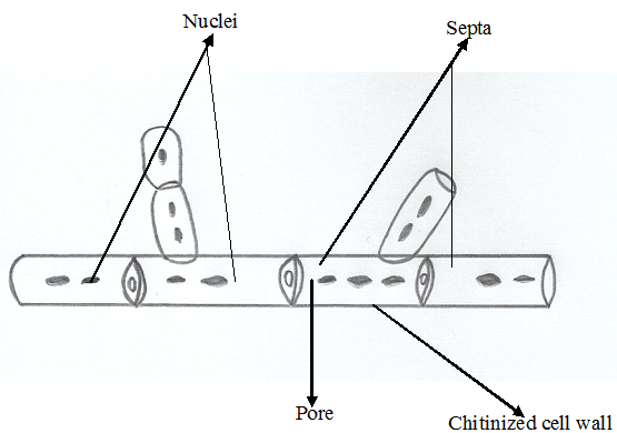
LPCB staining is especially useful for examining the major morphological characteristics of fungi, including hyphae, conidia, sporangia, and other reproductive structures. Hyphae may be assessed for their width, branching pattern, septation, and general organization, while reproductive structures can be examined according to their shape, arrangement, and mode of development. These features are important in distinguishing different fungal groups and supporting the recognition of characteristic structures associated with particular genera. For example, the arrangement of conidia, the presence or absence of septa, and the organization of specialized hyphae may provide useful morphological evidence during laboratory identification.
The procedure generally involves transferring a small quantity of fungal material into a drop of LPCB reagent on a clean glass slide, gently dispersing the specimen, and covering it with a coverslip before microscopic examination. Observation typically begins under low magnification to locate the fungal material, followed by higher magnification to assess finer structural details. Careful specimen preparation is essential because excessive fungal material, overlapping structures, or mechanical disruption may interfere with accurate interpretation.
Although LPCB staining remains a valuable method for routine fungal morphology, it does not independently establish the definitive identity of every fungal isolate. Closely related species may exhibit similar microscopic characteristics, and some diagnostic features may not be apparent in a single preparation. Consequently, observations obtained through LPCB staining should be interpreted alongside relevant cultural characteristics, additional laboratory tests, or molecular identification methods when necessary. The technique remains an important component of conventional mycological investigation because of its simplicity, accessibility, and capacity to reveal structural features essential for the preliminary examination and classification of fungi.
Principle of lactophenol cotton blue staining technique
LPCB staining is based on the combined action of four chemical components that facilitate the visualization and preservation of fungal structures during microscopic examination. The staining solution comprises lactic acid, phenol, glycerol, and cotton blue dye, each contributing a specific function to the preparation. Their complementary properties enable the differentiation of important morphological features, including hyphae, conidia, sporangia, and other reproductive structures, thereby supporting the preliminary identification and characterization of fungi.
Cotton blue, also known as aniline blue in certain formulations, serves as the principal staining agent. It has an affinity for chitin and other polysaccharide components of fungal cell walls, producing a blue coloration that enhances the contrast between fungal elements and the surrounding mounting medium. This coloration facilitates the examination of structural details, such as hyphal septation, branching patterns, spore arrangement, and the configuration of reproductive structures. The dye improves the visibility of fungal morphology under a light microscope.
Phenol functions primarily as a fungicidal agent, reducing the viability of fungal cells within the preparation. This property contributes to safer handling of the stained material, although the preparation should not be regarded as a substitute for appropriate laboratory biosafety procedures. Lactic acid acts as a preservative and clearing agent, helping to maintain the structural integrity of fungal elements while improving their visibility. By limiting excessive deterioration of the specimen, it supports the examination of characteristic morphological features that are useful in fungal differentiation.
Glycerol serves as a humectant and mounting component, retaining moisture within the preparation and slowing evaporation. This action helps prevent the rapid drying of the specimen, allowing sufficient time for microscopic observation. It also contributes to maintaining a stable mounting environment in which fungal structures can be examined with minimal distortion. The effectiveness of LPCB staining therefore depends on the coordinated action of its constituents rather than the activity of a single reagent.
The dye enhances contrast, phenol reduces fungal viability, lactic acid preserves and clears structural elements, and glycerol maintains the preparation in a moist state. Together, these components produce a suitable microscopic preparation for studying fungal morphology. However, LPCB staining primarily supports morphological assessment and does not independently establish species identity; accurate identification may require additional cultural, biochemical, molecular, or other confirmatory investigations.
Materials and reagents required for LPCB
LPCB staining technique requires a selection of laboratory materials, staining reagents, and microscopy equipment to facilitate the preparation and examination of fungal specimens. Each component contributes to the effective mounting, staining, preservation, and visualization of fungal structures, thereby supporting accurate morphological assessment.
1. Lactophenol cotton blue stain: This is the primary reagent used in the procedure. It comprises cotton blue dye, phenol, lactic acid, and glycerol, each serving a distinct function. Cotton blue imparts colour to fungal cell walls, improving the visibility of hyphae and reproductive structures. Phenol acts as a fungicidal component, while lactic acid helps preserve fungal morphology. Glycerol maintains moisture within the preparation and reduces rapid drying during microscopic examination.
2. Clean glass slides: Microscope slides provide a stable, transparent surface on which the fungal specimen and staining reagent are combined. They should be free from dust, grease, and residual chemicals that could interfere with specimen distribution or image clarity. Clean slides facilitate the production of an even preparation suitable for microscopic observation.
3. Coverslips: Thin glass coverslips are used to cover the mounted specimen. They help distribute the fungal material evenly, minimize movement during examination, and protect the microscope objective from direct contact with the preparation. Proper placement also reduces the formation of air bubbles that may obstruct the visualization of fungal structures.
4. Inoculating or mounting needle: A sterile inoculating needle, mounting needle, or suitable teasing instrument is required to transfer a small portion of the fungal specimen into the staining reagent. It can also be used to gently separate intertwined hyphae and distribute the material across the slide. Sterile instruments help minimize cross-contamination between specimens.
5. Fungal culture or specimen: The specimen serves as the biological material for examination. It may consist of fungal growth obtained from an appropriate culture medium or a suitable fungal sample collected for laboratory investigation. The specimen should be handled aseptically and in accordance with the relevant biosafety requirements to maintain sample integrity and reduce exposure to potentially harmful organisms.
6. Light microscope: A compound light microscope is essential for examining the stained preparation. Low-power magnification allows the general distribution of fungal material to be assessed, while higher magnification facilitates closer examination of hyphal characteristics, spore morphology, and reproductive structures. Together, these materials and reagents support the systematic preparation and preliminary morphological identification of fungi using the LPCB staining technique.
Procedure for lactophenol cotton blue staining
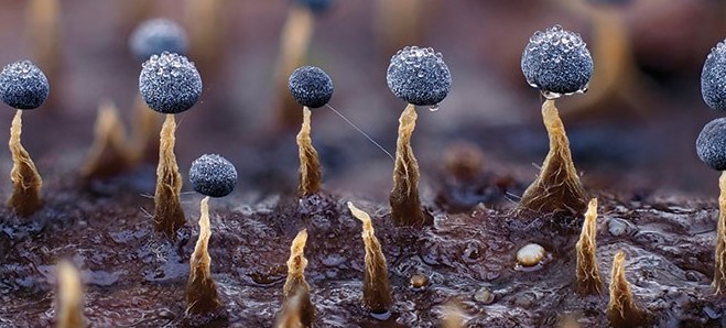
The LPCB staining technique is performed to prepare fungal specimens for microscopic examination and to reveal their characteristic morphological features. The procedure requires careful handling of the fungal material to preserve delicate structures, including hyphae, conidia, sporangia, and other reproductive elements (Figure 1). Accurate slide preparation is essential for obtaining a clear microscopic image and facilitating the preliminary identification of fungal isolates.
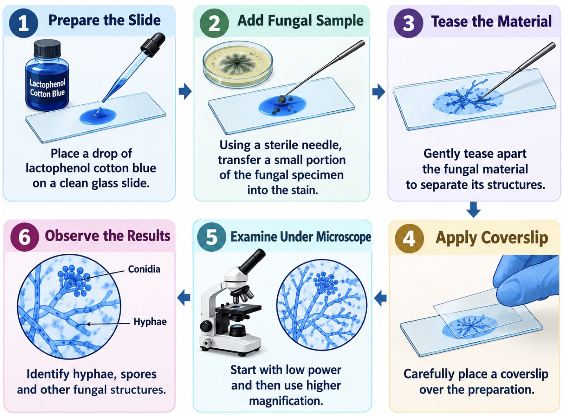
Step 1: Preparation of the microscope slide: A clean, dry glass slide is selected to provide a suitable surface for mounting the fungal specimen. A small drop of lactophenol cotton blue stain is placed at the centre of the slide using a dropper or applicator. The stain serves as the mounting medium and colouring agent, allowing the fungal structures to become more distinguishable during microscopic observation. Care is taken to ensure that the slide is free from dust, grease, and other contaminants that could interfere with the visibility of the specimen.
Step 2: Collection and transfer of the fungal specimen: A sterile inoculating needle or mounting needle is used to collect a small portion of the fungal growth from the culture. The specimen is transferred gently into the drop of lactophenol cotton blue on the slide. For filamentous fungi, a small fragment taken from the actively growing margin of the colony may provide well-preserved hyphae and developing reproductive structures. Excessive material should be avoided because a thick preparation can cause overlapping structures and reduce microscopic clarity. Appropriate laboratory precautions should be observed when handling fungal cultures to minimise exposure and cross-contamination.
Step 3: Dispersion and mounting of the fungal material: The fungal material is carefully teased apart within the stain using the needle. This action helps disperse tangled filaments and separate the individual components of the specimen, thereby improving the visibility of their structural arrangement. Excessive pressure or vigorous manipulation should be avoided because delicate hyphae and reproductive structures may become distorted or fragmented. Once the specimen has been adequately dispersed, a clean coverslip is lowered gently over the preparation at an angle to minimise the formation of air bubbles. The coverslip should rest evenly on the preparation without applying unnecessary pressure.
Step 4: Examination under the microscope: The prepared slide is placed on the microscope stage and secured appropriately. Examination begins with the low-power objective, which provides an overview of the specimen and helps locate regions containing clearly distributed fungal structures. The magnification is subsequently increased to examine finer morphological details. The medium- or high-power objective may be used, as appropriate, to investigate the characteristics of the hyphae, spores, conidiophores, and other diagnostic structures. Fine adjustment is performed carefully to achieve optimal focus, contrast, and resolution. The slide should be examined systematically rather than concentrating on a single field of view.
Step 5: Documentation of microscopic findings: The observable characteristics of the fungal specimen are recorded systematically. Particular attention is given to hyphal septation, branching patterns, spore shape, spore arrangement, and the morphology of specialised reproductive structures. The distribution and intensity of the blue staining are also noted where relevant. These observations provide morphological evidence that may assist in the preliminary identification of the fungus. Photomicrographs or labelled drawings may be prepared when required for laboratory records or academic reporting. Identification should be based on the combined morphological characteristics rather than on staining colour alone.
Step 6: Completion of the procedure and slide handling: Following microscopic examination, the preparation is handled and discarded according to the laboratory’s established biosafety procedures. Used needles and other contaminated materials are placed in their designated disposal containers, and the work surface is disinfected appropriately. The microscope stage and any contaminated equipment are cleaned according to laboratory guidelines. Proper documentation of the findings completes the procedure and supports reproducibility, accurate interpretation, and subsequent comparison with established fungal identification criteria.
Observation and interpretation of LPCB results
Following the preparation of the lactophenol cotton blue-stained slide, the specimen is examined under a light microscope to assess its microscopic characteristics. Observation begins with the low-power objective to locate the fungal material and establish the general distribution of its structures. The higher-power objective is then used to investigate specific morphological features in greater detail. The cotton blue dye enhances the visibility of fungal cell walls, allowing the structural organization of the specimen to be distinguished more clearly against the background.
During microscopic examination, attention is directed towards the presence, appearance, and arrangement of hyphae, spores, and specialized reproductive structures. Hyphae are assessed for their width, branching pattern, and septation, which may be described as septate or aseptate, depending on the presence or absence of cross-walls. Their orientation, continuity, and degree of branching are also documented, as these characteristics contribute to the morphological description of the fungus. Any variation in hyphal appearance, including irregular growth or fragmentation, is recorded where observed.
The reproductive features of the specimen are examined to identify the presence and distribution of spores or conidia. Their shape, size, pigmentation, and arrangement are noted alongside the structures responsible for their formation. Depending on the fungal organism, the preparation may reveal conidia arranged in chains or clusters, sporangia containing spores, or other distinctive reproductive formations. The relationship between these structures and the supporting hyphae is particularly important because their organization may provide useful clues for distinguishing between fungal groups.
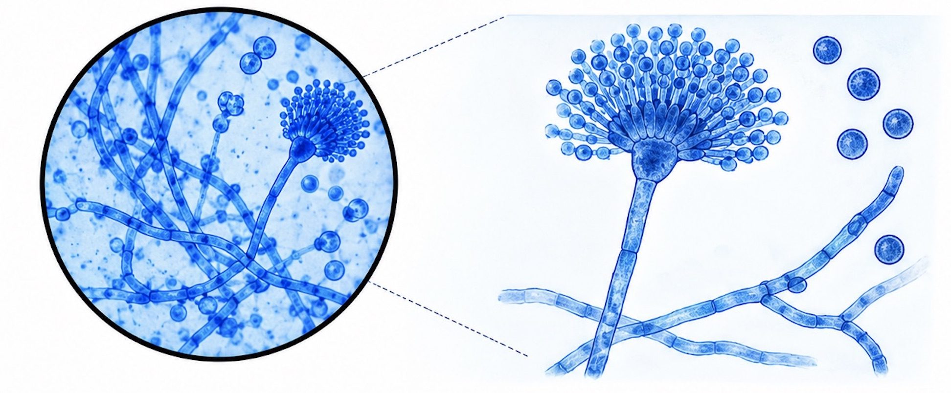
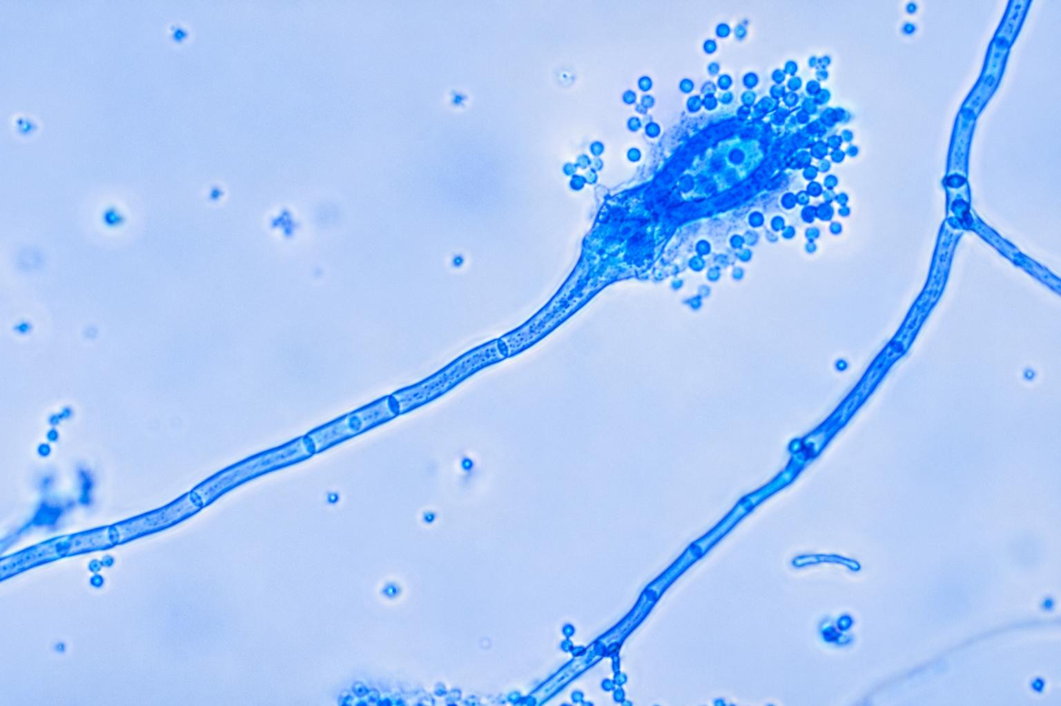
For example, the microscopic image of Aspergillus stained with LPCB reveals characteristic fungal structures, including hyphae, spores and conidia (Figure 2). The blue staining enhances the contrast and visibility of these structures, allowing detailed examination of hyphal morphology, branching patterns and conidial arrangement. The observed morphological features provide valuable preliminary evidence for the identification of Aspergillus and differentiation from other fungal genera. However, morphological observations alone may not be sufficient for definitive species-level identification, which may require complementary molecular or biochemical analyses.
Interpretation of a LPCB result is usually based on the combined morphological characteristics observed rather than on a single feature. The recorded findings are compared with established descriptions of fungal morphology to determine whether the specimen exhibits features consistent with a particular fungal group or genus. For example, differences in hyphal septation, spore arrangement, and the architecture of reproductive structures may help narrow the range of possible identifications. Nevertheless, LPCB staining primarily supports morphological examination and does not independently confirm the identity of every fungal species.
The final observation is documented in a clear and systematic manner, including the magnification used and the principal structures identified. Where possible, a labelled photomicrograph may be included to support the recorded findings. If the observed characteristics are insufficient for reliable identification, additional investigations, such as culture-based assessment, biochemical testing, or molecular methods, may be required. Accurate observation and cautious interpretation therefore ensure that the results reflect the actual microscopic features of the specimen without overstating the certainty of identification.
Precautions to take when carrying out LPCB staining technique
- Ensure that glass slides, coverslips, and mounting instruments are clean before use to minimize contamination and maintain the clarity of the microscopic preparation.
- Transfer only a small quantity of fungal material into the lactophenol cotton blue stain, as excessive specimen accumulation may produce overlapping structures and interfere with accurate morphological examination.
- Handle fungal cultures with appropriate laboratory precautions to reduce the risk of exposure to potentially harmful fungal spores and airborne contaminants.
- Avoid introducing air bubbles during coverslip placement, since they may obstruct the microscopic field and complicate the interpretation of fungal structures.
- Prevent unnecessary disturbance of the preparation after mounting to preserve the natural arrangement of hyphae, conidia, and other reproductive elements.
- Examine the stained specimen under suitable magnification, beginning with the low-power objective before progressing to higher magnification for detailed structural assessment.
- Dispose of contaminated materials according to established laboratory protocols, and observe appropriate precautions when handling phenol-containing staining reagents.
Applications of LPCB staining technique
- LPCB staining is employed in mycology laboratories to investigate the microscopic architecture of fungi, including hyphal morphology, spore formation, and reproductive structures.
- The technique facilitates the differentiation of septate and aseptate hyphae by improving the visibility of their structural characteristics.
- It supports the presumptive identification of fungal isolates through the examination of conidial arrangement, sporangia, branching patterns, and other diagnostic morphological features.
- LPCB preparations are useful in the routine examination of fungal cultures obtained during educational practicals, laboratory investigations, and microbiological research.
- The method provides a relatively simple approach for documenting fungal morphology and comparing observed characteristics with established identification criteria.
- It can assist in distinguishing among fungal genera when characteristic microscopic features are present and adequately preserved.
- Despite its usefulness, LPCB staining does not independently establish definitive species identity. Results should be interpreted alongside colony morphology, clinical or environmental findings, and additional identification methods when necessary.
References
Oliver, E. M., Bridge, P. D., & Hawksworth, D. L. (1987). The effect of selected mountants and stains on the measurement of fungal spores. Mycopathologia, 97, 165–172.
Leck, A. (1999). Preparation of lactophenol cotton blue slide mounts. Community Eye Health, 12(30), 24.
Khubnani, H., Sivarajan, K., & Khubnani, A. H. (1998). Application of lactophenol cotton blue for identification and preservation of intestinal parasites in faecal wet mounts. Indian Journal of Pathology & Microbiology, 41(2), 157–162.
Shamly, V., Kali, A., Srirangaraj, S., & Umadevi, S. (2014). Comparison of microscopic morphology of fungi using lactophenol cotton blue (LPCB), iodine glycerol and Congo red formaldehyde staining. Journal of Clinical and Diagnostic Research, 8(7), DL01–DL02.
Sangeetha, J., & Thangadurai, D. (2013). Staining techniques and biochemical methods for the identification of fungi. In V. Gupta, M. Tuohy, M. Ayyachamy, K. Turner, & A. O’Donovan (Eds.), Laboratory protocols in fungal biology. Springer.
Toma, T., Kováč, J., & Ďurkovič, J. (2024). Fast, easy, and comprehensive techniques for microscopic observations of fungal and oomycete organisms inside the roots of herbaceous and woody plants. Bio-protocol, 14(11), e5013.
Gray, P. (1941). Staining of bacteria and certain fungi. Nature, 147, 329.
Kowal, J., Arrigoni, E., & Lane, S. (2020). Acidified blue ink-staining procedure for the observation of fungal structures inside roots of two disparate plant lineages. Bio-protocol, 10(20), e3786.
Discover more from Microbiology Class
Subscribe to get the latest posts sent to your email.