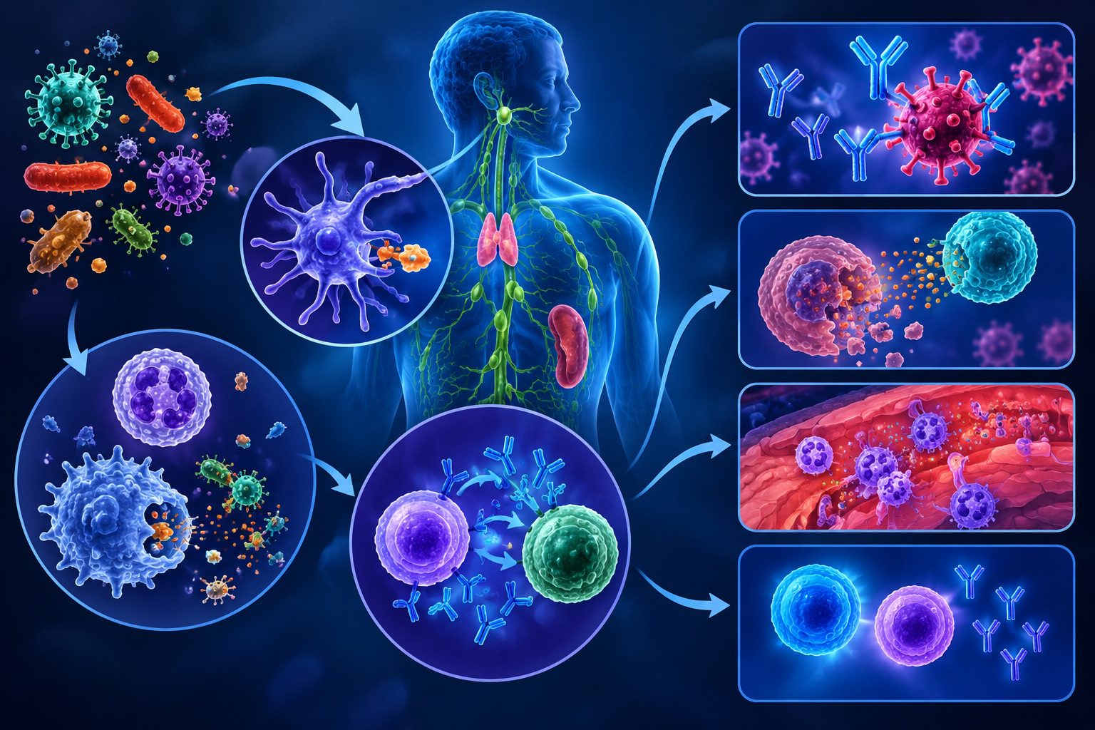

Antibodies, scientifically known as immunoglobulins (Igs), are specialised glycoproteins that constitute a fundamental component of the body’s adaptive immune defence. They are produced in response to the presence of foreign substances, collectively referred to as antigens, which may include bacteria, viruses, fungi, parasites, toxins, and other potentially harmful biological materials. Through their remarkable capacity to recognise and bind specific molecular structures, antibodies serve as highly selective biological recognition molecules, enabling the immune system to distinguish foreign materials from the body’s own cellular components.
Their activity represents one of the most sophisticated mechanisms through which the human body detects, targets, and eliminates infectious agents. Within the immune system, antibodies function as molecular mediators that translate antigen recognition into protective immune responses. Each antibody possesses a distinctive antigen-binding region that interacts with a particular antigenic determinant, also called an epitope. This interaction establishes the specificity of the antibody-antigen relationship and determines the nature of the subsequent immune response.
Following antigen recognition, antibodies can neutralize harmful substances by preventing pathogens or toxins from attaching to and entering host cells. They can also facilitate the removal of foreign particles through opsonization, a process in which pathogens are coated with antibodies to enhance their recognition and engulfment by phagocytic cells. Certain antibody classes initiate the complement cascade, a series of coordinated protein reactions that contributes to microbial destruction and the clearance of immune complexes.
The biological production of antibodies is primarily associated with B-lymphocytes, specialised white blood cells that develop and mature within the bone marrow. When a B-lymphocyte encounters an antigen corresponding to its specific receptor, it may undergo activation through a sequence of cellular and molecular interactions, often involving assistance from helper T-lymphocytes. Activated B-cells subsequently proliferate through clonal expansion, generating populations of cells with identical antigen specificity.
A proportion of these activated cells differentiates into plasma cells, which are specialized antibody-secreting cells responsible for releasing substantial quantities of immunoglobulins into the bloodstream, lymphatic circulation, and other body fluids. Other activated B-lymphocytes develop into memory B-cells, which persist within the body and facilitate a faster, more effective antibody response following subsequent exposure to the same antigen.
The protective significance of antibodies extends beyond the immediate elimination of invading microorganisms. They contribute to immunological surveillance, support communication between different components of the immune system, and establish a foundation for long-term adaptive protection. Their presence in blood, mucosal secretions, and other biological compartments enables the body to mount defensive responses at multiple anatomical sites.
Maternal antibodies transferred through the placenta and breast milk provide essential passive protection to infants during early development, when their immune systems are still undergoing maturation. Antibodies represent indispensable molecular effectors of adaptive immunity. Their antigen-specific recognition, diverse biological activities, and capacity to support immunological memory make them essential for host protection, resistance to infectious diseases, and the maintenance of immune homeostasis.
Structure of antibodies
Immunoglobulins, are specialised glycoproteins whose molecular architecture enables them to recognize and bind specific antigens. Their structural organisation is fundamental to their biological activity, as each component contributes to antigen recognition, immune regulation and the elimination of foreign substances. Although antibodies differ in their antigen-binding characteristics, they share a characteristic structural framework that gives them their distinctive Y-shaped configuration. This molecular arrangement provides both flexibility and functional precision, allowing antibodies to interact with a wide variety of antigens.
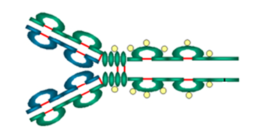
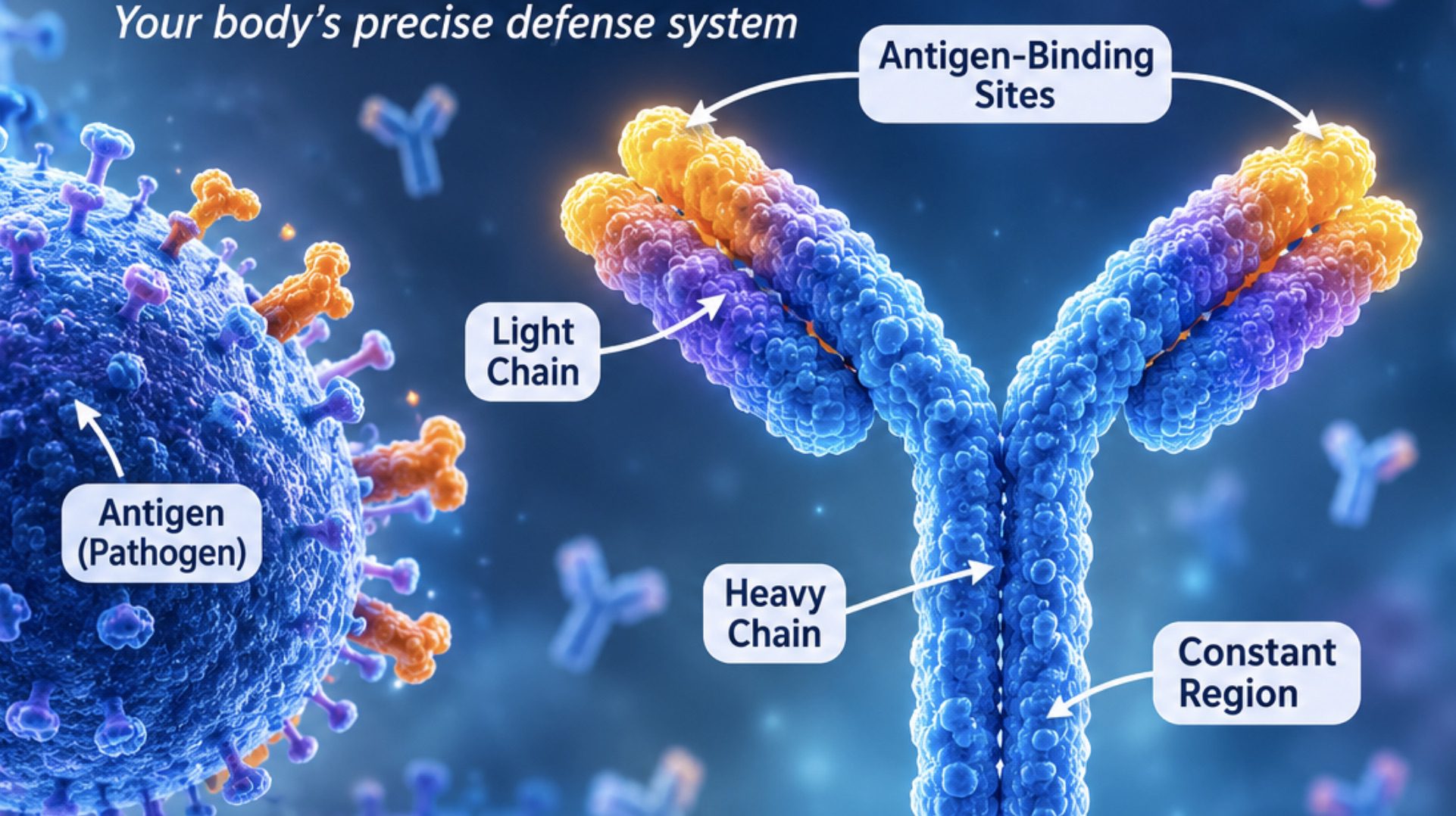
Basic structure of an immunoglobulin molecule: A typical antibody molecule consists of four polypeptide chains arranged in a symmetrical Y-shaped structure (Figure 1). These chains are held together by chemical bonds that maintain the stability and integrity of the molecule. The two upper arms of the Y-shaped structure are responsible primarily for antigen recognition, while the lower stem participates in immune effector activities. Each antibody possesses two antigen-binding sites, enabling it to attach to corresponding antigenic determinants, also known as epitopes.
The molecular framework is organized into distinct domains, which are compact regions of folded protein stabilised by internal disulphide bonds. This arrangement provides structural resilience while maintaining the conformational adaptability required for effective antigen binding. The overall architecture therefore combines stability with specificity, making antibodies highly efficient components of adaptive immunity.
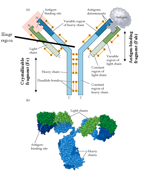
Heavy and light polypeptide chains: The four polypeptide chains forming an antibody molecule are classified into two heavy chains and two light chains. The heavy chains are comparatively larger, with molecular weights of approximately 50,000 daltons each, whereas the light chains weigh approximately 25,000 daltons each. Both pairs are arranged in an orderly configuration and connected through interchain disulphide bonds and non-covalent interactions.
Light chains occur in two principal forms: kappa (κ) and lambda (λ). An individual antibody contains either two kappa chains or two lambda chains, rather than a mixture of both. Heavy chains, on the other hand, determine the immunoglobulin class and are designated gamma (γ), mu (μ), alpha (α), epsilon (ε) and delta (δ), corresponding to IgG, IgM, IgA, IgE and IgD, respectively.
The association between heavy and light chains contributes to the formation of functional antigen-binding regions. Their coordinated arrangement ensures that the antibody maintains the appropriate three-dimensional configuration necessary for recognising specific molecular targets.
Variable and constant regions: Each heavy and light chain contains two major structural regions: the variable region and the constant region. The variable regions are located towards the amino-terminal ends of the polypeptide chains and exhibit considerable differences in amino acid sequences among antibodies. These variations account for the remarkable diversity of antigen recognition within the immune system.
Within the variable regions are specialised segments called complementarity-determining regions (CDRs). These regions form the principal antigen-contacting surfaces and determine the binding specificity of individual antibodies. The intervening framework regions provide structural support, maintaining the appropriate spatial arrangement of the CDRs.
In contrast, constant regions exhibit relatively conserved amino acid sequences within particular immunoglobulin classes. They contribute to the structural stability of antibodies and influence their biological functions, including interactions with complement proteins and Fc receptors on immune cells. The constant region of the heavy chain also determines the characteristic properties of each immunoglobulin class.
Antigen-binding sites and disulphide bonds: The antigen-binding sites are specialised molecular surfaces formed through the interaction of the variable regions of one heavy chain and one light chain. Each antibody molecule ordinarily possesses two identical antigen-binding sites, giving it the capacity to bind two corresponding epitopes. The binding process relies on non-covalent interactions, including hydrogen bonding, electrostatic attraction, hydrophobic interactions and van der Waals forces.
Disulphide bonds are essential structural linkages that maintain the organisation of antibody molecules. They form through the oxidation of sulphydryl groups in cysteine residues, connecting the two heavy chains and attaching each light chain to a heavy chain. Additional intrachain disulphide bonds stabilise individual immunoglobulin domains by maintaining their characteristic folded configurations.
The Y-shaped structure also contains a flexible hinge region in several immunoglobulin classes, particularly IgG, IgA and IgD. This region permits movement of the antibody arms, allowing the antigen-binding sites to accommodate epitopes positioned at different distances and orientations. Consequently, the combined arrangement of polypeptide chains, specialized binding regions and stabilizing chemical bonds establishes the structural foundation of antibody specificity and immune protection.
Classes (types) of immunoglobulins
Immunoglobulins constitute an essential component of humoral immunity, providing the body with a highly selective mechanism for identifying and eliminating foreign substances. Although all immunoglobulins share a characteristic molecular architecture consisting of heavy and light polypeptide chains, variations in their heavy-chain constant regions give rise to five major classes: Immunoglobulin G (IgG), Immunoglobulin M (IgM), Immunoglobulin A (IgA), Immunoglobulin E (IgE), and Immunoglobulin D (IgD).
Each class possesses distinctive structural properties, distribution patterns, and biological activities that enable it to perform specialized protective functions. the five immunoglobulin classes demonstrate the functional versatility of the antibody-mediated immune system. Their distinct structural configurations and biological specializations enable coordinated protection across the bloodstream, mucosal surfaces, and tissue environments, ensuring that immune responses remain appropriately adapted to different categories of infectious and foreign challenges.
Immunoglobulin G (IgG)
Immunoglobulin G is the most abundant antibody class in human blood plasma, accounting for approximately 75-80% of circulating immunoglobulins. It is a monomeric molecule characterized by two heavy chains and two light chains connected through disulfide bonds. IgG plays a fundamental role in sustained immune protection, particularly during the later stages of an immune response. Its principal activities include neutralizing bacterial toxins and viral particles, promoting phagocytosis through opsonization, and activating the classical complement pathway. An exceptional characteristic of IgG is its ability to cross the placental barrier, thereby transferring maternal antibodies to the developing fetus and providing passive immunity during early infancy. Its four subclasses, IgG1, IgG2, IgG3, and IgG4, exhibit differences in their capacity to activate complement and interact with immune cells.
Immunoglobulin M (IgM)
Immunoglobulin M is generally the first antibody produced during an individual’s initial exposure to a previously encountered antigen or a new infectious agent. In its predominant secreted form, IgM exists as a pentamer composed of five antibody units joined by a joining (J) chain. This arrangement provides multiple antigen-binding sites, giving IgM considerable avidity and making it particularly effective in agglutinating microorganisms and other particulate antigens. IgM is also a powerful activator of the classical complement pathway, contributing significantly to the early elimination of invading pathogens. Although it is predominantly confined to the bloodstream because of its relatively large molecular size, its membrane-bound form functions as an antigen receptor on naïve B-lymphocytes. IgM serves both as an early defensive antibody and an important participant in initiating adaptive immune responses.
Immunoglobulin A (IgA)
Immunoglobulin A is primarily associated with mucosal immunity, where it safeguards body surfaces exposed to the external environment. It is found in secretions such as saliva, tears, mucus, breast milk, and intestinal fluids. In the bloodstream, IgA commonly occurs as a monomer, whereas secretory IgA predominantly exists as a dimer containing two antibody units connected by a J chain and protected by a secretory component. This specialized structure enhances its resistance to enzymatic degradation within mucosal secretions. IgA protects epithelial surfaces by preventing microorganisms from attaching to host cells, neutralizing pathogens and toxins, and limiting microbial penetration into underlying tissues. Through these activities, it establishes an important immunological barrier without producing excessive inflammatory damage. Secretory IgA in breast milk also contributes to the protection of infants against gastrointestinal and respiratory infections.
Immunoglobulin E (IgE)
Immunoglobulin E is present in extremely low concentrations in normal blood plasma but possesses considerable biological significance in allergic reactions and defence against certain parasitic infections. Its principal mechanism of action involves binding to high-affinity receptors on mast cells and basophils. When an allergen subsequently cross-links the IgE molecules attached to these cells, it triggers degranulation and the release of inflammatory mediators, particularly histamine. These substances produce characteristic allergic manifestations, including itching, swelling, increased mucus secretion, and bronchoconstriction. IgE also participates in immune responses against helminths by facilitating the recruitment and activation of eosinophils and other effector cells. Despite its protective contribution to antiparasitic immunity, excessive or inappropriate IgE-mediated activity is implicated in allergic disorders such as asthma, allergic rhinitis, and anaphylaxis.
Immunoglobulin D (IgD)
Immunoglobulin D is one of the least abundant immunoglobulin classes in circulating blood, yet it performs an important regulatory function in B-cell immunity. It exists predominantly as a monomer and is expressed on the surface of mature naïve B-lymphocytes, where it functions alongside IgM as part of the B-cell antigen receptor complex. Through antigen recognition and subsequent intracellular signalling, membrane-bound IgD contributes to B-cell activation, maturation, and the initiation of appropriate immune responses. Although its precise functions are not as extensively characterized as those of the other immunoglobulin classes, evidence suggests that IgD also participates in immune surveillance at respiratory mucosal surfaces. Its significance therefore lies primarily in the regulation and coordination of B-lymphocyte activity rather than in direct antibody-mediated pathogen elimination.
Functions of antibodies
The protective activities of antibodies extend beyond the simple recognition of foreign substances, as they participate in several immunological mechanisms that facilitate the elimination of invading microorganisms, neutralization of harmful products, and destruction of infected or abnormal cells. The major functions of antibodies include neutralization, agglutination, precipitation, complement activation, opsonization, phagocytosis enhancement, and antibody-dependent cell-mediated cytotoxicity.
Neutralization of toxins and viruses: Antibodies protect the body by neutralizing bacterial toxins, viral particles, and other potentially harmful substances. This occurs when antibodies bind specifically to the biologically active regions of toxins or the surface structures of viruses, thereby preventing them from interacting with their target cells.
For instance, antibodies directed against bacterial exotoxins can obstruct the binding of these toxins to cellular receptors, preventing the cellular damage they would otherwise produce. Similarly, antiviral antibodies may interfere with viral attachment, penetration, or entry into susceptible host cells. Through this mechanism, antibodies restrict the establishment and spread of infections without necessarily destroying the microorganisms directly.
Agglutination and precipitation of antigens: Antibodies possess multiple antigen-binding sites that enable them to connect several antigenic particles simultaneously. This property facilitates agglutination, a process in which antibodies cross-link particulate antigens, such as bacteria and red blood cells, into visible clusters. Agglutination reduces the mobility and dispersal of microorganisms, making them easier for phagocytic cells to recognize and eliminate.
Precipitation, on the other hand, occurs when soluble antigens interact with their corresponding antibodies to form larger, insoluble antigen-antibody complexes. These complexes can subsequently be removed from body fluids through phagocytosis and other clearance mechanisms. Both reactions contribute to the sequestration and elimination of foreign antigens from the body.
Activation of the complement system: Antibodies participate in the activation of the complement system, an interconnected network of circulating and membrane-associated proteins that contributes to innate and adaptive immune defense. When particular antibodies, especially IgM and certain subclasses of IgG, bind to their corresponding antigens, their Fc regions become available for recognition by the complement protein C1q. This interaction initiates the classical complement pathway, resulting in a cascade of sequential protein activations.
Complement activation produces several protective outcomes, including enhanced opsonization, inflammatory recruitment, and direct microbial destruction. For example, the complement fragment C3b attaches to microbial surfaces and facilitates their recognition by phagocytes. Additionally, the terminal complement components assemble into the membrane attack complex (MAC), which creates pores in susceptible microbial membranes, resulting in osmotic disruption and cell lysis.
Opsonization and enhancement of phagocytosis: Opsonization refers to the process by which microorganisms are coated with molecules known as opsonins, which facilitate their recognition and ingestion by phagocytic cells. Antibodies, particularly IgG, function as effective opsonins by attaching their antigen-binding regions to microbial surfaces while exposing their Fc regions for recognition by Fc receptors on phagocytes, such as macrophages and neutrophils.
This interaction strengthens the attachment between phagocytes and microorganisms, thereby promoting engulfment and intracellular destruction. Following recognition, the phagocyte surrounds the microorganism with its cell membrane to form a phagosome. The phagosome subsequently fuses with lysosomes, producing a phagolysosome in which digestive enzymes and antimicrobial substances facilitate microbial degradation. Complement-derived C3b can further enhance this process by acting alongside antibody-mediated opsonization.
Antibody-dependent cell-mediated cytotoxicity (ADCC): Antibodies also promote antibody-dependent cell-mediated cytotoxicity (ADCC), an immune mechanism through which antibody-coated target cells are destroyed by specialized immune effector cells. In this process, antibodies bind to antigens expressed on the surface of infected, abnormal, or foreign cells. Their exposed Fc regions are subsequently recognized by Fc receptors on cytotoxic immune cells, particularly natural killer (NK) cells.
Following this interaction, NK cells become activated and release cytotoxic substances, including perforin and granzymes. Perforin facilitates the delivery of granzymes into the target cell, where they initiate programmed cell death (apoptosis). Unlike conventional antigen-specific recognition by T lymphocytes, ADCC allows effector cells to identify and destroy antibody-coated targets through Fc receptor-mediated recognition. This mechanism is particularly important in antiviral defence, tumour surveillance, and the therapeutic activity of certain monoclonal antibodies.
Clearance of antigen-antibody complexes: Antibodies contribute to the removal of foreign antigens by forming immune complexes that can be transported to sites where they are efficiently eliminated. These complexes are recognized by complement receptors and Fc receptors on various immune cells, facilitating their uptake and clearance. The spleen and liver play particularly important roles in removing circulating immune complexes through specialized phagocytic cells. Efficient clearance prevents excessive accumulation of antigen-antibody complexes in tissues, thereby reducing the likelihood of inflammation and immune-mediated tissue injury.
Mechanism of antibody production
Antibody production is a carefully coordinated biological process through which the immune system identifies foreign substances and develops specific defensive molecules against them. This process, known as the humoral immune response, is primarily mediated by B-lymphocytes, a specialized group of white blood cells responsible for producing immunoglobulins.
Antibodies are essential components of adaptive immunity because they recognize particular antigens and help eliminate invading microorganisms, neutralize harmful toxins, and prevent the spread of infection. The production of antibodies involves a sequence of cellular and molecular events, beginning with antigen recognition and progressing through B-cell activation, clonal expansion, and differentiation into specialized effector and memory cells.
Antibody production is a highly organized sequence of immunological events involving antigen recognition, B-cell activation, clonal expansion, affinity maturation, plasma-cell differentiation, and memory-cell formation. Through these interconnected mechanisms, the immune system transforms an initial encounter with a foreign antigen into a targeted and increasingly efficient protective response.
Recognition of antigens by B-lymphocytes
The initiation of antibody production begins when a B-lymphocyte encounters an antigen that corresponds to its specific antigen receptor. Each mature B-cell possesses membrane-bound immunoglobulins, principally IgM and IgD, which function as B-cell receptors (BCRs). These receptors are uniquely structured to recognize particular molecular features, known as epitopes, on foreign substances.
When an antigen enters the body, it may encounter B-lymphocytes within secondary lymphoid organs such as the lymph nodes, spleen, and mucosa-associated lymphoid tissues. The B-cell receptor binds directly to the antigen through complementary molecular interactions. This recognition is highly specific, meaning that a particular B-cell generally responds only to antigens that possess compatible epitopes.
Following antigen binding, the B-cell internalizes the antigen through receptor-mediated endocytosis. The antigen is subsequently broken down into smaller peptide fragments within intracellular vesicles. These fragments are then displayed on the B-cell surface in association with major histocompatibility complex class II (MHC II) molecules. This presentation enables helper T-lymphocytes, particularly follicular helper T-cells (Tfh), to recognize the antigen and provide additional activation signals.
Activation of B-lymphocytes
Antigen recognition alone is often insufficient to produce a strong and sustained antibody response. Full activation of most conventional B-cells requires additional stimulation, particularly signals from antigen-specific helper T-cells. The interaction begins when a Tfh cell recognizes the antigenic peptide presented by the B-cell through its T-cell receptor. This recognition establishes close communication between the two immune cells.
One of the most important interactions involves CD40, a protein expressed on the B-cell surface, and CD40 ligand (CD40L), expressed by the activated helper T-cell. In addition to this contact-dependent stimulation, helper T-cells release cytokines such as interleukin-4 (IL-4), interleukin-21 (IL-21), and other regulatory molecules. These chemical messengers influence B-cell proliferation, antibody class switching, and cellular differentiation.
The combined activation signals stimulate intracellular pathways that initiate changes in gene expression. Consequently, the B-cell becomes metabolically active, increases its size, and prepares for repeated cell division. Some antigens, particularly certain polysaccharides, can also activate B-cells through T-cell-independent pathways, although these responses generally produce less immunological memory.
Clonal expansion of activated B-cells
Following successful activation, the selected B-lymphocyte undergoes rapid multiplication through a process called clonal expansion. During this stage, the activated cell divides repeatedly by mitosis, generating numerous daughter cells that retain the same antigen specificity as the original B-cell.
The significance of clonal expansion lies in its ability to increase the population of antigen-specific lymphocytes. Before infection, only a relatively small number of B-cells may possess receptors capable of recognizing a particular antigen. Their multiplication ensures that sufficient immune cells become available to mount an effective defensive response.
In germinal centres of secondary lymphoid follicles, activated B-cells undergo further refinement through processes known as somatic hypermutation and affinity maturation. Somatic hypermutation introduces controlled mutations into the genes encoding the variable regions of immunoglobulins. These genetic alterations produce antibodies with slightly different antigen-binding properties.
B-cells whose receptors bind the antigen with greater affinity are preferentially selected for survival and further proliferation. This selection improves the overall quality of the antibody response, allowing the immune system to produce immunoglobulins with increasingly effective antigen recognition.
Differentiation into antibody-secreting plasma cells
As the immune response progresses, activated B-lymphocytes differentiate into specialized effector cells called plasma cells. These cells are highly adapted for immunoglobulin synthesis and secretion. During differentiation, plasma cells undergo substantial structural and functional modifications. Their rough endoplasmic reticulum becomes extensively developed to accommodate large-scale protein production, while their Golgi apparatus becomes prominent for antibody processing, modification, and packaging.
Unlike ordinary B-lymphocytes, which primarily display immunoglobulins on their surfaces, plasma cells release large quantities of soluble antibodies into the surrounding tissues and bloodstream. The secreted antibodies retain the antigen specificity established during the original B-cell recognition process.
Depending on the activating signals and cytokine environment, plasma cells can produce different immunoglobulin classes, including IgM, IgG, IgA, and IgE. Some plasma cells are short-lived and contribute to the immediate immune response, whereas others migrate to survival niches, particularly within the bone marrow, where they may continue secreting antibodies for extended periods.
Formation of memory B-cells
Not all activated B-lymphocytes become plasma cells. A proportion differentiates into long-lived memory B-cells, which are responsible for maintaining immunological memory after the initial encounter with an antigen. Memory B-cells possess antigen-specific receptors and remain within the body after the original infection has been controlled.
They may circulate through the blood and lymphatic system or reside within particular tissues, positioning them for rapid recognition of previously encountered antigens. When the same antigen enters the body again, memory B-cells can respond more rapidly than previously unexposed B-cells. They undergo renewed proliferation and differentiation, generating additional plasma cells that produce substantial quantities of specific antibodies.
This secondary immune response is generally faster, stronger, and more sustained than the primary response. It also frequently produces antibodies with higher antigen-binding affinity because of the selection processes that occurred during the initial immune response. The formation of memory B-cells therefore provides the biological foundation for long-term humoral protection and explains the effectiveness of many vaccination programmes.
Regulation and completion of the antibody response
Antibody production is regulated to ensure that immune activity remains proportional to the threat posed by an antigen. Regulatory signals, including inhibitory receptors and immunomodulatory cytokines, help control B-cell activation and prevent excessive antibody production.
As the antigen is eliminated, the stimulus for continued B-cell activation gradually decreases. Many short-lived plasma cells undergo programmed cell death, while selected memory B-cells and long-lived plasma cells remain in the body. The resulting balance allows the immune system to eliminate foreign substances efficiently without maintaining unnecessary immune activity. It also preserves antigen-specific memory, enabling a more effective response during subsequent exposure.
Antigen-antibody reactions
Antigen-antibody reactions are highly selective molecular interactions through which immunoglobulins recognize and bind to specific antigenic determinants, known as epitopes. These interactions constitute a fundamental mechanism of humoral immunity, enabling the immune system to distinguish foreign substances from normal body components and initiate appropriate defensive responses. The binding process involves the complementary association of an antibody’s antigen-binding site (paratope) with an epitope on the antigen.
This molecular recognition depends on structural compatibility, chemical affinity, and non-covalent forces, including hydrogen bonds, electrostatic interactions, hydrophobic forces, and van der Waals forces. Although these interactions are generally reversible, their specificity ensures that antibodies interact preferentially with their corresponding antigens. The formation of antigen–antibody complexes, also called immune complexes, occurs when antibodies attach to their corresponding antigens. A single antigen may possess several epitopes, allowing multiple antibody molecules to bind simultaneously.
Similarly, antibodies possess binding sites that facilitate the attachment of antigens and, under suitable conditions, the development of larger molecular networks. The strength of an individual antigen–antibody binding interaction is described as affinity, whereas avidity represents the combined binding strength of multiple interactions. These properties influence the stability of immune complexes and determine how effectively antigens are captured, neutralized, or eliminated. Following complex formation, the immune system may facilitate their removal through phagocytosis, complement activation, and recognition by specialised immune cells.
Several biological and physicochemical factors influence antigen–antibody reactions. Antigen concentration and antibody concentration are particularly important because their relative proportions determine the extent of complex formation. Optimal interaction occurs within a suitable proportion range, commonly described as the zone of equivalence. Antibody excess may produce the prozone phenomenon, while antigen excess may result in the postzone phenomenon, both of which can interfere with visible reaction outcomes. Other influential factors include temperature, pH, incubation duration, ionic strength, antibody class, and the structural accessibility of antigenic epitopes.
Changes in these conditions may alter binding efficiency and affect the sensitivity or reliability of immunological reactions. Antigen-antibody interactions produce several characteristic reactions with important biological and diagnostic applications. Agglutination occurs when antibodies cross-link particulate antigens, such as bacteria or red blood cells, resulting in visible clumping. This reaction forms the basis of blood grouping, compatibility testing, and certain microbial identification procedures. Precipitation occurs when soluble antigens combine with their corresponding antibodies to produce insoluble immune complexes that separate from the surrounding solution.
It is particularly useful in detecting and characterising soluble antigenic substances. Neutralization involves antibody binding to biologically active targets, such as bacterial toxins and viral surface proteins, thereby preventing their interaction with host cells or interfering with their biological activity. Antigen-antibody reactions provide the molecular foundation for specific immune recognition, pathogen control, and numerous laboratory diagnostic techniques. Their efficiency depends on precise molecular compatibility and favourable reaction conditions, making them indispensable to both immunological defence and modern biomedical investigations.
Factors affecting antigen-antibody reactions
Several physicochemical and biological conditions influence the efficiency and stability of antigen-antibody interactions.
Antigen and antibody concentration: Appropriate proportions of antigens and antibodies favour effective complex formation. Antibody excess may produce the prozone phenomenon, whereas antigen excess may result in the postzone phenomenon. Both conditions can interfere with visible reactions and produce misleading laboratory results.
Temperature: Temperature influences molecular movement and binding kinetics. Different antigen-antibody systems exhibit optimal reaction temperatures, depending on the nature of the interacting molecules.
pH: The acidity or alkalinity of the surrounding medium affects the electrical charges and structural stability of proteins. Extreme pH conditions may weaken antigen-antibody binding.
Ionic strength: Salt concentration influences electrostatic interactions between antigens and antibodies. Changes in ionic strength can either enhance or reduce the effectiveness of molecular association.
Incubation time: Adequate contact time allows the molecules to interact and establish stable complexes. Insufficient incubation may reduce the sensitivity of an immunological reaction.
Antigen structure and accessibility: The size, shape, valency, and spatial arrangement of antigenic determinants influence the ability of antibodies to bind and form larger molecular complexes.
Clinical and diagnostic importance of antibodies
Antibodies occupy a central position in clinical medicine because of their ability to recognize specific antigens and initiate targeted immune responses. Beyond their protective function, they serve as valuable biological indicators for identifying diseases, evaluating immune status, monitoring disease progression, and guiding therapeutic interventions. Their molecular specificity has also facilitated the development of advanced diagnostic technologies and antibody-based treatments.
Role of antibodies in immunity and disease prevention: Antibodies contribute significantly to the body’s defense against infectious agents, including bacteria, viruses, fungi, and parasites. They recognize and bind to foreign antigens, thereby preventing pathogens from attaching to or invading host cells. Through neutralization, antibodies can inactivate microbial toxins and interfere with viral entry into susceptible tissues. They also promote opsonization, a process that enhances the recognition and engulfment of pathogens by phagocytic cells. Furthermore, certain antibody classes activate the complement cascade, resulting in the destruction of susceptible microorganisms. Maternal antibodies transferred through the placenta and breast milk provide additional protection to infants during early development.
Application of antibodies in laboratory diagnosis: The specificity of antigen-antibody interactions forms the scientific foundation of several laboratory diagnostic procedures. Techniques such as enzyme-linked immunosorbent assay (ELISA), immunofluorescence, Western blotting, and rapid immunochromatographic tests utilize antibodies to detect specific biological substances. These methods can identify microbial antigens, circulating immunoglobulins, hormones, and other disease-associated molecules in blood, serum, plasma, and tissue specimens. Immunohistochemistry also employs labelled antibodies to identify particular cellular proteins within tissue sections, assisting in the classification and characterization of pathological conditions.
Detection of infectious and autoimmune diseases through antibody testing: Serological investigations measure specific antibodies in biological samples to establish evidence of previous exposure, ongoing immune responses, or immunological abnormalities. The detection of immunoglobulin M (IgM) may indicate a recent immune response, whereas immunoglobulin G (IgG) frequently reflects previous exposure, established immunity, or a later stage of infection. However, antibody results must be interpreted alongside clinical findings and other laboratory investigations because antibody production varies according to the pathogen and the individual’s immune status. In autoimmune disorders, autoantibody detection provides important diagnostic evidence. For instance, antinuclear antibodies (ANA) are associated with systemic autoimmune diseases, while rheumatoid factor and anti-cyclic citrullinated peptide antibodies support the investigation of rheumatoid arthritis.
Therapeutic applications of monoclonal antibodies: Monoclonal antibodies have transformed modern therapeutics through their capacity to selectively recognize defined molecular targets. Produced using specialized biotechnological techniques, these antibodies are designed to interact with specific receptors, proteins, or cellular antigens involved in disease processes. In oncology, certain monoclonal antibodies target tumour-associated antigens, inhibit growth-promoting signals, or facilitate immune-mediated destruction of malignant cells. In autoimmune and inflammatory conditions, they can block cytokines or immune receptors responsible for excessive inflammation. Additionally, monoclonal antibodies are employed in the management of selected infectious, allergic, and immunological disorders. Their increasing clinical application demonstrates the importance of antibody engineering in developing precise, targeted, and biologically informed treatment strategies.
Immunological disorders related to antibodies
Antibodies have numerous. However, abnormalities in their production, regulation, or activity can contribute to several immunological disorders. These conditions may arise from insufficient immunoglobulin synthesis, inappropriate immune recognition, excessive allergic responses, or uncontrolled proliferation of antibody-producing cells.
Hypogammaglobulinemia and antibody deficiency: Hypogammaglobulinemia is an immunological condition characterized by abnormally reduced concentrations of immunoglobulins in the blood. It may result from inherited genetic defects, impaired B-lymphocyte development, certain infections, or the effects of immunosuppressive medications. Individuals affected by this condition often experience recurrent bacterial infections, particularly of the respiratory and gastrointestinal tracts. Reduced antibody availability compromises antigen recognition and weakens the body’s capacity to eliminate invading microorganisms. Depending on its underlying cause, management may involve immunoglobulin replacement therapy and preventive antimicrobial treatment.
Autoimmune diseases caused by autoantibodies: Autoimmune disorders develop when the immune system loses its ability to distinguish self-antigens from foreign substances. Consequently, B-lymphocytes may produce autoantibodies that mistakenly recognize and attack the body’s own tissues. These antibodies can interfere with normal cellular activities, activate inflammatory pathways, or promote tissue destruction. Examples include systemic lupus erythematosus, rheumatoid arthritis, and myasthenia gravis. The resulting immunological damage may affect multiple organs, producing symptoms that vary according to the tissues involved.
Allergic reactions involving IgE antibodies: Immunoglobulin E (IgE) plays a central role in immediate hypersensitivity reactions. During initial exposure to an allergen, susceptible individuals may develop allergen-specific IgE antibodies, which attach to receptors on mast cells and basophils. Subsequent exposure triggers these cells to release histamine and other inflammatory mediators. This process produces manifestations such as itching, sneezing, skin eruptions, bronchoconstriction, and, in severe cases, anaphylaxis. The intensity of the reaction depends on individual sensitivity and the nature of the allergen.
Monoclonal gammopathies and multiple myeloma: Monoclonal gammopathies arise when a single population of antibody-producing plasma cells generates excessive quantities of an identical immunoglobulin, commonly referred to as a monoclonal protein or M-protein. Although some forms remain clinically insignificant for extended periods, others may progress into malignant conditions. Multiple myeloma is a plasma-cell cancer characterized by abnormal proliferation within the bone marrow. It can cause bone destruction, anaemia, renal impairment, and increased susceptibility to infections. These abnormalities demonstrate how uncontrolled immunoglobulin production can transform a protective immune mechanism into a source of serious disease.
References
Abbas A.K, Lichtman A.H and Pillai S (2010). Cellular and Molecular Immunology. Sixth edition. Saunders Elsevier Inc, USA.
Actor J (2014). Introductory Immunology. First edition. Academic Press, USA.
Alberts B, Bray D, Johnson A, Lewis J, Raff M, Roberts K and Walter P (1998). Essential Cell Biology: An Introduction to the Molecular Biology of the Cell. Third edition. Garland Publishing Inc., New York.
Barrett J.T (1998). Microbiology and Immunology Concepts. Philadelphia, PA: Lippincott-Raven Publishers. USA.
Jaypal V (2007). Fundamentals of Medical Immunology. First edition. Jaypee Brothers Medical Publishers (P) Ltd, New Delhi, India.
Levinson W (2010). Review of Medical Microbiology and Immunology. Twelfth edition. The McGraw-Hill Companies, USA.
Roitt I, Brostoff J and Male D (2001). Immunology. Sixth edition. Harcourt Publishers Limited, Spain.
Zon LI (1995). Developmental biology of hematopoiesis. Blood, 86(8): 2876–91.
Discover more from Microbiology Class
Subscribe to get the latest posts sent to your email.