
Pneumonic plague is an acute, highly dangerous infectious disease that primarily targets the respiratory system, particularly the lungs. It is caused by Yersinia pestis, a pathogenic bacterium historically associated with devastating plague outbreaks. Although plague is commonly recognized for its bubonic form, which is characterized by swollen lymph nodes, pneumonic plague represents a distinct and especially critical manifestation because of its rapid progression, severe pulmonary involvement, and potential for person-to-person transmission. Without timely medical intervention, the infection can deteriorate rapidly, leading to respiratory failure, systemic complications, and death.
Plague is generally classified into three principal clinical forms: bubonic, septicemic, and pneumonic plague. These forms differ in their predominant sites of infection and clinical presentation, although they may overlap during disease progression. Pneumonic plague occurs when Yersinia pestis infects the lungs. This may happen through the inhalation of infectious respiratory droplets or when an existing infection spreads to the pulmonary tissues. Pneumonic plague can arise as a primary infection, in which the lungs are affected directly, or as a secondary infection, in which bacteria reach the lungs from another infected site in the body. This distinction is important for understanding the disease’s origin, transmission, and clinical development.
The defining feature of pneumonic plague is its capacity to compromise respiratory function while creating a potential pathway for further transmission. Infected individuals may develop sudden fever, chills, profound weakness, chest discomfort, coughing, and difficulty breathing. As the infection advances, inflammation and tissue damage within the lungs can interfere with the normal exchange of oxygen and carbon dioxide. In severe cases, coughing may produce blood-stained sputum, indicating substantial pulmonary involvement. The rapid onset and progression of these manifestations make pneumonic plague a medical emergency requiring immediate assessment and treatment.
A particularly concerning aspect of the disease is its ability to spread between people through infectious respiratory droplets, especially during close contact with symptomatic individuals. Unlike the more familiar flea-associated transmission linked to many cases of bubonic plague, pneumonic plague can pass directly from an infected person to another person through respiratory secretions. This characteristic gives the disease considerable public-health significance, as delayed recognition may increase the likelihood of additional infections among household members, caregivers, and other close contacts.
Despite its severity, pneumonic plague is treatable when appropriate antibiotics are administered promptly. Early clinical suspicion, rapid laboratory investigation, suitable infection-control precautions, and coordinated public-health action are essential components of effective management. Treatment should begin as soon as the disease is suspected rather than being postponed until laboratory confirmation is complete. These measures are particularly important because the interval between the appearance of symptoms and the development of life-threatening complications can be short.
Causative agent of pneumonic plague
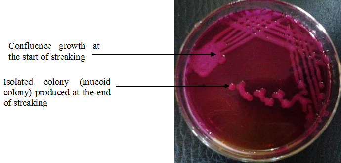
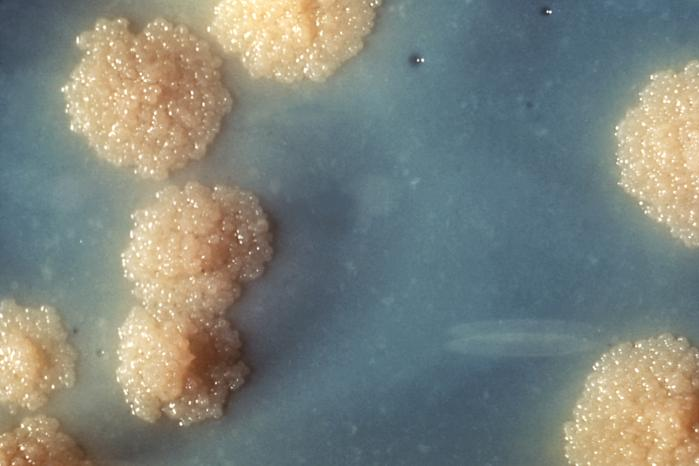
The principal causative agent of pneumonic plague is Yersinia pestis. Y. pestis is a Gram-negative, rod-shaped bacterium belonging to the family Yersiniaceae. This a Gram-negative, facultative anaerobic coccobacillus bacterium is renowned for its exceptional pathogenic potential and its ability to cause rapidly progressive infections in humans. Although it is naturally maintained within animal reservoirs, particularly certain wild rodent populations, it can cross species barriers and produce severe disease when introduced into the human body. Its capacity to invade host tissues, evade immune defences and multiply under favourable conditions makes it one of the most medically significant bacterial pathogens associated with plague.
A defining characteristic of Y. pestis is its Gram-negative cell envelope, which contains lipopolysaccharide and contributes to the bacterium’s interaction with the host immune system. The organism also possesses specialised virulence factors that facilitate survival within the host and interfere with normal immune responses. These mechanisms enable the bacterium to establish infection, resist immune-mediated elimination and contribute to the development of serious inflammatory damage. The severity of the resulting illness depends on several factors, including the route of infection, the bacterial burden, the host’s immune status and the timeliness of appropriate antimicrobial treatment.
In its natural ecological setting, Y. pestis circulates among susceptible mammals, particularly rodents, with fleas serving as important vectors in many transmission cycles. Human infection may occur following the bite of an infected flea, contact with infected animal tissues or inhalation of infectious respiratory droplets from an individual or animal with pneumonic plague. The route of entry influences the initial presentation of the disease. When the bacteria reach the lungs directly through inhalation, they may establish a primary pulmonary infection. Alternatively, bacteria introduced through another route can disseminate through the bloodstream and subsequently involve the lungs, resulting in secondary pneumonic plague.
The pulmonary environment provides an opportunity for Y. pestis to multiply and provoke an intense inflammatory response. As the infection advances, damage to lung tissue can interfere with normal gas exchange, producing cough, chest discomfort, breathing difficulties and, in severe cases, respiratory failure. The bacterium may also enter the bloodstream, contributing to systemic complications such as sepsis and circulatory instability. This combination of local pulmonary injury and potential systemic dissemination accounts for the considerable clinical danger associated with pneumonic plague.
The pathogenic significance of Y. pestis is further heightened by the rapid progression that may occur in untreated cases. Early manifestations can resemble other acute respiratory infections, making prompt clinical recognition particularly important. Laboratory confirmation may involve molecular testing, bacterial culture and other validated diagnostic methods performed under appropriate biosafety procedures. Because pneumonic plague can spread through infectious respiratory droplets during close contact, suspected cases require immediate medical evaluation and suitable infection-control measures.
Rodent fleas and the transmission of Y. pestis
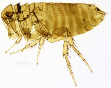
Rodent fleas are small, blood-feeding ectoparasites belonging to the order Siphonaptera. They live on hosts such as rats and mice and depend on blood meals for survival and reproduction. One important species is the Oriental rat flea (Xenopsylla cheopis), which is associated with the transmission of plague caused by the bacterium Yersinia pestis (Figure 1).
Fleas undergo complete metamorphosis, progressing through four life stages: egg, larva, pupa, and adult. Female fleas lay eggs on their hosts or in surrounding environments, such as nests and bedding. The eggs commonly fall into the environment, where larvae develop and feed on organic debris, including adult flea feces. They subsequently form pupae, from which adult fleas emerge under suitable conditions. Adult fleas locate hosts through cues such as body heat, vibrations, and carbon dioxide.
Rodents and their fleas play an important role in maintaining the natural transmission cycle of Y. pestis. Wild rodents, including rats and other small mammals, can carry the bacterium. When fleas feed on infected rodents, they may ingest the bacteria, which can multiply in the flea’s digestive tract and, under certain circumstances, interfere with feeding. Infected fleas may subsequently transmit the bacteria to another susceptible host through a bite. When infected rodents become severely ill or die, their fleas may seek alternative hosts, increasing opportunities for human exposure.
Although flea bites are commonly associated with bubonic plague, infection acquired through this route can sometimes progress to pneumonic plague if the bacteria spread to the lungs. However, flea bites do not directly cause pneumonic plague in every infected person. Pneumonic plague specifically involves infection of the lungs and can also develop when a person inhales infectious respiratory droplets from another person or animal with the disease.
Person-to-person transmission occurs particularly during close contact with an infectious individual. Unlike flea-mediated transmission, this respiratory route does not require an insect vector and can facilitate the spread of infection between people. Preventive measures include reducing contact with wild rodents, controlling fleas safely, avoiding handling sick or dead animals, and protecting pets from infected fleas. Anyone who develops sudden fever, cough, chest pain, or difficulty breathing after possible exposure should seek urgent medical attention.
Modes of transmission of pneumonic plague
Pneumonic plague is a highly dangerous bacterial infection that primarily affects the lungs and can spread through several distinct transmission pathways. Caused by Y. pestis, the disease (plague) is notable for its capacity to pass directly from an infected individual to another person under particular conditions.
1. Inhalation of infectious respiratory droplets
Direct inhalation of infectious respiratory droplets is the principal route of person-to-person transmission of pneumonic plague. When an infected individual coughs, sneezes, or produces respiratory secretions, droplets containing Y. pestis may be released into the surrounding environment. Individuals in close proximity may inhale these droplets, allowing the bacteria to enter the respiratory tract and establish infection in the lungs. Transmission is particularly concerning during prolonged or close contact with an untreated patient. Healthcare personnel, household members, and caregivers may face increased exposure risks if suitable protective precautions are not followed. Unlike infections that spread efficiently through tiny particles suspended in the air over long distances, pneumonic plague transmission is primarily associated with close contact and infectious respiratory droplets.
2. Development of secondary pneumonic plague
Pneumonic plague can develop as a secondary complication of another form of plague. Following the initial entry of Y. pestis into the body, the bacteria may multiply and spread through the lymphatic system or bloodstream. If the infection reaches the lungs, it can cause severe inflammation and pulmonary damage. This progression may occur in individuals initially affected by bubonic or septicemic plague. Once the lungs become infected, the patient may develop coughing, chest pain, breathing difficulties, and potentially bloody sputum. At this stage, the disease may become transmissible to others through infectious respiratory droplets, making early recognition and appropriate medical intervention particularly important.
3. Animal-to-human transmission
Animals can serve as sources of plague infection, particularly when they are infected with Y. pestis. Domestic cats are of particular concern because they may acquire the bacterium through contact with infected rodents or other animals. Human infection may occur through close exposure to an infected animal’s respiratory secretions, especially when the animal has developed pneumonic disease. Handling infected animals or providing veterinary care without appropriate protective equipment may increase exposure risk. Although animal-to-human transmission is possible, the likelihood depends on the animal’s infection status, the nature of contact, and the presence of respiratory involvement.
4. Transmission associated with infected fleas
Infected fleas are important vectors in the natural transmission cycle of plague. They acquire Y. pestis while feeding on infected rodents and may subsequently transmit the bacteria through bites to humans or other animals. This route generally causes bubonic plague rather than primary pneumonic plague. However, the infection may progress to the lungs, producing secondary pneumonic plague. Flea-mediated transmission represents an indirect pathway through which pneumonic disease can eventually emerge. Effective rodent and flea control therefore remains an important component of plague prevention.
5. Environmental and contact-related considerations
The risk of pneumonic plague transmission depends on the infectious source, proximity, duration of exposure, and effectiveness of protective measures. Ordinary contact with contaminated surroundings is not considered the principal transmission route. Prompt identification of suspected cases, appropriate respiratory precautions, rapid antibiotic treatment, and assessment of exposed contacts are essential for interrupting transmission and limiting outbreaks.
Signs and symptoms of pneumonic plague
Pneumonic plague is characterized by an abrupt onset of illness and rapid deterioration of respiratory function. Caused by Yersinia pestis, this severe pulmonary infection can progress within a short period from nonspecific systemic symptoms to extensive lung injury and life-threatening complications. Recognizing its early manifestations is essential because delayed diagnosis and treatment can significantly increase the risk of an adverse outcome.
1. Sudden fever and chills
The illness commonly begins with a sudden rise in body temperature accompanied by intense chills and shivering. Fever reflects the body’s inflammatory response to bacterial invasion. Patients may also experience sweating, generalized discomfort and a pronounced feeling of illness, indicating the rapid development of a systemic infection.
2. Headache, weakness and fatigue
Severe headache, profound weakness and unusual exhaustion are frequent early manifestations. Patients may report muscle aches, dizziness and difficulty performing ordinary activities. These symptoms arise as the infection triggers a widespread inflammatory response, reducing physical endurance and contributing to an overall decline in well-being.
3. Persistent and progressive cough
Cough is a prominent respiratory feature of pneumonic plague. It may initially be dry but can become productive as inflammation and damage within the lungs intensify. In advanced disease, sputum may contain blood, producing a characteristic warning sign of serious pulmonary involvement. The presence of a worsening cough should raise concern when accompanied by sudden fever and respiratory distress.
4. Chest pain and breathing difficulty
Inflammation of lung tissue can cause chest discomfort or pain, particularly during coughing or deep inspiration. As the infection progresses, patients may develop breathlessness because damaged lung tissue becomes less effective at transferring oxygen into the bloodstream. Increasing respiratory discomfort may indicate substantial pulmonary involvement and requires urgent medical evaluation.
5. Rapid breathing and elevated heart rate
Tachypnea, or abnormally rapid breathing, may develop as the body attempts to compensate for impaired oxygen exchange. Tachycardia, characterized by an increased heart rate, can occur in response to fever, physiological stress and declining oxygenation. These signs may indicate worsening disease and the need for immediate supportive care.
6. Severe complications in advanced disease
In severe cases, pneumonic plague can progress to acute respiratory failure, in which the lungs cannot maintain adequate oxygenation. The bacteria may also enter the bloodstream, resulting in sepsis and potentially septic shock. These complications can impair vital organ function and lead to death if effective treatment is not initiated promptly.
The clinical presentation of pneumonic plague is distinguished by its sudden onset, rapid progression and prominent respiratory involvement. Any suspected case requires emergency medical assessment, immediate infection-control precautions and prompt antibiotic treatment.
Pathogenesis and disease development of pneumonic plague
Entry of the bacterium into the respiratory tract
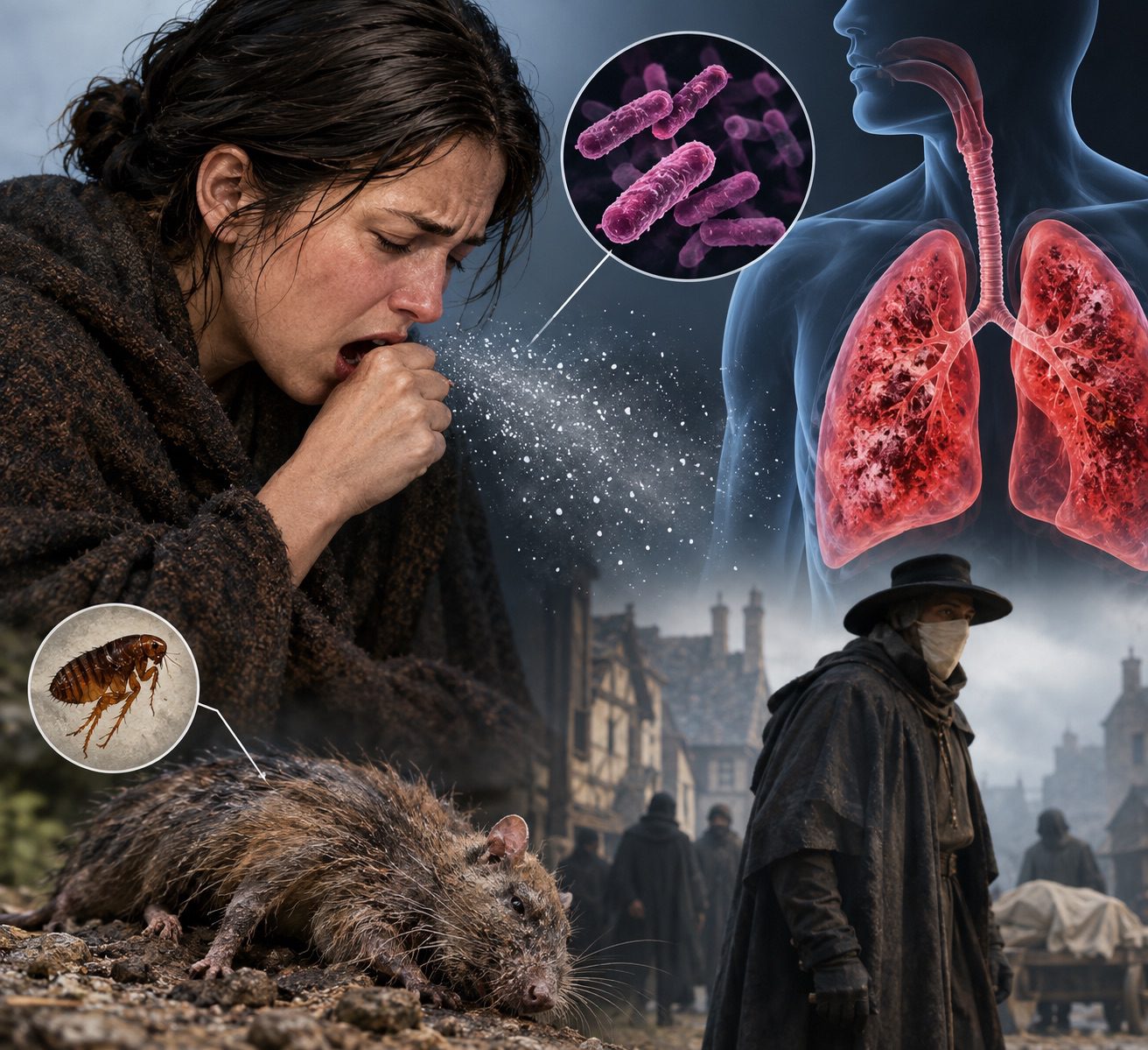
The pathogenesis of pneumonic plague begins when Yersinia pestis, the causative bacterium, enters the respiratory system through the inhalation of infectious respiratory droplets (Figure 2). Following inhalation, the bacteria travel through the upper respiratory passages and reach the lower respiratory tract, where they can establish infection in the lungs. The inhaled bacterial cells encounter several natural defence mechanisms, including mucus, ciliary movement and immune cells that normally protect the respiratory system from invading microorganisms. However, Y. pestis possesses specialised virulence factors that help it evade immune recognition and establish itself within lung tissue. The ability of the bacterium to overcome these initial barriers is a critical step in the development of pneumonic plague.

Bacterial multiplication and colonization of lung tissue
After reaching the lungs, Y. pestis begins to multiply within the respiratory environment. The bacterium uses several virulence mechanisms to interfere with the activity of immune cells, particularly phagocytes responsible for engulfing and destroying pathogens. Its type III secretion system delivers effector proteins into host cells, disrupting immune signalling and cellular functions. These mechanisms enable the bacteria to survive and proliferate while the body’s initial defences struggle to contain the infection. As the bacterial population increases, the infection becomes established within the lung tissue, creating conditions for progressive inflammation and tissue injury. The speed of bacterial multiplication contributes to the unusually rapid clinical progression associated with pneumonic plague.
Inflammatory response and pulmonary tissue damage
The growing bacterial burden stimulates an inflammatory response as the immune system attempts to eliminate the invading pathogen. Immune cells release inflammatory mediators that recruit additional defensive cells to the infected lung tissue. Although this response is intended to restrict bacterial growth, excessive or poorly regulated inflammation can contribute to tissue damage. The delicate structures responsible for gas exchange, including the alveolar walls and surrounding capillaries, may become impaired. Inflammation increases the permeability of pulmonary blood vessels, allowing fluid, proteins and inflammatory cells to accumulate within the air spaces. These pathological changes interfere with the lungs’ ability to transfer oxygen into the bloodstream and remove carbon dioxide efficiently.
Development of pneumonia and respiratory dysfunction
As infection and inflammation intensify, the affected lungs develop severe pneumonia. The alveoli may become filled with inflammatory fluid, cellular debris and blood, reducing the effective surface area available for gas exchange. This process can produce coughing, chest pain, fever, rapid breathing and progressive breathlessness. In advanced cases, patients may develop haemoptysis, characterised by coughing up blood-stained sputum. The resulting impairment of oxygen exchange can cause hypoxaemia, in which the oxygen concentration in the blood falls below normal levels. As more pulmonary tissue becomes affected, respiratory distress may worsen rapidly. Without effective treatment and appropriate supportive care, extensive lung injury can culminate in acute respiratory failure.
Spread into the bloodstream and systemic complications
In severe infection, Y. pestis may cross damaged tissue barriers and enter the bloodstream, producing bacteraemia and potentially leading to sepsis. The presence of bacteria in the circulation can trigger a widespread inflammatory response that disrupts normal blood-vessel function and circulation. Increased vascular permeability, abnormal clotting and reduced tissue perfusion may develop, compromising the delivery of oxygen and nutrients to vital organs. The kidneys, liver, cardiovascular system and other organs may consequently become impaired. When these disturbances become severe, the patient may develop septic shock, a life-threatening condition characterised by circulatory failure and inadequate tissue perfusion.
6. Rapid progression and possible fatal outcome
The defining feature of pneumonic plague is its potential for exceptionally rapid deterioration. The combined effects of bacterial proliferation, pulmonary inflammation, impaired oxygen exchange and systemic infection can overwhelm the body’s compensatory mechanisms within a short period. The likelihood of severe complications increases substantially when diagnosis and effective antibiotic treatment are delayed. Advanced disease may involve respiratory failure, septic shock, multiple-organ dysfunction and death. Progression is not inevitable: prompt recognition, immediate appropriate antimicrobial therapy and intensive supportive management can substantially improve the chances of survival.
Diagnosis of pneumonic plague
The diagnosis of pneumonic plague requires a coordinated approach that combines clinical evaluation, exposure assessment, respiratory specimen analysis, and specialized laboratory investigations. Because the disease can deteriorate rapidly and may become fatal without timely treatment, healthcare professionals must recognize its characteristic features early. Diagnostic decisions should be guided by the patient’s symptoms, epidemiological background, and the possibility of exposure to Y. pestis, the bacterium responsible for plague. A strong clinical suspicion is particularly important because waiting for definitive laboratory results may delay life-saving intervention.
Clinical assessment and symptom evaluation. The diagnostic process begins with a detailed assessment of the patient’s clinical presentation. Healthcare professionals examine the onset, duration, and progression of symptoms, paying particular attention to sudden fever, chills, profound weakness, chest discomfort, coughing, and difficulty breathing. The production of blood-stained sputum is an especially concerning feature in a patient with suspected pneumonic plague. Physical examination may reveal rapid breathing, an elevated heart rate, reduced oxygen saturation, and signs of severe respiratory distress. Clinicians also investigate the possibility of contact with an infected individual, exposure to potentially infected animals, or residence in or travel to an area where plague has been reported. These findings help establish the level of suspicion and determine the urgency of further investigations.
Respiratory specimen examination. Respiratory samples, particularly sputum, are important in investigating suspected pneumonic plague because the infection primarily affects the lungs. When appropriate, specimens are collected by trained healthcare personnel using suitable infection-control precautions. Laboratory specialists may examine the material microscopically for bacterial characteristics consistent with Y. pestis. However, microscopic appearance alone cannot reliably establish the diagnosis, as other microorganisms may produce similar findings. Respiratory specimen analysis must therefore be interpreted alongside the clinical presentation and more specific laboratory investigations.
Blood investigations. Blood tests provide additional information about the severity of infection and the patient’s overall condition. A complete blood count may reveal changes in white blood cell numbers associated with a serious bacterial infection. Other investigations, including blood cultures, can help detect bacteria in the bloodstream when systemic spread is suspected. Measurements of kidney function, liver function, oxygenation, and other physiological parameters may help clinicians assess organ involvement and identify complications such as sepsis. Although routine blood tests can indicate severe infection, they do not independently confirm pneumonic plague. Their primary value lies in supporting the diagnostic assessment and guiding clinical management.
Microbiological and molecular confirmation. Definitive diagnosis depends on specialized laboratory methods capable of identifying Y. pestis. Bacterial culture can demonstrate the presence of the organism in an appropriate clinical specimen, while molecular techniques, including polymerase chain reaction (PCR), can detect specific bacterial genetic material. Depending on the circumstances, laboratories may also employ additional validated identification methods. Specimens must be handled and processed according to appropriate biosafety procedures, and suspected cases should be communicated promptly to the receiving laboratory and relevant public-health authorities. Confirmation through specialized testing helps distinguish plague from other severe respiratory infections that may initially produce similar symptoms.
Urgent clinical response and diagnostic coordination. Suspected pneumonic plague constitutes a medical and public-health emergency. Appropriate infection-control measures, including droplet precautions, should be initiated promptly, with additional precautions determined by the clinical setting and applicable guidance. Healthcare professionals must arrange urgent assessment, notify relevant public-health authorities, and collect suitable specimens when this can be done safely. Most importantly, antibiotic treatment should begin immediately when pneumonic plague is strongly suspected; clinicians must not postpone treatment while awaiting laboratory confirmation. Rapid communication between clinical teams, diagnostic laboratories, and public-health services is essential for improving patient outcomes, identifying potentially exposed contacts, and preventing further transmission.
Treatment and medical management of pneumonic plague
The treatment of pneumonic plague requires immediate medical intervention, carefully selected antimicrobial therapy, and continuous clinical supervision. Since the disease can deteriorate rapidly and cause life-threatening respiratory complications, healthcare professionals must initiate treatment as soon as pneumonic plague is suspected, without waiting for definitive laboratory confirmation. Timely management is essential to limit bacterial multiplication, prevent systemic complications, and improve the likelihood of recovery.
In general, the successful management and treatment of pneumonic plague depends on rapid recognition of the disease, immediate antibiotic administration, appropriate respiratory support, and coordinated infection-control measures. Delays in treatment can substantially increase the risk of death, whereas early, evidence-based intervention offers the best opportunity for survival and helps prevent further transmission.
Antibiotic therapy forms the cornerstone of treatment. Appropriate antibiotics are selected according to the patient’s clinical condition, age, disease severity, potential contraindications, and current public-health recommendations. Depending on the circumstances, treatment may involve agents such as gentamicin, ciprofloxacin, levofloxacin, or doxycycline. The choice of medication, route of administration, and duration of therapy must be determined by qualified healthcare professionals in accordance with established clinical guidelines. In critically ill patients, intravenous antimicrobial treatment may be necessary to ensure prompt and reliable drug delivery.
Hospitalization and supportive care are particularly important for patients experiencing severe respiratory distress, persistent fever, circulatory instability, or other signs of deterioration. Oxygen therapy may be administered to maintain adequate blood oxygen levels, while intravenous fluids can help support circulation when clinically indicated. Patients with advanced respiratory failure may require mechanical ventilation and admission to an intensive care unit. Supportive interventions must be individualized to the patient’s physiological needs and response to treatment.
Continuous clinical monitoring enables healthcare teams to recognize complications at an early stage. Respiratory rate, oxygen saturation, blood pressure, heart rate, temperature, and mental status should be assessed regularly. Laboratory investigations may also be used to evaluate organ function, identify systemic infection, and monitor the patient’s response to therapy. Particular attention is required to detect sepsis, septic shock, acute respiratory distress syndrome, and multiple-organ dysfunction, all of which may develop in severe cases.
Infection prevention and public-health coordination are integral components of medical management. Patients with suspected or confirmed pneumonic plague should be managed using appropriate isolation procedures and droplet precautions. Healthcare workers must follow recommended personal protective measures, while public-health authorities should be notified promptly. Close contacts must be identified and assessed so that monitoring and preventive antibiotic treatment can be provided when indicated.
Prevention and control of pneumonic plague
The prevention and control of pneumonic plague depend on rapid case recognition, effective infection-control practices, timely medical intervention, and coordinated public-health action. Since the disease can progress rapidly and spread through infectious respiratory droplets during close contact, preventive measures must be implemented as soon as an infection is suspected. A well-organized control strategy aims to interrupt transmission, protect healthcare personnel, safeguard exposed individuals, and reduce the risk of community outbreaks.
1. Avoidance of exposure and close contact with people suspected of having the disease
Individuals should avoid close, unprotected contact with people suspected of having pneumonic plague, particularly those experiencing persistent coughing, fever, chest pain, or breathing difficulties. Suspected patients should seek urgent medical assessment and inform healthcare providers of possible exposure before arrival whenever feasible. Maintaining an appropriate distance and avoiding direct exposure to respiratory secretions can reduce the likelihood of transmission while professional assistance is arranged.
2. Respiratory protection and infection-control practices
Healthcare workers and caregivers must follow established infection-prevention protocols when attending suspected or confirmed cases. Appropriate personal protective equipment, including a well-fitting medical mask or respirator according to the exposure risk and applicable guidance, should be used. Gloves, protective clothing, and eye protection may also be necessary when contact with respiratory secretions is anticipated. Hand hygiene, careful handling of contaminated materials, and environmental cleaning further reduce the possibility of infection.
3. Isolation and management of suspected cases
Prompt isolation is an essential component of outbreak containment. Individuals with suspected pneumonic plague should be separated from others in accordance with public-health recommendations, with droplet precautions implemented promptly. Healthcare facilities should minimize unnecessary exposure, ensure suitable ventilation, and restrict access to designated care areas. Appropriate precautions should continue until healthcare and public-health professionals determine that the risk of transmission has been adequately controlled.
4. Identification and Protection of Close Contacts
Public-health teams should rapidly identify people who have had significant exposure to an infected individual, particularly through close, unprotected contact with respiratory secretions. Exposed contacts require assessment, clear instructions about symptoms, and appropriate monitoring. Preventive antibiotic treatment may be recommended for individuals meeting established exposure criteria. Early identification enables prompt intervention if symptoms develop and helps prevent further transmission within households, healthcare facilities, and other affected settings.
5. Rodent and flea control
Although pneumonic plague can spread directly between people, controlling infected animals and their fleas remains important in preventing plague infections more broadly. Rodent infestations should be managed through safe sanitation practices, secure food storage, waste disposal, and appropriate pest-control measures. People should avoid handling sick or dead rodents and other potentially infected animals without suitable protection. Flea-control measures should be coordinated carefully because disturbing rodents before controlling their fleas may increase human exposure to infected fleas.
6. Community education and public-health coordination
Public awareness strengthens prevention by helping communities recognize warning signs and understand how transmission occurs. Educational initiatives should emphasize early medical consultation, avoidance of unprotected contact, and cooperation with public-health authorities. Healthcare services, laboratories, and public-health agencies must coordinate case reporting, diagnostic testing, contact tracing, and appropriate preventive interventions. Clear communication and timely action are essential for limiting transmission and protecting vulnerable populations.
References
Durden, L. A., & Hinkle, N. C. (2019). Fleas (Siphonaptera). In G. R. Mullen & L. A. Durden (Eds.), Medical and veterinary entomology (3rd ed., pp. 145–169). Academic Press.
Rajerison, M., Ratsitorahina, M., & Andrianaivoarimanana, V. (2014). Plague. In J. Farrar, P. J. Hotez, T. Junghanss, G. Kang, D. Lalloo, & N. J. White (Eds.), Manson’s tropical infectious diseases (23rd ed., pp. 404–409). W. B. Saunders.
Salam, A. P., Rojek, A., Cai, E., Raberahona, M., & Horby, P. (2020). Deaths associated with pneumonic plague, 1946–2017. Emerging Infectious Diseases, 26(10), 2432–2434.
Evans, C. (2022). Pneumonic plague: Incidence, transmissibility and future risks. Hygiene, 2(1), 14–27.
Pechous, R. D., Sivaraman, V., Stasulli, N. M., & Goldman, W. E. (2016). Pneumonic plague: The darker side of Yersinia pestis. Trends in Microbiology, 24(3), 190–197.
Wagle, P. M., & Bedarkar, M. K. (1948). Pneumonic plague and its treatment. Indian Medical Gazette, 83(9), 406–409.
Jalpota, Y. P., Charati, S. C., Sharma, S. C., & Raghunath, D. (1997). Pneumonic plague—Autopsy findings: A case report. Medical Journal Armed Forces India, 53(1), 56–58.
Lathem, W. W., Crosby, S. D., Miller, V. L., & Goldman, W. E. (2005). Progression of primary pneumonic plague: A mouse model of infection, pathology, and bacterial transcriptional activity. Proceedings of the National Academy of Sciences, 102(49), 17786–17791.
MacDermot, H. E. (1927). A treatise on pneumonic plague. Canadian Medical Association Journal, 17(11), 1410.
Meyer, K. F. (1961). Pneumonic plague. Bacteriological Reviews, 25(3), 249–261.
Ansari, I., Grier, G., & Byers, M. (2020). Deliberate release: Plague—A review. Journal of Biosafety and Biosecurity, 2(1), 10–22.
Centers for Disease Control and Prevention. (n.d.). About plague.
World Health Organization. (n.d.). Plague.
Discover more from Microbiology Class
Subscribe to get the latest posts sent to your email.