NanoSIMS is the acronym for Nanoscale Secondary Ion Mass Spectrometry. NanoSIMS is a high-resolution analytical technique designed to measure the elemental, isotopic, and in some cases molecular composition of materials at the nanometer scale. It is one of the most spatially precise forms of mass spectrometry currently available, bridging a long-standing gap between high-sensitivity chemical analysis and ultrastructural imaging. Its defining feature is the ability to generate quantitative or semi-quantitative chemical maps with spatial resolution approaching tens of nanometers, enabling direct interrogation of heterogeneity at the scale of individual cells, organelles, mineral grains, and subcellular compartments.
NanoSIMS emerged as an evolution of conventional Secondary Ion Mass Spectrometry (SIMS), which itself dates back to the mid-20th century. Traditional SIMS instruments already offered sensitive surface chemical analysis by sputtering a sample with a focused primary ion beam and analyzing emitted secondary ions. However, their spatial resolution was typically limited to the micrometer scale, which constrained their applicability in biological and environmental systems where critical processes occur at sub-micron and nanometer dimensions. NanoSIMS was developed to overcome this limitation by refining ion optics, improving beam stability, and integrating multi-collector detection systems capable of high-precision isotopic measurements.
Mechanisms of NanoSIMS analysis: How NanoSIMS works
NanoSIMS is built upon three tightly coupled physical and instrumental principles:
(1) Ion beam sputtering of the sample surface
A finely focused primary ion beam (commonly Cs⁺ or O⁻/O₂⁺) is directed onto the sample surface under ultra-high vacuum conditions. The energetic impact of these ions transfers momentum to atoms in the surface lattice, causing a localized ejection of atoms, atomic clusters, and molecular fragments in a process known as sputtering. A small fraction of these sputtered species is ionized, producing secondary ions. The choice of primary ion species is critical: cesium enhances the yield of negative secondary ions (e.g., C⁻, CN⁻, O⁻), while oxygen enhances positive ion formation (e.g., Na⁺, K⁺, metal ions). This ionization efficiency directly determines analytical sensitivity and elemental coverage.
(2) Mass spectrometric detection of secondary ions
Once generated, secondary ions are extracted from the sample surface using electrostatic fields and introduced into a high-resolution mass analyzer. NanoSIMS typically employs a double-focusing magnetic sector mass spectrometer, which combines electrostatic and magnetic analyzers. This configuration enables high mass resolving power, sufficient to distinguish isobaric interferences (ions with nearly identical mass-to-charge ratios), which is essential for accurate isotopic measurements. The system can separate isotopes such as ¹²C⁻ and ¹³C⁻ or ¹⁴N⁻ and ¹⁵N⁻ with high precision, even in chemically complex matrices.
(3) High-resolution imaging through raster scanning
NanoSIMS does not simply measure bulk composition; it produces spatially resolved chemical images. The focused primary ion beam is rastered across the sample surface in a pixel-by-pixel fashion. At each point, secondary ion intensities are recorded by multiple detectors simultaneously. This scanning approach converts ion counts into spatially resolved maps of elemental and isotopic distributions. Because beam diameters can be reduced to ~50 nm or less under optimal conditions, the resulting images reveal chemical heterogeneity at near-subcellular scales. In addition, repeated sputtering cycles can be used to progressively remove layers of material, enabling three-dimensional compositional reconstructions.
The integration of these three components: (1) sputtering, (2) mass spectrometry, and (3) high-resolution scanning, allows NanoSIMS to uniquely combine chemical specificity with nanoscale spatial resolution. Unlike many other imaging modalities, it does not rely on fluorescence labeling or optical contrast. Instead, it directly detects elemental and isotopic signatures, making it particularly powerful for systems where labeling is intrinsic (e.g., stable isotope probing) or where fluorescent markers are not feasible.
A key conceptual advantage of NanoSIMS lies in its ability to perform simultaneous multi-isotope detection. Modern instruments typically house multiple (often up to seven) parallel detectors, enabling concurrent measurement of several isotopes or molecular fragments. This capability reduces analytical uncertainty associated with sequential scanning and allows direct calculation of isotopic ratios at the pixel level. As a result, NanoSIMS is especially powerful for tracer studies involving stable isotopes such as ¹³C, ¹⁵N, ²H, and ³⁴S, where small differences in incorporation can indicate major functional differences between cells or microenvironments.
From a conceptual standpoint, NanoSIMS is not merely an imaging tool but a quantitative analytical platform for linking structure and function. In biological systems, it enables researchers to directly correlate metabolic activity with individual cells or even subcellular regions. In environmental systems, it allows the mapping of nutrient fluxes across heterogeneous matrices such as soil aggregates or mineral surfaces. In materials science, it provides insight into dopant distributions, diffusion processes, and nanoscale compositional gradients.
NanoSIMS is widely applied across several major scientific domains
In microbial ecology and environmental microbiology, it is used to determine which individual microbial cells actively assimilate labeled substrates in complex communities. This is particularly important for uncultured organisms, where activity cannot be inferred from genomic data alone. Coupled with techniques such as fluorescence in situ hybridization (FISH), NanoSIMS enables direct linkage between microbial identity and metabolic function at the single-cell level. In geochemistry and cosmochemistry, NanoSIMS is used to analyze isotopic anomalies in meteorites, minerals, and planetary materials. These measurements provide insights into early solar system processes, planetary differentiation, and geochemical cycling over geological timescales.
In materials science, NanoSIMS is applied to study thin films, semiconductor devices, corrosion layers, and nanostructured materials. Its ability to detect trace dopants and impurities with spatial precision is critical for understanding material performance and failure mechanisms. In cell biology and biomedical research, NanoSIMS enables visualization of nutrient uptake, drug distribution, lipid metabolism, and protein turnover at subcellular resolution. It is particularly valuable in studies involving stable isotope labeling, where dynamic biological processes can be traced without perturbing native structure.
NanoSIMS occupies a unique analytical niche defined by the intersection of ultra-high spatial resolution, isotopic sensitivity, and quantitative imaging capability. Its conceptual foundation is rooted in the controlled sputtering of matter and precise mass analysis of emitted ions, but its scientific impact extends far beyond instrumentation. It provides a methodological bridge between molecular biology, environmental chemistry, and materials science, enabling direct observation of processes that were previously inferable only indirectly.
Principles of NanoSIMS
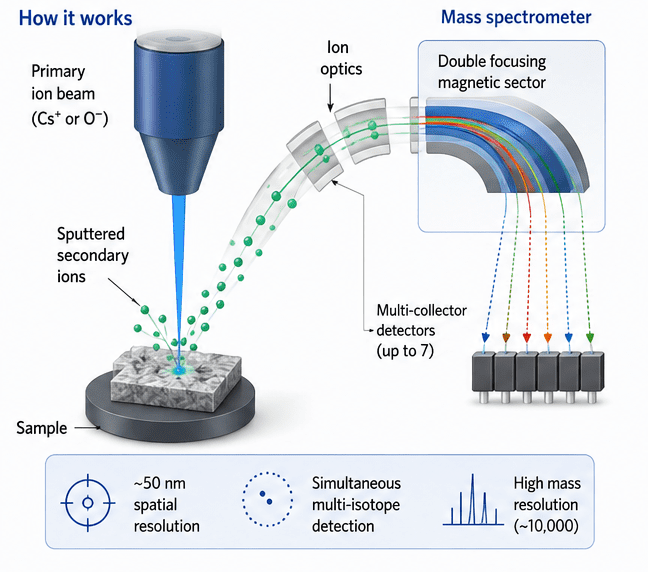
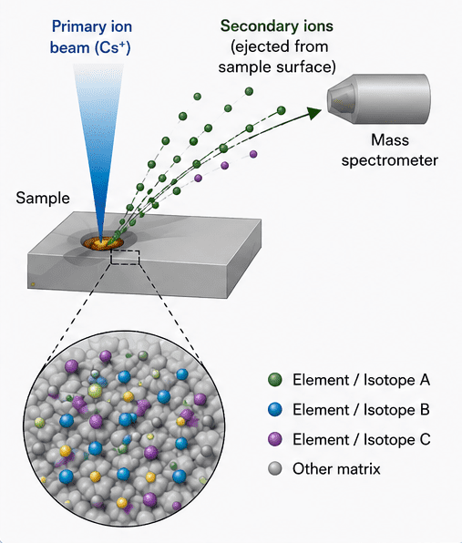
NanoSIMS is based on the principle of secondary ion mass spectrometry, where a focused primary ion beam (commonly Cs⁺ or O⁻ ions) is directed onto the surface of a solid sample under ultra-high vacuum conditions (Figure 1). The energetic primary ions collide with the sample surface and initiate a process called sputtering, in which atoms, clusters, and molecular fragments are ejected from the top few nanometers of the material. A small fraction of these sputtered species is ionized, forming so-called secondary ions.
These secondary ions are extracted from the sample surface and guided into a high-performance mass spectrometer, where they are separated according to their mass-to-charge (m/z) ratios. NanoSIMS uses a double-focusing mass analyzer combining electrostatic and magnetic sectors, enabling extremely high mass resolution and the ability to distinguish ions with very similar masses (isobaric species). Multiple detectors allow simultaneous measurement of several isotopes or elements, significantly improving precision and efficiency.
By rastering the primary ion beam across the sample surface in a controlled pattern, NanoSIMS constructs high-resolution spatial maps of elemental and isotopic distributions. Each pixel in the resulting image corresponds to the intensity of a specific ion signal, enabling quantitative chemical imaging at a spatial resolution down to ~50 nanometers. This principle allows NanoSIMS to link chemical composition with spatial context, making it uniquely powerful for studying heterogeneous systems such as microbial cells, soil particles, and subcellular structures. It is especially valuable for stable isotope tracing, where incorporation of labeled substrates (e.g., ¹³C or ¹⁵N) can be visualized at the single-cell level to reveal metabolic activity and functional heterogeneity.
Instrumentation and components of NanoSIMS
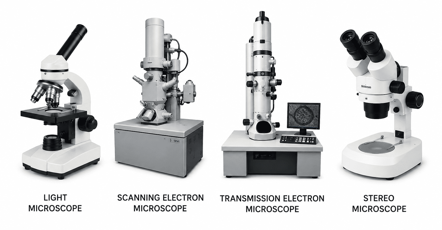
The NanoSIMS instrument is a highly sophisticated and large-scale analytical platform composed of multiple integrated systems, including high-energy ion sources, ultra-high vacuum chambers, ion optics, magnetic sector mass analyzers, and multi-collector detectors. Due to the requirement for nanometer-scale spatial resolution and extremely high mass sensitivity, the instrument operates under stringent vacuum and electromagnetic stability conditions, making its engineering and maintenance technically complex. A typical NanoSIMS setup occupies an entire laboratory room and requires specialized environmental controls such as vibration isolation, temperature regulation, and stable electrical supply (Figure 2). The operation and calibration of the NanoSIMS demand highly trained personnel because precise alignment of the ion beam, detector tuning, and mass resolution optimization are critical for obtaining accurate isotopic and elemental measurements. NanoSIMS instrumentation is a tightly integrated system designed to optimize three competing requirements which are:
(1) Spatial resolution,
(2) Mass resolution, and
(3) Detection sensitivity.
Each subsystem in NanoSIMS is engineered to preserve lateral chemical information from the sample surface while enabling high-precision isotopic separation. The core architecture consists of the primary ion source, ultra-high vacuum chamber, ion extraction optics, a double-focusing mass analyzer, and a multi-collector detection system. The NanoSIMS instrumentation suite is a highly optimized integration of ion generation, ultra-stable vacuum conditions, precision ion optics, high-resolution mass separation, and parallel detection. Each component contributes to maintaining the central requirement of the technique: preserving nanoscale spatial information while delivering accurate isotopic and elemental quantification.
The major components of NanoSIMS comprises:
- A primary ion source
- Sample chamber and vacuum system
- Secondary ion extraction optics
- Mass analyzer
- Multi-collector detection system
1. Primary ion source
The primary ion source is the analytical engine of NanoSIMS. It generates a focused beam of energetic ions that bombard the sample surface, initiating sputtering and secondary ion emission. Two primary ion species are commonly used: cesium (Cs⁺) and oxygen (O⁻ / O₂⁺). Each of these primary ion species are selected based on the ionization characteristics of target elements.
Cesium ion (Cs⁺) source
The Cs⁺ beam is predominantly used to enhance the yield of negative secondary ions. Cesium has a low ionization potential and readily donates electrons, which increases the probability that sputtered species will leave the surface as negative ions. This makes Cs⁺ particularly effective for electronegative elements such as:
- Carbon (C⁻)
- Nitrogen (often detected as CN⁻ or C₂N⁻ clusters)
- Oxygen (O⁻)
- Sulfur (S⁻)
This configuration is especially important in biological and environmental applications where light elements dominate the chemical signature.
Oxygen ion (O⁻ / O₂⁺) source
The oxygen primary beam is used to enhance positive secondary ion production. Oxygen implantation into the sample surface increases the likelihood of electron removal from sputtered atoms, thus favoring positive ion formation. This is particularly effective for:
- Metals (Fe⁺, Mg⁺, Ca⁺, Cu⁺)
- Alkali and alkaline earth elements
- Certain trace inorganic species
The choice between Cs⁺ and O⁻ beams is therefore dictated by the ionization efficiency of the analyte matrix and the polarity of ions of interest.
Key beam parameters
There are two critical operational parameters define performance of the NanoSIMS:
- Beam current: Beam current controls the trade-off between sensitivity and spatial resolution. Higher currents increase sputtering rate and ion yield, improving detection sensitivity but degrading spatial resolution due to beam broadening and increased interaction volume. Lower currents produce finer beams suitable for subcellular imaging but reduce signal intensity.
- Beam diameter: The beam diameter directly defines the achievable lateral spatial resolution, often reaching ~50 nm under optimal conditions. Beam focusing systems, including electrostatic lenses and apertures, are used to minimize aberrations and maintain beam coherence.
2. Sample chamber and vacuum system
The sample chamber operates under ultra-high vacuum (UHV) conditions, typically around 10-10 mbar, which is essential for preserving analytical fidelity.
At such low pressures:
- Contamination from residual gases (e.g., hydrocarbons, water vapor) is minimized
- Secondary ion scattering is significantly reduced
- Surface chemistry remains stable during analysis
The UHV environment also prevents oxidation or adsorption artifacts that would otherwise distort isotopic or elemental measurements. In biological applications, this requirement often necessitates rigorous sample dehydration or cryo-fixation prior to insertion into the chamber. The chamber itself is equipped with precision stages that allow nanometer-scale sample positioning and raster scanning, enabling systematic surface interrogation.
3. Secondary ion extraction optics
Once sputtering occurs, emitted secondary ions must be efficiently collected without compromising spatial information. This is achieved using electrostatic extraction optics, which perform three key functions:
- Ion collection: Secondary ions emitted from the surface are drawn into the extraction field.
- Acceleration: Ions are accelerated to a defined kinetic energy for mass separation.
- Beam focusing: Electrostatic lenses maintain spatial correlation between sputtered location and detected signal.
A critical design requirement is preserving lateral spatial fidelity, ensuring that ions originating from a specific nanoscale region are correctly mapped back to that region in the final image. Any distortion in this stage directly degrades image resolution and quantitative accuracy.
4. Mass analyzer: double-focusing magnetic sector
The NanoSIMS mass analyzer is based on a double-focusing magnetic sector configuration, combining an electrostatic analyzer (ESA) with a magnetic sector field.
- Electrostatic Analyzer (ESA): The ESA filters ions based on their kinetic energy. Because sputtering produces ions with a range of energies, this stage ensures that only ions within a narrow energy band proceed further, improving mass resolution and reducing spectral noise.
- Magnetic Sector: The magnetic field then separates ions according to their mass-to-charge ratio (m/z). Heavier ions experience less deflection, while lighter ions are more strongly curved, enabling spatial separation onto different detector channels.
Analytical advantages of the ESA and magnetic sector
This dual-stage design provides:
- High mass resolving power (m/Δm up to ~10,000 or higher): Essential for resolving isobaric interferences (e.g., distinguishing ¹²C¹⁵N⁻ from ¹³C¹⁴N⁻ in biological systems).
- Accurate isotopic ratio determination: Critical for tracer studies involving subtle isotopic enrichments.
- Stable long-term signal acquisition: Important for slow raster imaging and depth profiling experiments.
5. Multi-collector detection system
One of the defining innovations of NanoSIMS is its multi-collector detector array, capable of simultaneously recording up to seven ion species in parallel. In terms of its functional design, each detector channel is independently tuned to a specific mass, allowing concurrent measurement of multiple isotopes without sequential scanning. This eliminates temporal drift effects and improves statistical precision.
Advantages of parallel detection
- Reduced acquisition time: Multiple isotopes measured simultaneously rather than sequentially
- Improved precision: Eliminates inter-scan variability
- Enhanced isotopic ratio accuracy: Ratios are derived from synchronized signals
Example of multi-isotope detection
A common configuration in biological and environmental studies includes simultaneous monitoring of:
- ¹²C⁻ → bulk carbon distribution
- ¹³C⁻ → isotope-labeled carbon uptake
- ¹²C¹⁴N⁻ → proxy for total biomass or protein content
- ¹²C¹⁵N⁻ → nitrogen assimilation and metabolic activity
This combination is especially powerful in stable isotope probing (SIP) experiments, where metabolic incorporation of labeled substrates must be resolved at the single-cell level.
Workflow of NanoSIMS
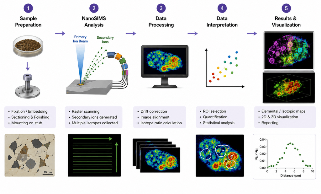
The NanoSIMS workflow involves a sequence of carefully coordinated steps designed to obtain high-resolution elemental and isotopic information from complex samples. The process begins with sample preparation, which may include fixation, embedding, sectioning, polishing, and mounting onto a conductive sample holder to ensure compatibility with ultra-high vacuum conditions (Figure 3).
During NanoSIMS analysis, a focused primary ion beam (commonly Cs⁺ or O⁻) raster-scans the sample surface, causing sputtering and release of secondary ions from the uppermost nanometers of the material. These emitted ions are extracted, separated according to their mass-to-charge ratio in the mass spectrometer, and simultaneously detected using multiple collectors for parallel isotope measurement. The acquired raw data then undergo processing steps such as image alignment, drift correction, dead-time correction, and isotope ratio calculation to improve analytical accuracy.
In the interpretation stage, regions of interest (ROIs) are selected for quantitative and statistical analysis of elemental or isotopic distributions. Finally, the processed data are visualized as elemental maps, isotope ratio images, line profiles, or three-dimensional depth reconstructions, enabling detailed investigation of microbial activity, nutrient cycling, cellular metabolism, and nanoscale material heterogeneity.
Output of NanoSIMS
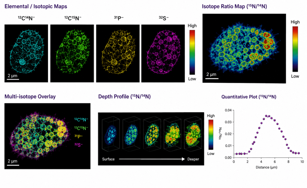
NanoSIMS produces high-resolution elemental and isotopic images that reveal the spatial distribution of chemical components within biological, environmental, and material samples at the nanometer scale. During analysis, a focused primary ion beam sputters the sample surface, generating secondary ions that are separated according to their mass-to-charge ratio and simultaneously detected by multiple collectors.
The resulting outputs include single-isotope maps, isotope ratio maps, multi-isotope overlays, quantitative line profiles, and three-dimensional depth reconstructions (Figure 4). Elemental and isotopic maps provide detailed information on the localization and relative abundance of specific ions or isotopes, while isotope ratio images enable quantitative assessment of isotopic enrichment and metabolic activity. Multi-isotope overlays combine several ion signals into a single composite image to visualize spatial relationships among different chemical species.
Depth profiling further allows visualization of isotopic or elemental distributions beneath the sample surface, generating three-dimensional compositional information. Quantitative plots derived from selected regions of interest enable statistical analysis of isotope incorporation and elemental variation. These outputs make NanoSIMS a powerful tool for studying microbial activity, nutrient cycling, cellular metabolism, contaminant distribution, and nanoscale heterogeneity in complex systems.
Isotopic analysis and quantification
One of the most powerful capabilities of NanoSIMS is its ability to perform highly sensitive isotopic analysis at subcellular and nanometer spatial scales. Unlike bulk isotope ratio mass spectrometry, which provides only averaged isotopic compositions for an entire sample, NanoSIMS enables the visualization and quantification of isotopic enrichment within individual cells, organelles, mineral phases, or microenvironments. This capability has transformed studies in microbial ecology, environmental science, geochemistry, and cell biology by allowing researchers to directly connect isotopic composition with spatial organization and biological function.
Stable isotope measurements
NanoSIMS is particularly effective for measuring stable isotope ratios with exceptional spatial resolution and sensitivity. Common isotopic systems analyzed include:
- ¹³C/¹²C
- ¹⁵N/¹⁴N
- ²H/¹H
- ³⁴S/³²S
- ¹⁸O/¹⁶O
- ²⁹Si/²⁸Si
These isotopic measurements provide insight into metabolic activity, nutrient assimilation, biogeochemical cycling, and elemental transformations within highly heterogeneous systems. Because NanoSIMS can detect isotopic enrichments at the level of individual microbial cells or microscopic structures, it has become a central tool for tracing nutrient flow in complex biological and environmental samples.
The instrument achieves this sensitivity through simultaneous multi-collector detection, which allows several isotopes to be measured concurrently from the same sputtered region. Simultaneous detection minimizes temporal fluctuations in primary ion beam intensity and improves the precision of isotope ratio measurements. For example, NanoSIMS can simultaneously collect signals for ¹²C⁻ and ¹³C⁻ or for ¹²C¹⁴N⁻ and ¹²C¹⁵N⁻, enabling highly accurate determination of carbon and nitrogen isotopic enrichment.
In biological systems, isotopic ratios are frequently used to quantify nutrient assimilation and metabolic turnover. An increase in the ¹³C/¹²C ratio within a microbial cell, for instance, indicates incorporation of a ¹³C-labeled substrate into cellular biomass. Similarly, enrichment in ¹⁵N can reveal nitrogen uptake pathways or rates of protein synthesis. Because NanoSIMS measurements are spatially resolved, isotopic heterogeneity among neighboring cells can also be visualized, revealing functional diversity within microbial communities.
In geochemistry and cosmochemistry, stable isotope analysis using NanoSIMS is applied to investigate mineral formation histories, isotopic anomalies in meteorites, and early solar system processes. The high mass resolving power of NanoSIMS is particularly important in these applications because it enables separation of isotopes from isobaric interferences that differ only slightly in mass.
Isotopic labeling experiments
Stable isotope probing (SIP) combined with NanoSIMS is one of the most important experimental approaches in modern microbial and environmental research. In these experiments, samples are incubated with isotopically labeled substrates such as:
- ¹³C-glucose
- ¹³C-bicarbonate
- ¹⁵N-ammonium
- ¹⁵N-nitrate
- ²H-labeled water
Microorganisms or cells that actively metabolize these compounds incorporate the isotopic label into their biomass. NanoSIMS can then detect and quantify this incorporation at single-cell resolution, allowing researchers to identify metabolically active organisms directly within complex communities.
This approach is especially valuable because it links microbial identity with physiological function. When combined with fluorescence in situ hybridization (FISH), NanoSIMS enables taxonomic identification of specific microbial groups while simultaneously measuring isotope incorporation. Such FISH-NanoSIMS workflows have revolutionized the study of uncultured microorganisms by revealing which taxa actively participate in carbon fixation, nitrogen cycling, methane oxidation, or pollutant degradation.
In environmental microbiology, isotopic labeling experiments are widely used to study nutrient cycling in soils, sediments, marine systems, and wastewater environments. In biomedical research, NanoSIMS-based isotope tracing has been applied to examine lipid turnover, protein synthesis, drug uptake, and cellular metabolism at subcellular resolution.
The technique is also increasingly important in antimicrobial resistance and antibiotic-response studies. For example, incorporation of ¹³C- or ¹⁵N-labeled substrates can distinguish metabolically active antibiotic-resistant cells from inactive or damaged populations. This allows researchers to investigate physiological heterogeneity within microbial communities exposed to antibiotics or antibiotic mixtures.
NanoSIMS quantification challenges
Despite its exceptional analytical power, quantitative isotopic analysis using NanoSIMS presents several technical challenges. One of the major limitations is the occurrence of matrix effects, where the secondary ion yield depends strongly on the chemical and physical composition of the sample. Identical isotopic concentrations may therefore produce different signal intensities in different matrices, complicating direct quantification. Another major issue is instrumental mass fractionation (IMF). During sputtering and ionization, isotopes are not always emitted or transmitted equally, leading to systematic deviations between measured and true isotope ratios. IMF varies depending on instrumental settings, sample composition, and analytical conditions, making correction essential for accurate measurements.
To overcome these limitations, NanoSIMS analyses require careful calibration using standards of known isotopic composition that closely resemble the sample matrix. Matrix-matched standards are particularly important because they minimize uncertainties arising from differences in ionization efficiency. Additional corrections for detector dead time, quasi-simultaneous arrival effects, and signal drift are also often necessary during data processing. Accurate quantification therefore depends not only on instrument performance but also on rigorous experimental design, calibration protocols, and statistical analysis. Despite these challenges, NanoSIMS remains one of the most precise and informative techniques available for spatially resolved isotopic analysis at the nanoscale.
Sample preparation for NanoSIMS
Sample preparation is one of the most critical steps in NanoSIMS analysis because the quality of the preparation directly influences spatial resolution, ion yield, signal stability, and analytical accuracy. Since NanoSIMS operates under ultra-high vacuum conditions and analyzes only the uppermost layers of a sample surface, improper preparation can introduce artifacts, contamination, charging effects, or structural distortions.
General requirements for sample preparations in NanoSIMS experimentation
Regardless of sample type, NanoSIMS specimens must satisfy several fundamental requirements. First, samples must be vacuum compatible, meaning they should not release volatile compounds under ultra-high vacuum conditions. Materials containing water or volatile solvents therefore require careful dehydration or cryogenic preservation before analysis. Second, samples should be electrically conductive or coated with a conductive material such as gold, platinum, or carbon to minimize surface charging during ion bombardment.
Surface charging can distort secondary ion trajectories, reduce signal stability, and compromise image quality. Third, the sample surface must be as flat and smooth as possible. Uneven or rough surfaces affect ion extraction efficiency and reduce spatial accuracy. Consequently, polishing and surface leveling are often essential steps, particularly for geological and environmental samples.
For NanoSIMS experimentation, there is usually variations in sample preparations as follows:
- Biological samples: Biological sample preparation is particularly demanding because cellular structures are highly sensitive to dehydration and chemical alteration. Common preparation workflows include chemical fixation using aldehydes or cryo-fixation, which rapidly immobilizes cellular structures while preserving metabolic organization. Following fixation, samples are dehydrated through graded ethanol or acetone series to remove water. The dehydrated material is then embedded in a resin, such as epoxy or acrylic resin, to provide mechanical stability. Ultrathin sections are subsequently produced using ultramicrotomy, typically ranging from 100 nm to several micrometers in thickness. Cryo-preservation methods, including high-pressure freezing and freeze substitution, are increasingly preferred because they minimize redistribution of diffusible compounds and reduce preparation-induced artifacts. These approaches are especially important in studies investigating intracellular metabolites or isotopic tracer incorporation.
- Environmental and soil samples: Environmental and soil samples present additional challenges due to their extreme heterogeneity and the presence of complex organic-mineral interfaces. Soil aggregates often contain pores, minerals, organic matter, and microbial cells within the same microscopic region, making structural preservation difficult. To maintain sample integrity, soil materials are commonly resin-embedded before sectioning and polishing. Thin-section preparation enables visualization of microbial localization and microscale nutrient distribution. Careful polishing is required to obtain smooth surfaces suitable for high-resolution ion imaging while avoiding smearing or redistribution of fine particles.
- Geological samples: Geological samples generally require less complex preparation than biological specimens because minerals are inherently vacuum stable and mechanically robust. Standard preparation typically involves producing polished thin sections or grain mounts with highly smooth surfaces. These samples are frequently analyzed for isotopic geochemistry, mineral zoning, and elemental distribution studies. High-quality polishing is essential to achieve accurate isotope ratio measurements and minimize topographic effects during sputtering.
Applications of NanoSIMS
NanoSIMS has emerged as a powerful analytical tool with broad applications across biological, environmental, geological, and materials sciences. Its unique ability to provide nanoscale spatial resolution together with highly sensitive elemental and isotopic analysis enables investigation of complex systems at the single-cell and subcellular levels. Unlike conventional bulk analytical techniques, NanoSIMS can directly link chemical composition, isotopic enrichment, and spatial organization within heterogeneous samples. Consequently, it has become indispensable for studying microbial activity, nutrient cycling, cellular metabolism, advanced materials, and planetary materials.
The following are some of the fields were NanoSIMS is applied:
1. Microbial ecology: NanoSIMS has revolutionized microbial ecology by enabling the investigation of microbial activity, nutrient assimilation, and interspecies interactions at the single-cell level. Traditional molecular approaches such as sequencing and bulk isotope analyses provide community-wide information but cannot directly connect microbial identity with metabolic function. NanoSIMS bridges this gap through high-resolution isotopic imaging.
2. Single-cell activity measurements: One of the most significant applications of NanoSIMS is the detection of stable isotope incorporation into individual microbial cells. By supplying isotopically labeled substrates such as ¹³C-glucose, ¹⁵N-ammonium, or ²H-labeled water, researchers can quantify metabolic activity at subcellular resolution. This allows discrimination between metabolically active, dormant, and inactive microorganisms within highly heterogeneous environments such as soil, sediments, or biofilms. NanoSIMS has been extensively used to study carbon fixation, nitrogen assimilation, and substrate utilization in uncultured microorganisms.
3. Coupling with FISH (FISH-NanoSIMS): The integration of fluorescence in situ hybridization (FISH) with NanoSIMS represents a major advance in microbial ecology. FISH identifies microorganisms based on phylogenetic markers, whereas NanoSIMS determines isotopic enrichment associated with metabolic activity. This combined approach enables direct linkage between microbial identity and ecological function. FISH-NanoSIMS has been applied to study syntrophic microbial interactions, nutrient exchange within microbial consortia, and the ecological roles of specific taxa in environmental systems.
4. Antibiotic resistance studies: NanoSIMS is increasingly important in antimicrobial resistance (AMR) research because it can track metabolic responses of individual microbial cells exposed to antibiotics. Using isotope-labeled substrates or antibiotics, researchers can identify resistant subpopulations that remain metabolically active during antimicrobial exposure. Furthermore, NanoSIMS can be integrated with metagenomics or ARG profiling to associate antimicrobial resistance genes with physiological activity. This provides valuable insight into how antibiotic mixtures influence microbial community dynamics, resistance selection, and functional resilience in environmental matrices such as soil and wastewater systems.
5. Soil and environmental sciences: In environmental research, NanoSIMS is widely used to investigate biogeochemical cycling and contaminant fate. It enables visualization of carbon sequestration processes, nitrogen and sulfur transformations, and microbe–mineral interactions at microscale resolution. NanoSIMS has also been applied to track the distribution and transformation of pollutants, including antibiotics and heavy metals, within soil aggregates and rhizosphere environments.
6. Cell biology: In cell biology, NanoSIMS provides detailed spatial mapping of metabolites and biomolecules within cells and tissues. Applications include studying protein turnover, lipid metabolism, nutrient trafficking, and intracellular drug distribution. Its nanoscale resolution allows investigation of metabolic heterogeneity among organelles and individual cells.
7. Materials science: NanoSIMS is extensively employed in materials science for elemental and isotopic characterization of advanced materials. Applications include semiconductor doping analysis, thin-film characterization, corrosion studies, and nanoscale compositional mapping of engineered materials and nanostructures.
8. Geochemistry and cosmochemistry: In geochemistry and cosmochemistry, NanoSIMS is essential for ultra-high-resolution isotopic analysis of minerals and extraterrestrial materials. It has been widely used to determine isotopic compositions of meteorites, reconstruct mineral formation histories, and investigate processes associated with the early solar system and planetary evolution.
Strengths of NanoSIMS
NanoSIMS possesses several unique analytical advantages that distinguish it from conventional imaging and spectrometric techniques. One of its most significant strengths is its ultra-high spatial resolution, which enables chemical and isotopic imaging at the nanometer scale. With lateral resolutions reaching approximately 50 nm, NanoSIMS allows researchers to visualize elemental and isotopic distributions within individual microbial cells, subcellular compartments, mineral interfaces, and nanoscale material structures. This capability is particularly valuable in heterogeneous environmental and biological systems where microscale interactions govern overall system behavior. In addition to spatial precision, NanoSIMS exhibits exceptionally high sensitivity for detecting trace elements and rare isotopes, even at extremely low concentrations. Such sensitivity makes it highly suitable for stable isotope probing experiments involving isotopes such as ¹³C, ¹⁵N, and ³⁴S.
Another major advantage of NanoSIMS is its ability to perform simultaneous multi-isotope detection using multiple parallel detectors. Unlike sequential detection systems, NanoSIMS can measure several isotopes concurrently, thereby improving analytical precision, reducing acquisition time, and minimizing errors caused by instrumental fluctuations. This feature is especially important in tracer studies where accurate isotope ratio determination is essential for quantifying metabolic activity and nutrient assimilation. NanoSIMS also provides highly quantitative isotopic analysis when calibrated with appropriate standards, making it indispensable in ecological, geochemical, and biomedical investigations. Furthermore, the technique is remarkably versatile and interdisciplinary, with applications spanning microbial ecology, antimicrobial resistance studies, soil science, cosmochemistry, cell biology, and materials engineering. Its capacity to integrate structural, chemical, and isotopic information within a single analytical framework makes NanoSIMS one of the most powerful tools currently available for nanoscale analytical research.
Limitations and challenges of NanoSIMS
Despite its exceptional analytical capabilities, NanoSIMS also has several important limitations and technical challenges that can restrict its broader application. One of the primary drawbacks is that it is an inherently destructive technique. During analysis, the primary ion beam continuously sputters material from the sample surface, permanently removing the analyzed region. Consequently, the exact same area cannot be reanalyzed after measurement, limiting opportunities for repeated experiments or subsequent analyses using alternative techniques. Another major challenge is the occurrence of matrix effects, where the ionization efficiency of secondary ions varies depending on the surrounding chemical environment. These variations can significantly influence ion yields and complicate quantitative interpretation, particularly when comparing chemically heterogeneous samples such as soils or biological tissues. Accurate quantification therefore requires carefully matched calibration standards and sophisticated correction procedures.
Sample preparation for NanoSIMS can also be technically demanding and time-consuming. Biological and environmental samples often require fixation, dehydration, resin embedding, ultrathin sectioning, polishing, and conductive coating prior to analysis. Improper preparation may introduce artifacts, alter isotope distributions, or damage delicate structures. Additionally, NanoSIMS provides limited molecular information because it primarily detects elemental ions and small molecular fragments rather than intact biomolecules. As a result, it cannot fully replace molecular characterization techniques such as liquid chromatography-mass spectrometry (LC-MS) or nuclear magnetic resonance spectroscopy (NMR). Finally, the extremely high cost of NanoSIMS instrumentation and maintenance represents a substantial barrier to accessibility. The instruments require specialized infrastructure, ultra-high vacuum systems, and highly trained operators, while only a limited number of facilities worldwide possess operational NanoSIMS platforms. These factors collectively constrain routine access and large-scale adoption of the technique.
References
He, C., Fong, L. G., Young, S. G., & Jiang, H. (2017). NanoSIMS imaging: An approach for visualizing and quantifying lipids in cells and tissues. Journal of Investigative Medicine, 65(3), 669–672.
Jiang, H., Favaro, E., Goulbourne, C., Rakowska, P., Hughes, G., Ryadnov, M., Fong, L., Young, S., Ferguson, D., & Harris, A. (2014). Stable isotope imaging of biological samples with high resolution secondary ion mass spectrometry and complementary techniques. Methods, 68, 317–324.
Wilson, R. G., Stevie, F. A., & Magee, C. W. (1989). Secondary ion mass spectrometry: A practical handbook for depth profiling and bulk impurity analysis. Wiley.
Slodzian, G., Daigne, B., Girard, F., Boust, F., & Hillion, F. (1992). Scanning secondary ion analytical microscopy with parallel detection. Biology of the Cell, 74(1), 43–50.
Pett-Ridge, J., & Weber, P. K. (2022). NanoSIP: NanoSIMS applications for microbial biology. In Methods in Molecular Biology (Vol. 2349, pp. 91–136). Humana.
Nuñez, J., Renslow, R., Cliff, J. B., III, & Anderton, C. R. (2017). NanoSIMS for biological applications: Current practices and analyses. Biointerphases, 13(3), 03B301.
Guerquin-Kern, J. L., Wu, T. D., Quintana, C., & Croisy, A. (2005). Progress in analytical imaging of the cell by dynamic secondary ion mass spectrometry (SIMS microscopy). Biochimica et Biophysica Acta (BBA) – Molecular Cell Research, 1724(3), 228-238.
Nuñez, J., Renslow, R., Cliff, J. B., III, & Anderton, C. R. (2018). NanoSIMS for biological applications: Current practices and analyses. Biointerphases, 13(3).
Kraft, M. L., & Klitzing, H. A. (2014). Imaging lipids with secondary ion mass spectrometry. Biochimica et Biophysica Acta (BBA) – Molecular and Cell Biology of Lipids, 1841(8), 1108–1119.
Kilburn, M. R., & Wacey, D. (2014). NanoSIMS imaging in geosciences. In K. Grice (Ed.), Principles and practice of analytical techniques in geosciences (pp. 1-34). Royal Society of Chemistry.
www.cameca.com/products/sims/nanosims
Discover more from Microbiology Class
Subscribe to get the latest posts sent to your email.