The morphology of viruses refers to the structural organization, shape, size, and physical components of viral particles. Understanding viral morphology is fundamental in virology because structure determines how viruses attach to host cells, penetrate tissues, replicate, and evade immune defenses. The structural design of a virus also plays a critical role in viral classification, pathogenesis, vaccine development, antiviral drug targeting, and diagnostic identification.
A complete, infectious viral particle is known as a virion. Although viruses are acellular and lack independent metabolic machinery, they possess highly organized structural components that enable them to infect host cells efficiently. Despite their simplicity, viruses exhibit remarkable diversity in shape, symmetry, genome type, and structural complexity.
The morphology of viruses reflects an elegant balance between structural simplicity and functional sophistication. Despite lacking cellular organization, viruses possess highly ordered architectures optimized for genome protection, host attachment, and replication.
Virions consist fundamentally of:
- A nucleic acid genome (DNA or RNA)
- A protective capsid
- An optional lipid envelope
Based on capsid symmetry, viruses are classified into:
- Icosahedral viruses
- Helical viruses
- Complex viruses
Representative examples include Adenoviridae, Orthomyxoviridae, Poxviridae, Tobacco mosaic virus, and Bacteriophage T4.
A comprehensive understanding of viral morphology is essential in virology, epidemiology, immunology, and biotechnology. Structural insights continue to inform antiviral drug design, vaccine development, and the study of viral evolution. Viral morphology provides the architectural blueprint that underlies infectivity, transmission, and pathogenic potential – making it a foundational concept in the study of viruses.
Basic Structural Components of a Virion
A typical virion consists of three principal structural elements:
- The nucleic acid genome (DNA or RNA)
- The capsid (protein coat)
- The envelope (in some viruses)
Each of these components contributes uniquely to viral infectivity and survival.
1. Viral Nucleic Acid Genome
The viral genome contains the genetic information necessary for viral replication and infection. Unlike cellular organisms, viruses possess only one type of nucleic acid—either DNA or RNA, but never both.
Viral genomes can exist in several configurations:
- Double-stranded DNA (dsDNA)
- Single-stranded DNA (ssDNA)
- Double-stranded RNA (dsRNA)
- Single-stranded RNA (ssRNA)
Genome organization may also vary in polarity (positive-sense or negative-sense RNA), segmentation (segmented vs non-segmented), and topology (linear or circular).
For example, viruses in the family Adenoviridae possess double-stranded DNA genomes enclosed within an icosahedral capsid. In contrast, members of Orthomyxoviridae, which include influenza viruses, contain segmented single-stranded RNA genomes.
The viral genome is not merely structural. It encodes proteins responsible for:
- Viral replication
- Host cell attachment
- Immune evasion
- Structural assembly
- Enzymatic functions (e.g., polymerases, proteases)
The size of viral genomes varies widely, ranging from approximately 3 kilobases in small RNA viruses to over 300 kilobases in large DNA viruses.
2. The Capsid
The capsid is a protein shell that surrounds and protects the viral genome. It is composed of repeating protein subunits called capsomeres, which self-assemble into highly ordered structures.
The capsid performs several essential functions:
- Protects the viral genome from enzymatic degradation
- Facilitates attachment to host cells
- Determines host specificity (tropism)
- Provides antigenic determinants recognized by the immune system
When the genome and capsid are considered together, they form the nucleocapsid.
The structural arrangement of capsid proteins gives rise to distinct morphological categories of viruses.
Classification of Viruses Based on Capsid Symmetry
From a morphological perspective, viruses are commonly classified into three major structural symmetry groups:
- Icosahedral (cubic) symmetry
- Helical symmetry
- Complex symmetry
Additionally, viruses may be enveloped or non-enveloped.
Icosahedral (Cubic) Viruses
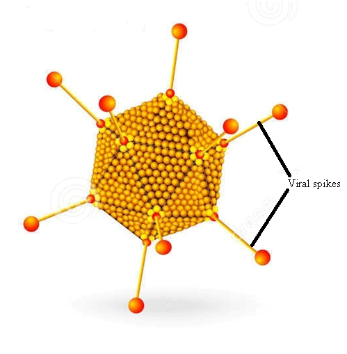
Icosahedral viruses exhibit a symmetrical, closed-shell structure composed of 20 triangular faces, 12 vertices, and 30 edges (Figure 1). This geometric arrangement provides maximal internal volume with minimal protein subunits, making it energetically efficient.
Icosahedral symmetry occurs in both DNA and RNA viruses. The resulting structure often appears spherical under the electron microscope, although it is geometrically polyhedral.
A classical example of an icosahedral virus is Adenoviridae, which causes respiratory infections, conjunctivitis, and gastroenteritis. These viruses lack envelopes and rely on capsid proteins for host cell attachment.
Other characteristics of icosahedral viruses include:
- Stable environmental resistance (especially non-enveloped forms)
- Efficient genome packaging
- Strong antigenic properties due to exposed capsid proteins
Icosahedral viruses may be either enveloped or non-enveloped. Non-enveloped icosahedral viruses tend to be more resistant to environmental stressors such as heat, drying, and detergents.
The icosahedral symmetry is exhibited by both DNA and RNA viruses. Some viruses have a spherical symmetry (Figure 2).

Helical Viruses
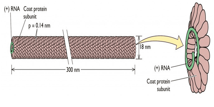
Helical viruses possess capsid proteins arranged in a spiral or helical configuration around the viral genome. In this arrangement, the nucleic acid binds to protein subunits in a repeating pattern, forming a rod-like or filamentous structure. The length of a helical virus is typically determined by the length of its genome, while its diameter remains relatively constant. A classic plant example is Tobacco mosaic virus, which forms rigid rod-shaped particles (Figure 3). Among animal viruses, members of the family Orthomyxoviridae exhibit helical nucleocapsids and include influenza viruses.
Key features of helical viruses include:
- Flexible or filamentous morphology
- Often enveloped in animal viruses
- Genome length directly influencing particle size
- Efficient assembly due to repetitive protein subunits
Most animal viruses with helical symmetry are enveloped. The envelope surrounds the nucleocapsid and provides additional functionality in host cell entry.
Complex Viruses
Complex viruses do not conform strictly to icosahedral or helical symmetry. Instead, they possess elaborate and unique structural architectures.
Two prominent examples include:
- Poxviridae
- Bacteriophage T4
Poxviruses
Members of the Poxviridae exhibit large, brick-shaped virions with:
- A complex outer membrane
- Lateral bodies
- A central core containing DNA
Unlike most DNA viruses, poxviruses replicate in the cytoplasm and carry their own transcription machinery.
Bacteriophages
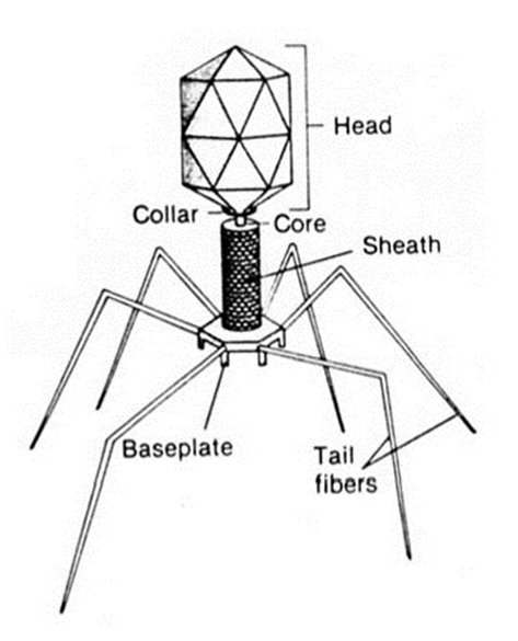
Bacteriophages (viruses that infect bacteria) often display highly complex structures. Bacteriophage T4 is a well-characterized example.
Its morphology includes:
- An icosahedral head containing DNA
- A contractile tail sheath
- Tail fibers for attachment
- A base plate for anchoring
During infection, the tail contracts, injecting viral DNA into the bacterial host cell. This injection mechanism is structurally sophisticated and resembles a molecular syringe.
Enveloped Viruses
Some viruses possess an outer lipid membrane known as an envelope. The envelope is derived from host cell membranes during viral budding.
The envelope typically contains:
- Lipid bilayer
- Viral glycoproteins (spikes)
- Matrix proteins beneath the membrane
These glycoproteins are essential for:
- Host cell recognition
- Membrane fusion
- Viral entry
- Immune system interaction
Enveloped viruses are generally more sensitive to:
- Heat
- Drying
- Detergents
- Organic solvents
This sensitivity arises because disruption of the lipid membrane compromises viral infectivity.
In contrast, non-enveloped viruses rely solely on their capsid for stability and tend to be more environmentally resilient.
Size and Structural Diversity of Viruses
Viruses exhibit significant variation in size. Most range between 20 and 300 nanometers, although some giant viruses exceed 400 nanometers.
Size influences:
- Mode of entry
- Immune detection
- Transmission dynamics
- Environmental persistence
Structural diversity also affects tissue tropism and pathogenicity. For instance, surface proteins determine receptor specificity, which in turn dictates host range.
Functional Significance of Viral Morphology
Viral morphology is not merely descriptive; it has direct clinical and biological implications.
1. Host Cell Attachment
Capsid proteins or envelope glycoproteins bind to specific cellular receptors. Structural compatibility determines host range and tissue specificity.
2. Antigenicity
Capsid and envelope proteins serve as antigenic determinants. Vaccines often target these structural proteins to elicit protective immune responses.
3. Environmental Stability
Non-enveloped viruses are generally more resistant to harsh environmental conditions than enveloped viruses.
4. Replication Strategy
Morphology often correlates with replication mechanisms. For example:
- Complex viruses such as the T4 bacteriophage (Figure 4) may carry their own enzymes.
- Segmented RNA viruses (such as Orthomyxoviridae) enable genetic reassortment.
5. Diagnostic Identification
Electron microscopy, immunofluorescence, and molecular assays rely on morphological and structural features for viral identification.
Advanced Structural Organization
Beyond basic symmetry, viruses may exhibit:
- Segmented genomes
- Matrix proteins
- Tegument layers (in herpesviruses)
- Surface projections
- Contractile appendages
Each structural adaptation enhances survival, transmission, and replication efficiency.
Icosahedral symmetry is a prominent form of cubic symmetry adopted by many viruses, reflecting an efficient strategy for packaging viral genetic material within a protein shell. This structural arrangement is characterized by a polyhedral geometry with 20 triangular faces, 12 vertices, and 30 edges, providing maximal internal volume while using a minimal number of protein subunits. Such an organization allows viruses to maintain structural stability and protects their nucleic acid contents, whether DNA or RNA, from environmental stressors and host immune defenses. Both DNA-containing and RNA-containing viruses exhibit icosahedral symmetry, underscoring the versatility and evolutionary advantage of this architectural design across diverse viral families.
Representative examples of viruses exhibiting icosahedral symmetry span multiple viral families, including Caliciviridae, Astroviridae, Picornaviridae, Birnaviridae, Reoviridae, Parvoviridae, Polyomaviridae, Papillomaviridae, Adenoviridae, Hepadnaviridae, and Bornaviridae. Within these families, the icosahedral capsid not only ensures structural integrity but also plays a critical role in host-virus interactions, influencing infectivity, antigenicity, and immune recognition. The uniformity of the icosahedral structure enables the repetitive presentation of surface proteins, which can enhance the binding efficiency of viral ligands to specific host cell receptors.
A key feature of many icosahedral viruses is the presence of viral spikes, which are specialized polypeptide projections extending from the capsid surface. These spikes are essential for the initial stages of infection, as they mediate attachment, adsorption, and often entry into host cells by recognizing specific cellular receptors. The molecular composition of these spikes can vary among viral families, but they consistently function as critical determinants of host specificity and tissue tropism. In addition to viral spikes, other polypeptide components contribute to the virion structure and function. Membrane proteins, for instance, play roles in viral assembly and budding, particularly in enveloped viruses. Haemagglutinins, commonly found in some RNA viruses, facilitate binding to host cell surface carbohydrates and are central to viral entry mechanisms. The nucleocapsid, composed of structural proteins complexed with viral nucleic acids, protects the genome and assists in the precise assembly of new virions.
The combination of icosahedral symmetry and associated polypeptide structures illustrates the sophisticated design of viral particles. The structural regularity of the icosahedron minimizes genetic coding requirements while maximizing stability, and the external polypeptides, including spikes, haemagglutinins, and membrane proteins, provide functional versatility necessary for successful infection. This architectural strategy exemplifies how viruses have evolved to balance efficiency, protection, and infectivity, enabling them to thrive across a wide range of host organisms. Understanding these structural and functional aspects is fundamental to virology, informing the development of antiviral strategies, vaccines, and diagnostic tools.
References
Acheson N.H (2011). Fundamentals of Molecular Virology. Second edition. John Wiley and Sons Limited, West Sussex, United Kingdom.
Alan J. Cann (2005). Principles of Molecular Virology. 4th edition. Elsevier Academic Press, Burlington, MA, USA.
Alberts B, Bray D, Johnson A, Lewis J, Raff M, Roberts K and Walter P (1998). Essential Cell Biology: An Introduction to the Molecular Biology of the Cell. Third edition. Garland Publishing Inc., New York.
Barrett J.T (1998). Microbiology and Immunology Concepts. Philadelphia, PA: Lippincott-Raven Publishers. USA.
Black, J.G. (2008). Microbiology: Principles and Explorations (7th ed.). Hoboken, NJ: J. Wiley & Sons.
Brian W.J Mahy and Mark H.C van Regenmortel (2010). Desk Encyclopedia of Human and Medical Virology. Elsevier Academic Press, San Diego, USA.
Brooks G.F., Butel J.S and Morse S.A (2004). Medical Microbiology, 23rd edition. McGraw Hill Publishers. USA.
Cann A.J (2011). Principles of Molecular Virology. Fifth edition. Academic Press, San Diego, United States.
Carter J and Saunders V (2013). Virology: Principles and Applications. Second edition. Wiley-Blackwell, New Jersey, United States.
Champoux J.J, Neidhardt F.C, Drew W.L and Plorde J.J (2004). Sherris Medical Microbiology: An Introduction to Infectious Diseases. 4th edition. McGraw Hill Companies Inc, USA.
Dimmock N (2015). Introduction to Modern Virology. Seventh edition. Wiley-Blackwell, New Jersey, United States.
Dimmock N.J, Easton A.J and Leppard K.N (2001). Introduction to modern virology. 5th edition. Blackwell Science publishers. Oxford, UK.
Discover more from Microbiology Class
Subscribe to get the latest posts sent to your email.