DIRECT PLATE COUNTING
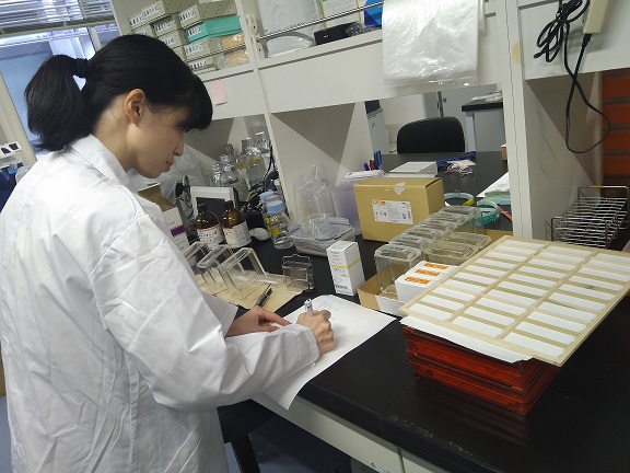
Direct plate counting is a microbiological technique used to evaluate the actual bacterial content of a product or specimen; and it gives an estimate of viable or living cells present in a sample. It is usually carried out by plating an aliquot of the sample on solid agar medium that supports the growth of the bacteria being sought for. The number of viable cells in the sample of interest is assessed from the number of colonies which develop on the solid agar medium after incubation.
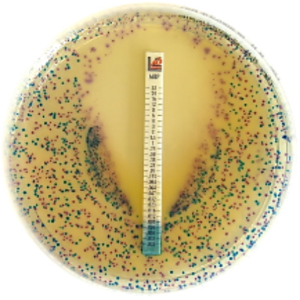
The colony counts of bacteria may range from < 1000 to > 106 colonies per ml (m-1) of the diluted sample or product. And the counted organisms or colonies on the solid agar plate is usually expressed as colony forming units per ml (cfu/ml); and cfu is used to express a single cell of bacteria that is capable of producing a single colony because it is generally assumed that each colony on the agar plate developed from a single bacterial cell.
CFU is the microbiological expression of the number of bacteria or fungi in a microbial population (which is usually seen as distinct colonies on solid agar plates); and it is a measure of the viable cells (bacteria and fungi inclusive). While direct microscopy of cells under the microscope may give an estimate of both viable cells and dead cells; CFU (which is obtained via direct plating on solid culture media) measures only viable or living bacterial cells, and the theory behind it is based on the fact that a single bacterium can grow on agar medium and become a colony especially through binary fission. Direct plate count can also be used for counting fungi and other microbial cells excluding viruses. Electron microscopy is used for viral counts, and the counted viral particles are expressed as plaque forming units (pfu).
Spread plating and pour plating are the two methods of direct plate counting techniques for microorganisms (with the exception of viruses). But the pour plate method is more preferable to the spread plate technique when counting microbes because the later (in which the diluted sample is spread over an agar plate) usually produces colonies that only form on the surface of the agar medium. In the pour plate technique, colonies do not just form on the surface of the agar but throughout the agar medium (i.e. in the substratum of the agar medium).
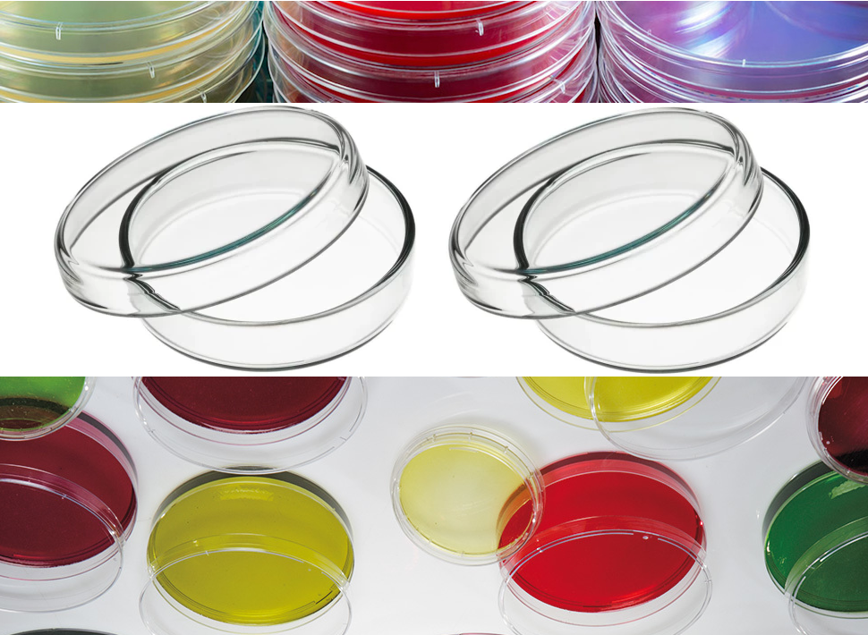
The pour plate technique involves the dispensing of an aliquot of the test sample (usually in a diluted form in the range of 0.1 – 1.0 ml) on a clean Petri dish; and then the sterile molten agar is poured into the plate and the Petri dish is swirled in order to ensure an even mixture of the medium and the inoculum and/or diluted sample. The plate is allowed to set or dry prior to incubation at the appropriate temperature conditions. Other techniques available for the counting of microbes in the microbiology laboratory include microscopy and turbidometric method.
IDENTIFICATION TECHNIQUE
Identification technique is used to discover and categorize isolated microbial cultures in the microbiology laboratory. This technique is used to identify microorganisms to their precise species or strain level. Identification of microorganisms in the microbiology laboratory is often carried out using specific biochemical tests which is unique for a particular organism.
Some biochemical tests or identification techniques employed in the microbiology laboratory for the identification of microbial pathogens include indole test, citrate test, urease test, motility test, Gram staining, sugar fermentation test, Voges Proskauer (VP) test, coagulase test, catalase test, optochin test, nitrate reduction test, oxidase test, starch hydrolysis test, determination of hydrogen sulphide production and methyl red test amongst others.
Identification tests help microbiologists to identify a particular microorganism from a mixture of organisms; and they also assist microbiologists to put a name to a given pathogen.
MOLECULAR TECHNIQUES
Molecular techniques used in the microbiology laboratory include polymerase chain reaction (PCR) based tests which are employed for the identification of microorganisms from clinical specimens and other samples based on the identification of nucleotide sequences peculiar to each microbe. Molecular detection techniques usually employ DNA probes, gene chips and other gene amplification and sequencing techniques that allow microbiologists to detect pathogen-specific nucleotide sequences directly from clinically relevant specimen and other samples.
They are important tool for the amplification of genes responsible for antimicrobial resistance in pathogenic microorganisms. Molecular techniques can also be used for the prompt identification of microorganisms especially bacteria from culture plates. Other molecular methods used for microbial characterization include nucleic acid sequencing and restriction fragment length polymorphism (RFLP) amongst others.
REFERENCES
Atlas R.M (2010). Handbook of Microbiological Media. Fourth edition. American Society of Microbiology Press, USA.
Balows A, Hausler W, Herrmann K.L, Isenberg H.D and Shadomy H.J (1991). Manual of clinical microbiology. 5th ed. American Society of Microbiology Press, USA.
Basic laboratory procedures in clinical bacteriology. World Health Organization (WHO), 1991. Available from WHO publications, 1211 Geneva, 27-Switzerland.
Black, J.G. (2008). Microbiology: Principles and Explorations (7th ed.). Hoboken, NJ: J. Wiley & Sons.
Garcia L.S (2010). Clinical Microbiology Procedures Handbook. Third edition. American Society of Microbiology Press, USA.
Garcia L.S (2014). Clinical Laboratory Management. First edition. American Society of Microbiology Press, USA.
Ira R (1995). Bacteriology, Standard Operative procedure manual for microbiology laboratories, National Institute of Biologicals. Pp. 73-97.
Madigan M.T., Martinko J.M., Dunlap P.V and Clark D.P (2009). Brock Biology of Microorganisms, 12th edition. Pearson Benjamin Cummings Inc, USA.
Woods GL and Washington JA (1995). The Clinician and the Microbiology Laboratory. Mandell GL, Bennett JE, Dolin R (eds): Principles and Practice of Infectious Diseases. 4th ed. Churchill Livingstone, New York.
Discover more from Microbiology Class
Subscribe to get the latest posts sent to your email.


