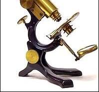
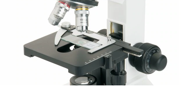
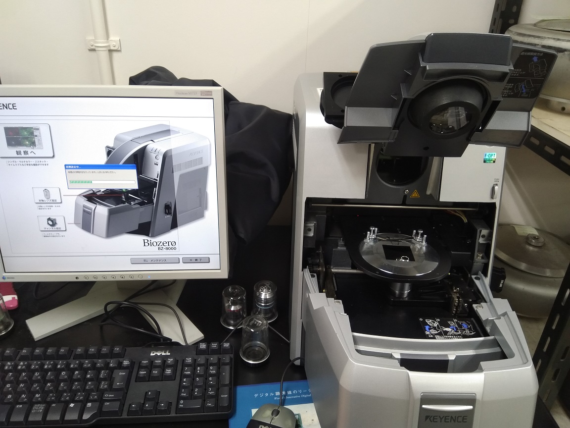
Before the advent of microscopes and its actual application in viewing and making clear the microbial world which our eyes are not competent enough to see, mankind in time past have been using shiny stones and glasses to view objects; and they discovered that the objects become enlarged when viewed under such objects they termed “magnifying objects or magnifiers”. Top among those that experimented on the use of such glasses in the first century (100 AD) to view objects were the Romans who discovered that viewing objects using the magnifiers made them to appear larger. They named these magnifying objects “lenses” because their shapes resemble that of the lentil bean seeds.
The term “lens” was not adopted and used until the end of the 13th century when the eye glasses (also known as spectacles) was developed by an Italian called Salvino D’Armate who invented the world’s first wearable eye glasses. During this period, other spectacle/glass makers continued experimenting on different glasses (or lenses) and they discovered magnifying glasses (with about 6x-10x magnification) which were only used as at the time to observe small/tiny insects such as fleas. These magnifying glasses were the earlier microscopes, and they were simply small tubes made of a plate at one end for the object and, a lens at the other end for magnification of the object being viewed.
The first compound microscope (i.e. a microscope that uses two or more lenses) was actually invented in 1590 by Hans Janssen and his son, Zacharias Janssen who were both Dutch spectacle/eye glass makers and, who co-experimented on the earlier lenses used previously for viewing objects (e.g. tiny insects). The Janssen’s microscopes were able to provide a much larger and enlarged image of objects than the previously discovered magnifying glasses (simple microscopes). Their experiments and breakthrough in this aspect gave impetus to Galileo (the father of modern physics and astronomy) who developed the telescope with a much better focusing device in 1609.
Galileo’s work helped describe the working principles of lenses and light rays and, this helped to improve the microscopes and even the telescope that he discovered. In 1660, Robert Hooke (1635-1703), an English Physicist observed some biological materials with the best compound microscopes as at the time, and he discovered small pores in them which he termed “cells”. Hooke’s findings were published in a journal known as the “Micrographia”- which showed detailed drawings of his discoveries. The first real microscope was actually discovered and invented in the late 17th century (precisely 1676) by Anthony Von Leeuwenhoek (1632-1723), a Dutch draper (clothing and dry goods dealer) from Holland who developed one of the finest simple microscopes (with a magnification of about 270x) as at the time.
Anthony Von Leeuwenhoek is regarded as the father of microscopy and the field of microbiology because of his many biological discoveries that was made possible with his new improved microscope which enabled him to see some living things that was never discovered as at the time. He was the first person to see and describe bacteria, blood cells, yeast cells and other tiny forms of life. Leeuwenhoek’s work together with that of Hooke’s helped to further study in to the field of microbiology and other biological sciences.
Carl Zeiss and Ernst Abbe pioneered the development of immersion lenses in 1883. Both helped to improve the basic understanding about the optical principles and quality of the microscope. The first transmission electron microscope was constructed by Ernst Ruska in 1933. Very little was made in the development of the microscope until the late 19th century that ushered in great innovations in the field of microscopy which lead to the development of very fine optical lenses and equipment, thus making the use of microscope a very important tool for scientists as at the time even till date.
Top among the scientists that improved the technical workings of the microscope was the American scientist; Charles A. Spencer (1813-1881) who was America’s first microscope maker and, who was famously noted for his development of achromatic objectives with great aperture angle. Joseph Jackson showed in 1830 that several weak lenses used together at certain distances gave good magnification without blurring the image of the object. In 1872, Ernst Abbe developed a mathematical formular called the “Abbe Sine Equation” which made possible the maximum resolutions of microscope lenses. The phase contrast microscope was invented in 1936 by Frits Zernike while the scanning microscope was invented by Gerd Binnig and Heinrich Rohrer in 1981.
REFERENCES
Beck R.W (2000). A chronology of microbiology in historical context. Washington, D.C.: ASM Press.
Cheesbrough, M (2006). District Laboratory Practice in Tropical countries Part I Cambridge
Chung K.T, Stevens Jr., S.E and Ferris D.H (1995). A chronology of events and pioneers of microbiology. SIM News, 45(1):3–13.
Dictionary of Microbiology and Molecular Biology, 3rd Edition. Paul Singleton and Diana Sainsbury. 2006, John Wiley & Sons Ltd. Canada.
Glick B.R and Pasternak J.J (2003). Molecular Biotechnology: Principles and Applications of Recombinant DNA. ASM Press, Washington DC, USA.
Goldman E and Green L.H (2008). Practical Handbook of Microbiology, Second Edition. CRC Press, Taylor and Francis Group, USA.
Madigan M.T., Martinko J.M., Dunlap P.V and Clark D.P (2009). Brock Biology of microorganisms. 12th edition. Pearson Benjamin Cummings Publishers. USA.
Nester E.W, Anderson D.G, Roberts C.E and Nester M.T (2009). Microbiology: A Human Perspective. Sixth edition. McGraw-Hill Companies, Inc, New York, USA.
Prescott L.M., Harley J.P and Klein D.A (2005). Microbiology. 6th ed. McGraw Hill Publishers, USA.
Willey J.M, Sherwood L.M and Woolverton C.J (2008). Harley and Klein’s Microbiology. 7th ed. McGraw-Hill Higher Education, USA.
Discover more from Microbiology Class
Subscribe to get the latest posts sent to your email.


