The history of cell culture cannot be complete without the mention of Henrietta Lacks, whose immortal cell lines (HeLa cells) from cervical carcinoma helped revolutionize the field of cell culture. HeLa cells are immortal cell lines that was derived from a deadly cervical tumor from a cancer patient (named Henrietta Lacks) in 1951 after she died from her cancer disease. They were the first human cell lines to grow well in the laboratory (i.e. in vitro) and, they were found to be very productive. HeLa cells contributed significantly in some major scientific/biomedical research development and discoveries.
HeLa cells actually descended from a biopsy that was taken from a visible lesion on the cancerous cervix of Madam Henrietta Lacks in 1951. HeLa cells are called immortal cell culture lines because they are infinite cell lines. This implies that HeLa cells can divide an unlimited number of times in vitro as long as the environmental growth conditions and nutrients are maintained and adequately sustained. Many strains of the HeLa cells have been developed and propagated. They have been used in plethora of cancer research and even in the testing of putative anticancer agents.
Henrietta Lacks (though an unsung hero) immortal cells has made unparallel innovations and contribution to the biomedical and medical sciences since 1951 after her death from a cervical cancer. Doctors at John Hopkins hospital, Baltimore, USA discovered that Madam Lack’s cervical cancer cells continued to divide indefinitely even while the regular cells of a human being divide only 50 times before they eventually die.
This very noteworthy discovery by doctors at the John Hopkins hospital, Baltimore, USA in the 1950s paved way for the development of various vaccines (e.g. polio vaccines) which have over the years helped to close down on some stubborn diseases through immunization. HeLa cells have also been very critical in major research techniques in the cell culture laboratory.
Cell culture as a basic molecular science has been in existence for over 10 decades now. Its development even till date has been spurred by the innovative works of scientists in time past. Sydney Ringer in the early 19th century developed a salt solution called Ringers solution which contains sodium, potassium, calcium and magnesium chlorides. This salt solution was used to maintain the heart of an animal outside of its body. Wilhelm Roux, a German zoologist showed in 1885 that the neural plate from chicken embryos could be removed and maintained in warm saline solution for several days outside the chicken’s embryo.
Cell culture was successfully undertaken by the work of the American born scientist, Ross Granville Harrison who in 1907 showed that the nerve fibers of frog/amphibians can be removed and maintained outside the cell in conditions that can favour its proliferation. In 1913, the French medical scientist, Alexis Carrel successfully showed that the heart of a chicken embryo could be removed and kept for a period much longer than the normal lifespan of a chicken. These achievements set the foundation for the development of cell culture which advanced greatly in the 1940s and 1950s.
The fields of virology was also driven further in the area of vaccine development as a result of cell culture techniques which allowed the culturing of viruses for the production of vaccines. Earle and his colleagues in 1943 developed mouse lymphocyte cell lines. In 1949, John Franklin Enders and colleagues successfully showed that viruses can be grown in cell culture. They used monkey kidney for this purpose and was awarded the Nobel Prize for the discovery of a method for growing the virus in monkey kidney cell cultures, a technique which was used for the mass production of the Salk polio vaccine. In 1952 the HeLa cell lines was developed by George Otto Gey and colleagues. Harry Eagle in 1955 developed a defined cell culture media for cell culture techniques.
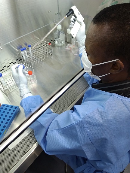
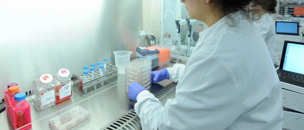
Their works developed a well-defined nutrient mixture containing amino acids, vitamins, carbohydrates, salts, and serum which is used even till date. This replaced the tissue extracts previously used to grow cells. This medium helped to maintain pure cell lines indefinitely. It gave impetus to the studying of the cellular processes specific to particular organs or tissues of organisms with greater simplicity. Kleinsmith and Pierce discovered the pluripotential stem cells in 1964, and in 1970 the laminar flow cabinets for cell culture was developed. The laminar flow cabinets are very important in cell culture laboratory because they provide a sterile environment for cell culture techniques to take place.
Innovations and discoveries continued in the field of cell culture until the early 1970s when the hybridoma cell lines was developed. The development of hybridoma cell lines was made possible by the works of Cesar Milstein, Georges Köhler and Niels Kaj Jerne. This earned them the Nobel Prize in Medicine and Physiology in 1984. Hybridoma cell lines are immortalized cell lines formed by the fusion of a normal cell with an immortal (tumor) cell. They are used for the production of monoclonal antibodies.
Monoclonal antibodies are antibodies produced from a single clone of B cells. Another ground breaking discovery and development in cell culture occurred in 1998 when James Thomson and John Gearhart independently showed the first culturing of human embryonic stem cells in culture. This brief history on cell culture has incompletely enumerated some of the important discoveries in this field. Nevertheless, there are plethora of advances and discoveries in the field of cell culture especially in the area of stem cell research (which is gradually growing) which are spurring the medical and biomedical sciences into a grater height.
References
Alberts B, Bray D, Johnson A, Lewis J, Raff M, Roberts K andWalter P (1998). Essential Cell Biology: An Introduction to the Molecular Biology of the Cell. Third edition. Garland Publishing Inc., New York.
Alberts B, Bray D, Lewis J, Raff M, Roberts K and Watson J.D (2002). The molecular Biology of the Cell. Fourth edition. New York, Garland, USA.
Ausubel, F.M., Brent, R., Kingston, R.E., Moore, D.D., Seidman, J.G., Smith, J.A., Struhl, K., eds (2002). Short Protocols in Molecular Biology, 5th edn. John Wiley & Sons, New York.
Caputo J.L (1996). Safety Procedures. In: Freshney, R.I., Freshney, M.G., eds., Culture of Immortalized Cells. New York, Wiley-Liss, Pp. 25-51.
Cooper G.M and Hausman R.E (2004). The cell: A Molecular Approach. Third edition. ASM Press.
Davis J.M (2002). Basic Cell Culture, A Practical Approach. Oxford University Press, Oxford, UK.
Freshney R.I (2005). Culture of Animal Cells, a Manual of Basic Technique, 5th Ed. Hoboken NJ, John Wiley and Sons Publishers.
Health Services Advisory Committee (HSAC) (2003). Safe Working and the Prevention of Infection in Clinical Laboratories. HSE Books: Sudbury
Lodish H, Berk A, Matsudaira P, Kaiser C.A, Kreiger M, Scott M.P, Zipursky S.L and Darnell J (2004). Molecular Cell Biology. Fifth edition. Scientific American Books, Freeman, New York, USA.
Marcovic O and Marcovic N (1998). Cell cross-contamination in cell cultures: the silent and neglected danger. In Vitro Cell Dev Biol. 34:108.
Mather J and Barnes D (1998). Animal cell culture methods, Methods in cell biology. 2rd eds, Academic press, San Diego.
Verma P.S and Agarwal V.K (2011). Cytology: Cell Biology and Molecular Biology. Fourth edition. S. Chand and Company Ltd, Ram Nagar, New Delhi, India.
Discover more from Microbiology Class
Subscribe to get the latest posts sent to your email.


